. 2018 Nov 14;38(46):9829-9839.
doi: 10.1523/JNEUROSCI.0154-18.2018. Epub 2018 Sep 24.

Interactive Repression of MYRF Self-Cleavage and Activity in Oligodendrocyte Differentiation by TMEM98 Protein

Affiliations

Interactive Repression of MYRF Self-Cleavage and Activity in Oligodendrocyte Differentiation by TMEM98 Protein

Hao Huang et al. J Neurosci. .

Abstract

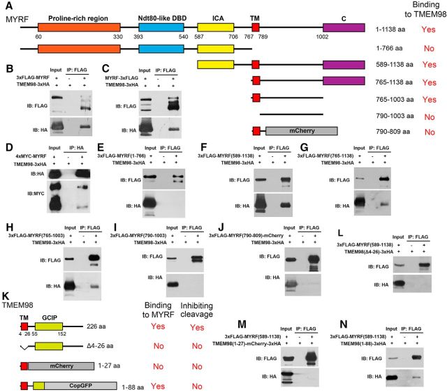
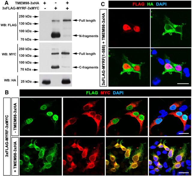
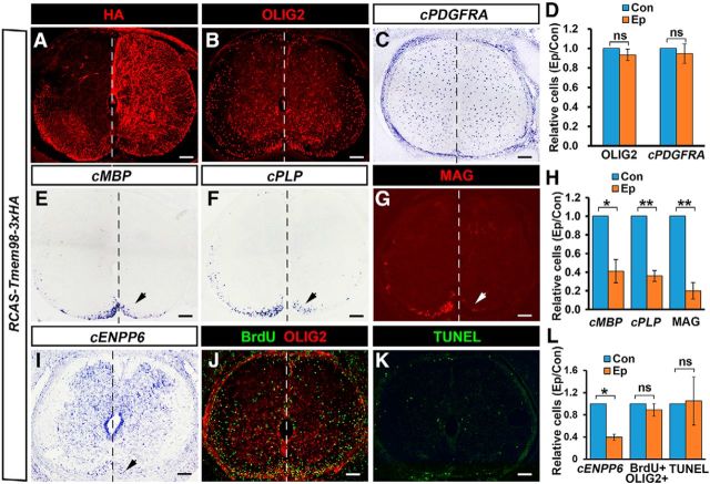
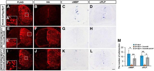
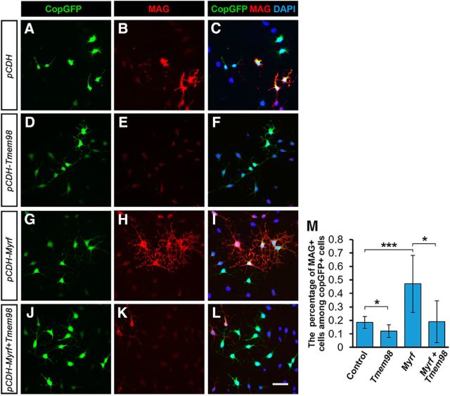
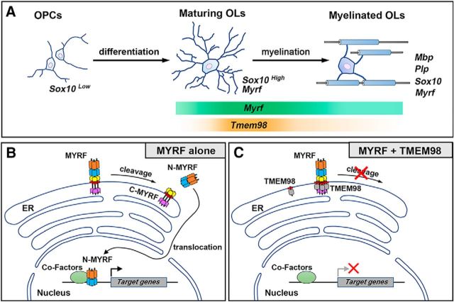
Myelin sheath formed by oligodendrocytes (OLs) is essential for the rapid propagation of action potentials in the vertebrate CNS. Myelin regulatory factor (MYRF) is one of the critical factors that control OL differentiation and myelin maintenance. Previous studies showed that MYRF is a membrane-bound transcription factor associated with the endoplasmic reticulum (ER). After self-cleavage, the N-fragment of MYRF is released from the ER and translocated into the nucleus where it functions as a transcription factor to activate myelin gene expression. At present, it remains unknown whether MYRF self-cleavage and functional activation can be regulated during OL differentiation. Here, we report that TMEM98, an ER-associated transmembrane protein, is capable of binding to the C-terminal of MYRF and inhibiting its self-cleavage and N-fragment nuclear translocation. In the developing CNS, TMEM98 is selectively expressed in early maturing OLs in mouse pups of either sex. Forced expression of TMEM98 in embryonic chicken spinal cord of either sex suppresses endogenous OL differentiation and MYRF-induced ectopic expression of myelin genes. These results suggest that TMEM98, through inhibiting the self-cleavage of MYRF, functions as a negative feedback regulator of MYRF in oligodendrocyte differentiation and myelination.SIGNIFICANCE STATEMENT MYRF protein is initially synthesized as an ER-associated membrane protein that undergoes autoproteolytic cleavage to release the N-fragment, which is then transported into the nucleus and activates the transcription of myelin genes. To date, the molecular mechanisms that regulate the self-cleavage and function of MYRF in regulating oligodendrocyte differentiation have remained unknown. In this study, we present the molecular and functional evidence that TMEM98 membrane protein physically interacts with MYRF in the ER and subsequently blocks its self-cleavage, N-terminal nuclear translocation, and functional activation of myelin gene expression. To our knowledge, this is the first report on the regulation of MYRF self-proteolytic activity and function by an interacting protein, providing new insights into the molecular regulation of OL differentiation and myelinogenesis.

Keywords: MYRF; TMEM98; oligodendrocyte differentiation; self-cleavage.


Figures

Figure 1.
Figure 1.
Developmental expression pattern of Tmem98 in the CNS. A–D, In situ hybridization for Tmem98 was performed on the sections of spinal cord from E18.5 (A), P4 (B), P7 (C), and P15 (D) wild-type mice. E, F, Sections from P15 corpus callosum (E) and cerebellum (F) of wild-type mice were subjected to ISH with Tmem98 riboprobes. G, H, Transcriptional level of Tmem98 and Myrf mRNA at different developmental stages was quantified by RT-qPCR. Representative Tmem98-positive cells are indicated by arrows. *p < 0.05, **p < 0.01, ***p < 0.001. Scale bars, 100 μm.
Figure 2.
Figure 2.
Selective expression of Tmem98 in differentiated oligodendrocytes. A–I, Spinal cord sections from P7 wild-type mice were subject to Tmem98 ISH, followed by immunohistochemical staining with anti-SOX10 (A, B), anti-CC1 (C, D), anti-MYRF (E, F), or anti-PDGFRA (G–I). B, D, and F are higher magnifications of the outlined areas in A, C, and E, respectively. J–M, Tmem98 expression in Nkx2.2-CKO (J, K) and Myrf-CKO (L, M) mutant spinal cords is dramatically reduced at P3 stages. Scale bars, 100 μm.
Figure 3.
Figure 3.
Colocalization of TMEM98 and MYRF C-fragments in ER. A–H, Both C-fragments of MYRF (3xMYC-tagged; A–D) and TMEM98 (E–H) were colabeled with V5-tagged Calnexin, an ER marker. I–L, C-fragments of MYRF were colabeled with TMEM98 in HEK293T cells. Scale bars, 10 μm.
Figure 4.
Figure 4.
Physical interactions between TMEM98 and MYRF proteins. A, Schematic of various truncated MYRF proteins. B–D, TMEM98 proteins physically bind to full-length MYRF proteins. E, TMEM98 does not bind to the cytoplasmic fragments (1–766) of MYRF. F–H, TMEM98 protein binds to the C-fragments of MYRF (589–1138, 765–1138, and 765–1003). I, Deletion of the TM domain in MYRF abolishes the interaction. J, Deletion of the region C terminal to the TM domain in MYRF abolishes the efficient binding between MYRF and TMEM98. K–N, The N-terminal region of TMEM98, including the TM domain, is involved in the MYRF–TMEM98 interaction, whereas the C terminal of TMEM98 is required for inhibiting MYRF cleavage. DBD, DNA-binding domain; C, unknown conserved domain; IB, immunoblot.
Figure 5.
Figure 5.
Inhibition of MYRF self-cleavage by TMEM98. A, The self-cleavage of MYRF was suppressed when TMEM98 was coexpressed. B, The nuclear translocation of MYRF N-fragments released from full-length proteins was blocked when TMEM98 was coexpressed. C, TMEM98 does not interfere with the nuclear translocation of N-fragments of MYRF (1–588). Scale bars, 20 μm. WB, Western blot.
Figure 6.
Figure 6.
Suppression of OL differentiation by TMEM98 overexpression. A, Detection of TMEM98 expression at the electroporated side (right side) of chicken spinal cords at cE10 stages by anti-HA staining. B–D, The expression of OLIG2 and OPC marker PDGFRA were not impacted at the electroporated side. E–I, As indicated by arrows, the expression of MBP, PLP, MAG, and ENPP6 at the electroporated side was dramatically reduced. J–L, Cell proliferation and apoptosis were not affected by TMEM98 overexpression. *p < 0.05; **p < 0.01; ns, no significant difference; n = 3. Scale bars, 100 μm. Con, control side (left side). Ep, electroporated side.
Figure 7.
Figure 7.
Inhibition of MYRF-induced myelin gene expression by TMEM98 in chicken spinal cords. A–D, Overexpression of MYRF induced ectopic expression of MBP and PLP in chicken spinal cords at cE7 stages. E–H, MYRF-induced MBP/PLP expression was almost completely blocked by TMEM98 coexpressoin. I–L, TMEM98-ΔGCIP was not capable of inhibiting MYRF self-cleavage and repressing MYRF functions. M, Quantitative analyses of MBP+ and PLP + cells. A′, I′, N-fused Flag (N-fragments of MYRF) was located in the nucleus when MYRF was expressed alone or with TMEM98-ΔGCIP. E′, N-fused Flag was detected in cell processes, but not in the nucleus when TMEM98 was coexpressed in chicken spinal cords. *p < 0.05, **p < 0.01. Scale bar, 100 μm. EP, electroporating.
Figure 8.
Figure 8.
Inhibition of MYRF-induced myelin gene expression by TMEM98 in CG4 cells. A–L, TMEM98 overexpression reduces spontaneous OL differentiation (A–F) and inhibits MYRF-induced MAG expression in CG4 cells (G–L). M, Quantitative analyses of MAG+ cells. *p < 0.05, ***p < 0.001. Scale bar, 25 μm.
Figure 9.
Figure 9.
Model for TMEM98 function in inhibiting MYRF self-cleavage and activity in the oligodendrocyte differentiation. A, Tmem98 is transiently expressed in newly differentiated oligodendrocytes. B, When expressed alone, N-fragments of MYRF are released from the ER membrane after self-cleavage and translocate into the nucleus to drive target-gene expression. C, When coexpressed with TMEM98, the release and nuclear translocation of MYRF N-fragments are suppressed, and the induction of target-gene expression by MYRF is turned off.

Similar articles

Cited by

References

    1. Andachi Y. (2008) A novel biochemical method to identify target genes of individual microRNAs: identification of a new Caenorhabditis elegans let-7 target. RNA 14:2440–2451. 10.1261/rna.1139508 - DOI - PMC - PubMed
    1. Aubourg P. (1993) The leukodystrophies: a window to myelin. Nat Genet 5:105–106. 10.1038/ng1093-105 - DOI - PubMed
    1. Awadalla MS, Burdon KP, Souzeau E, Landers J, Hewitt AW, Sharma S, Craig JE (2014) Mutation in TMEM98 in a large white kindred with autosomal dominant nanophthalmos linked to 17p12–q12. JAMA Ophthalmol 132:970–977. 10.1001/jamaophthalmol.2014.946 - DOI - PubMed
    1. Barañano KW. (2016) Leukodystrophies. Semin Neurol 36:362–366. 10.1055/s-0036-1585455 - DOI - PubMed
    1. Baumann N, Pham-Dinh D (2001) Biology of oligodendrocyte and myelin in the mammalian central nervous system. Physiol Rev 81:871–927. 10.1152/physrev.2001.81.2.871 - DOI - PubMed

Publication types

Substances