. 2018 Sep 24;9(10):982.
doi: 10.1038/s41419-018-0962-6.

SP1-induced lncRNA-ZFAS1 contributes to colorectal cancer progression via the miR-150-5p/VEGFA axis

Affiliations

SP1-induced lncRNA-ZFAS1 contributes to colorectal cancer progression via the miR-150-5p/VEGFA axis

Xiaoxiang Chen et al. Cell Death Dis. .

Abstract

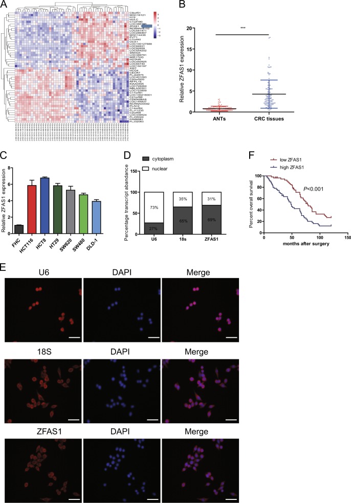
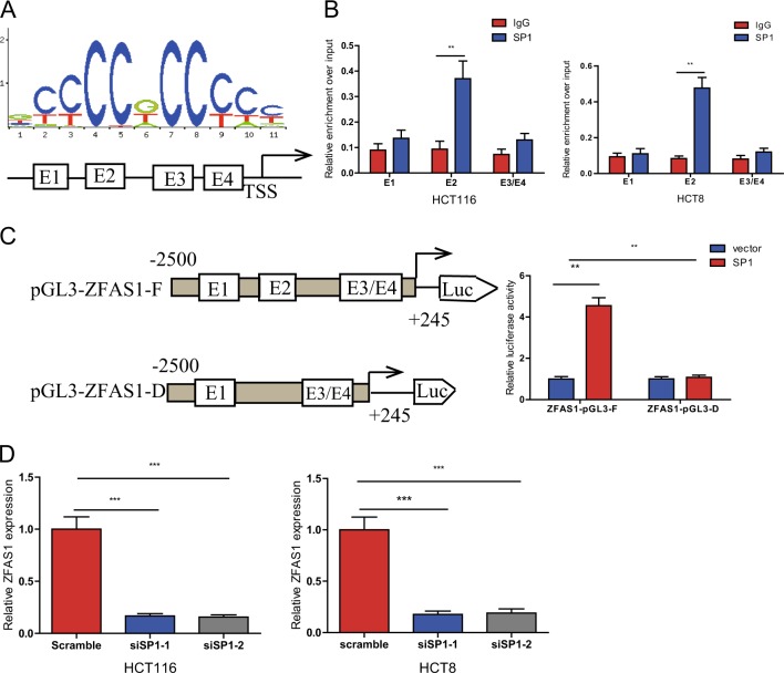
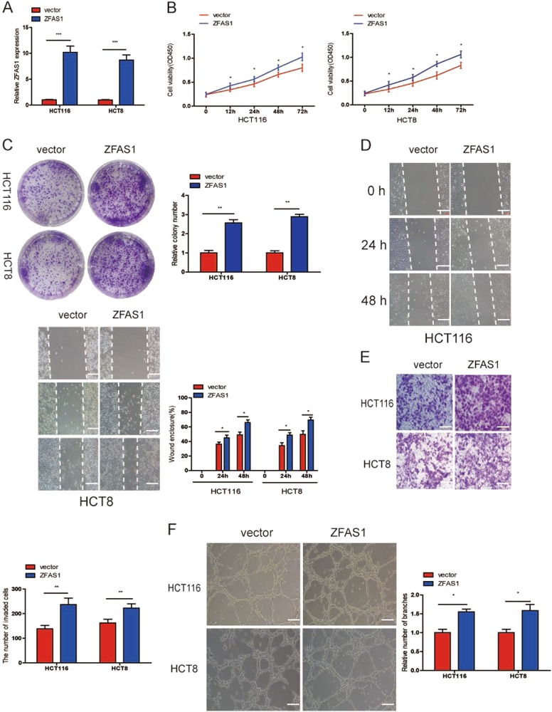
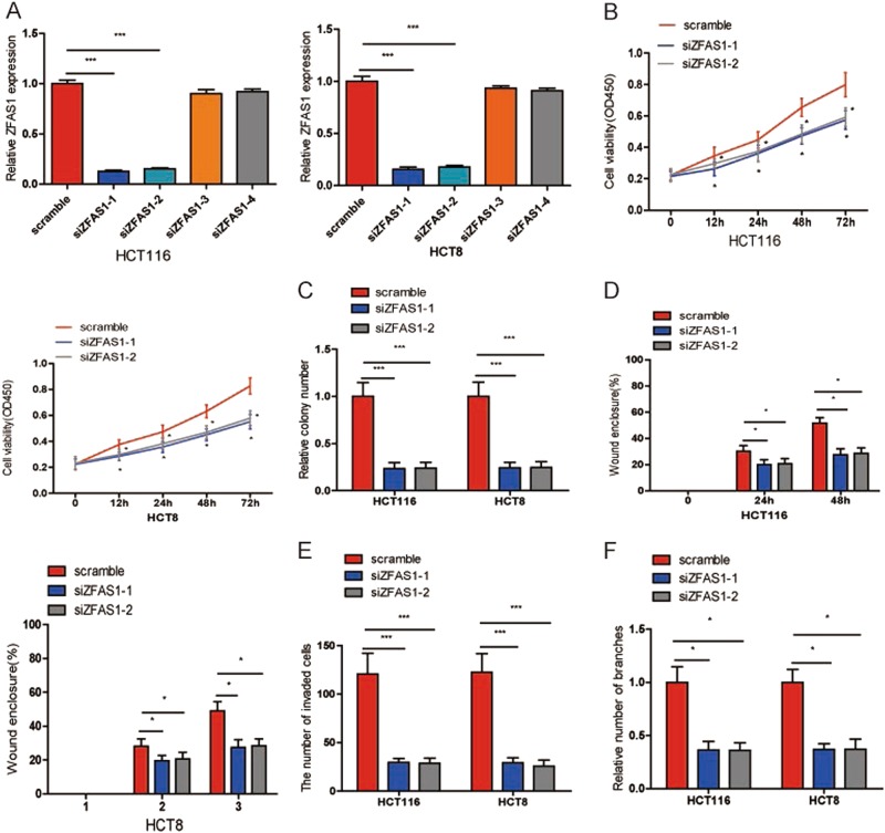
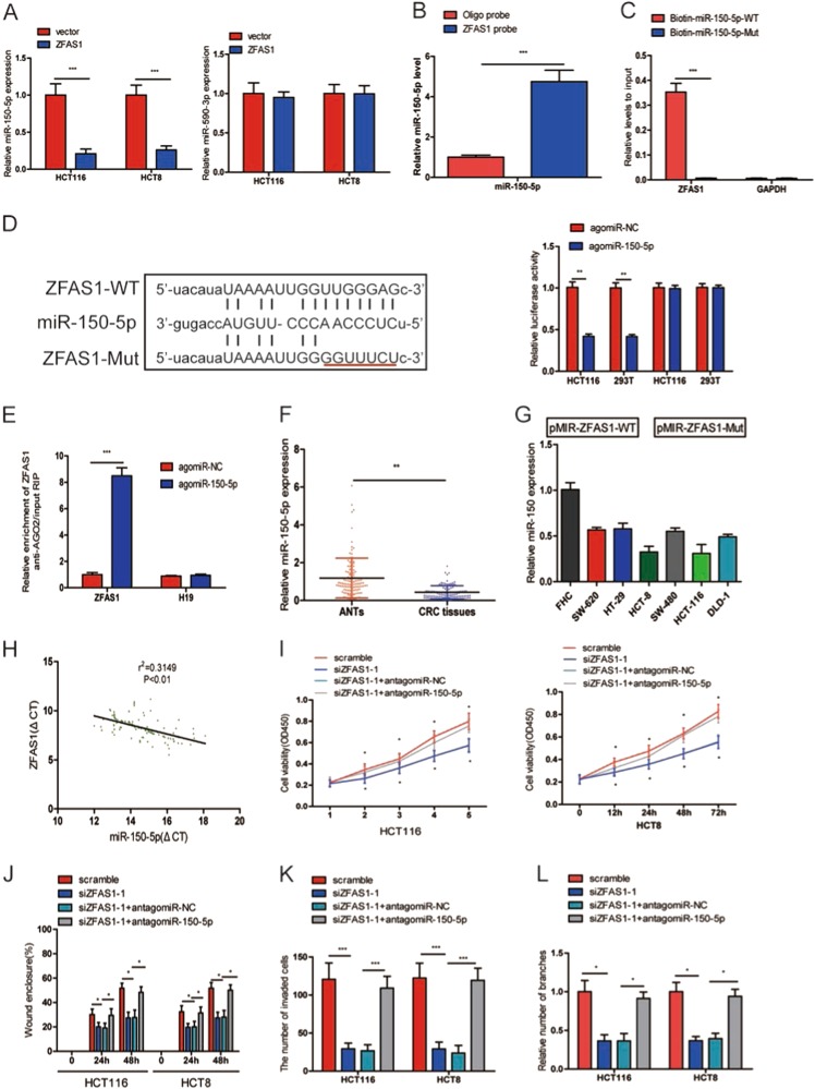
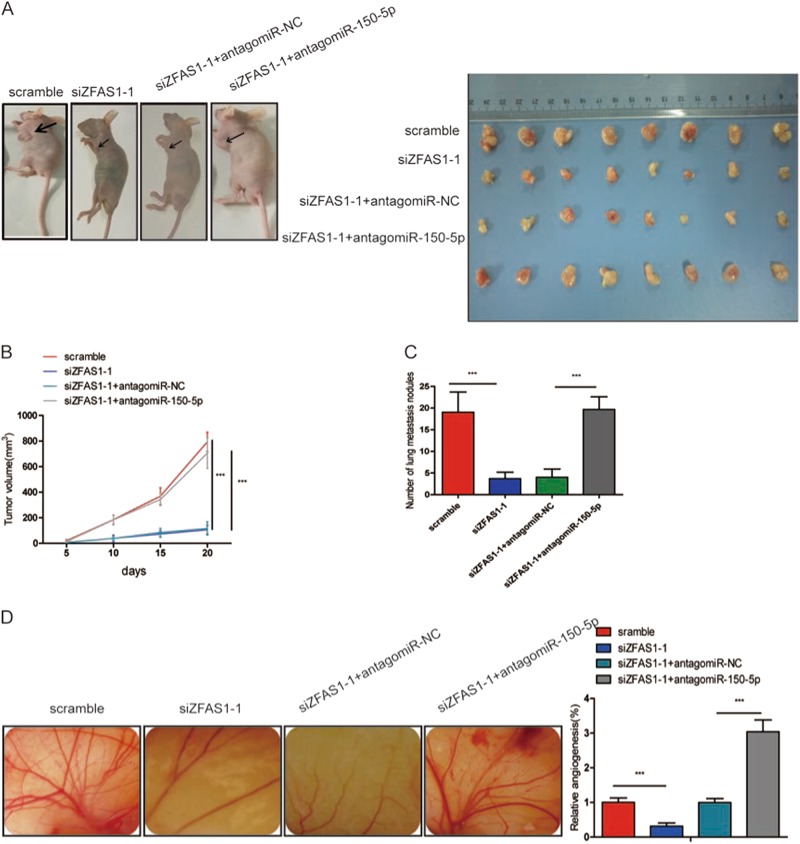
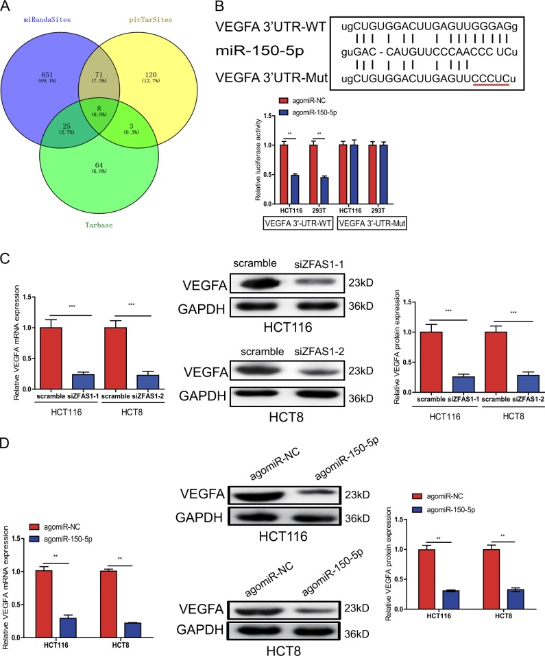
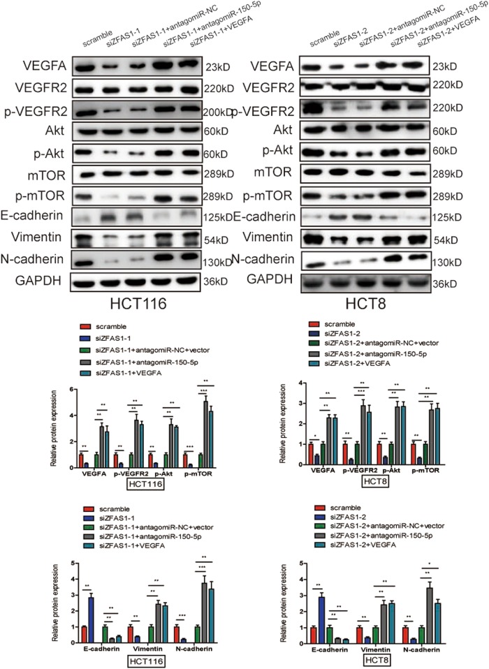
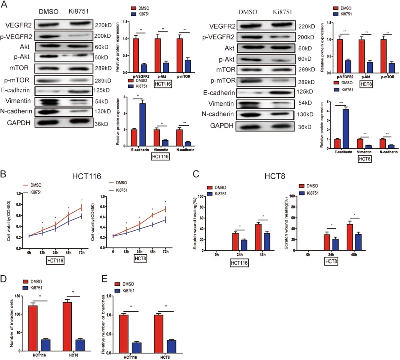
Increasing long non-coding RNAs (lncRNAs) have been reported to play key roles in the development and progression of various malignancies. ZNFX1 antisense RNA1 (ZFAS1) has been reported to be aberrant expression and suggested as a tumor suppressor or oncogene in many cancers. However, the biological role and underlying molecular mechanism of ZFAS1, especially the miRNA sponge role of which in CRC remain largely unknown. We found that ZFAS1 expression was higher in CRC tissues, where it was associated with poor overall survival (OS), we also showed that ZFAS1 upregulation was induced by nuclear transcription factor SP1. Moreover, ZFAS1 and VEGFA are both targets of miR-150-5p, while ZFAS1 binds to miR-150-5p in an AGO2-dependent manner. Additionally, ZFAS1 upregulation markedly promoted as well as ZFAS1 knockdown significantly suppressed CRC cell proliferation, migration, invasion and angiogenesis, and the inhibitory effect caused by ZFAS1 knockdown could be reversed by antagomiR-150-5p. Lastly, we demonstrated that ZFAS1 knockdown inhibited EMT process and inactivated VEGFA/VEGFR2 and downstream Akt/mTOR signaling pathway in CRC. Our data demonstrated that SP1-induced ZFAS1 contributed to CRC progression by upregulating VEGFA via competitively binding to miR-150-5p, which acts as a tumor suppressor by targeting VEGFA in CRC.


Conflict of interest statement

The authors declare that they have no conflict of interest.

Figures

Fig. 1
Fig. 1. LncRNA-ZFAS1 was overexpressed in CRC tissues and cells, and is associated with poor overall survival.
a A heat map revealed the differentially expressed lncRNAs between 25 CRC samples and corresponding adjacent normal tissues from TCGA database. b qRT-PCR analysis of ZFAS1 expression in 112 paired CRC tissues and corresponding adjacent normal tissues. GAPDH was used as an internal control. c qRT-PCR analysis of ZFAS1 expression in HCT116, HCT8, HT29, SW620, SW480, DLD-1, and FHC cells. d Nuclear and cytoplasmic RNA fractions were isolated from HCT116 cells. ZFAS1 was located mainly in the cytoplasm, 18S and U6 were used as controls. e RNA-FISH were performed to further verified that ZFAS1 was located mainly in the cytoplasm, 18S and U6 were used as controls. f Association of ZFAS1 expression with OS (Kaplan-Meier plot). ***P < 0.001
Fig. 2
Fig. 2. The transcription factor SP1 is involved in ZFAS1 upregulation.
a The predicted positions of puative SP1 binding motif in −2500 bp human ZFAS1 promoter. b Quantitative ChIP assays were performed to show direct binding of SP1 to endogenous ZFAS1 promoter regions. The primers designed for ChIP were provided in supplementary materials and methods. c A luciferase reporter assay was used by cotransfecting the full ZFAS1 promoter (ZFAS1-pGL3-F) or deleted ZFAS1 promoter fragment E2 (ZFAS1-pGL3-D) with SP1 expression plasmid or blank vector in 293T cells. Luciferase activities were expressed as relative to that of the pGL3 vector. d qPCR analysis of ZFAS1 expression levels following the treatment of siSP1-1, siSP1-2 in HCT116 and HCT8 cells. Data were shown as mean ± SD of three independent experiments. **P < 0.01, ***P < 0.001
Fig. 3
Fig. 3. ZFAS1 upregulation promoted CRC cells growth, migration, invasion, and HUVECs tube formation.
a Relative ZFAS1 expression was assessed after transfection with pcDNA3.1-ZFAS1 (ZFAS1) or blank vector (vector). b, c The proliferation of HCT116 and HCT8 cells transfected with ZFAS1 or blank vector were measured using CCK-8 (b) and colony formation (c). d, e Wound healing (d) and transwell invasion (e) assays were performed to evaluate the ability of migration and invasion in HCT116 and HCT8 cells transfected with ZFAS1 or blank vector, respectively. f HUVECs were cultured in TCM derived from HCT116 and HCT8 cells transfected with pcDNA3.1-ZFAS1 or blank vector, the relative number of tube branches were measured in random 8 photographic fields. Each experiment were performed three times. Data were shown as mean ± SD. *P < 0.05, **P < 0.01
Fig. 4
Fig. 4. ZFAS1 knockdown inhibited CRC cells proliferation, migration, invasion, and HUVECs tube formation.
a The relative expression of ZFAS1 were detected in HCT116 and HCT8 cells after transfecting with siZFAS1-1, siZFAS1-2, siZFAS1-3, siZFAS1-4 or scramble. b, c Cells proliferation were assessed in ZFAS1 knockdown HCT116 and HCT8 cells using CCK-8 (b) and colony formation (c). d, e. Wound healing (d) and transwell invasion (e) assays were used to evaluate the ability of ZFAS1 knockdown CRC cells. f HUVECs were cultured in TCM derived from ZFAS1 knockdown HCT116 and HCT8 cells. The relative number of tube branches were measured in random 8 photographic fields. Each experiment were performed three times. Data were shown as mean ± SD. *P < 0.05, ***P < 0.001
Fig. 5
Fig. 5. ZFAS1 functioned as a competing endogenous RNA (ceRNA) by sponging miR-150-5p.
a The expression of miR-150-5p and miR-590–3p were detected in HCT116 and HCT8 cells after transfecting with pcDNA3.1-ZFAS1 or blank vector. b The expression of miR-150-5p was detected using qRT-PCR after the biotinylated-ZFAS1 pull down assay in HCT116 cells. c The biotinylated wild-type/mutant miR-150-5p was transfected into HCT116 cells with ZFAS1 overexpression. The expression levels of ZFAS1 were measured by qRT-PCR after streptavidin capture. d Wild and mutant ZFAS1 sequences were cloned into pmirGLO reporter, Luciferase activity in HCT116 and 293T cells cotransfected with agomiR-150-5p or agomiR-NC and pmirGLO-ZFAS1-WT or pmirGLO-ZFAS1-Mut. Luciferase activities were normalized to renilla luciferase. e Anti-AGO2 RIP was used in HCT116 cells overexpressing agomiR-150-5p, followed by qRT-PCR to evaluate the expression of ZFAS1 or H19 (control) associated with AGO2. The data are shown as the mean ± SD of three independent experiments. f miR-15-5p was downregulated in CRC tissues compared to paired adjacent normal tissues. g The expression of miR-150-5p in HCT116, HCT8, HT29, SW620, SW480, DLD-1, and FHC. h The correlation between ZFAS1 level and miR-150-5p level in CRC tissues. i CCK-8 proliferation assays in siZFAS1-1 and antagomiR-150-5p transfected HCT116 and HCT8 cells. j Wound healing assays in siZFAS1-1 and antagomiR-150-5p transfected HCT116 and HCT8 cells. k Transwell invasion assays in siZFAS1-1 and antagomiR-150-5p transfected HCT116 and HCT8 cells. l HUVECs tube formation in siZFAS1-1 and antagomiR-150-5p transfected HCT116 and HCT8 cells. Each experiment were performed three times. Data were shown as mean ± SD. *P < 0.05, **P < 0.01, ***P < 0.001
Fig. 6
Fig. 6. ZFAS1 knockdown inhibited tumor growth, metastasis, and angiogenesis in vivo.
a, b Subcutaneous implant model was established using HCT116 cells. The volume of xenograft tumors in four groups (n = 8). Data are presented as the mean ± SD. c The number of metastatic nodules in the lungs of mice (three sections evaluated per lung) from four groups (n = 8). d Chicken embryos were incubated with conditioned medium (CM) from the four groups (n = 8) for 4 days, then photographed with a camera and quantified by vessel count. Each experiment were performed three times. Results are presented as mean ± SD. ***P < 0.001
Fig. 7
Fig. 7. VEGFA was identified as a direct target of miR-150-5p in CRC cells.
a The direct target genes of miR-150-5p were predicted using PicTarSites, miRandaSites, and Tarbase databases. b Wild and mutant VEGFA-3′UTR sequences were cloned into luciferase reporter, Luciferase activity in HCT116 and 293T cells cotransfected with agomiR-150-5p or agomiR-NC and pmirGLO-VEGFA-3′UTR-WT or pmirGLO-VEGFA-3′UTR-Mut. Luciferase activities were normalized to renilla luciferase. c, d QRT-PCR and Western blot analysis showed that both VEGFA mRNA and protein expression levels were dramatically suppressed by siZFAS1(c) or agomiR-150-5p (d) in HCT116 and HCT8 cells. **P < 0.01, ***P < 0.001
Fig. 8
Fig. 8. ZFAS1 knockdown inhibited CRC progression via inhibiting EMT process and inactivating Akt/mTOR signaling pathway.
Western blot was used to measure the expression of VEGFA, VEGFR2, p-VEGFR2, Akt, p-Akt, mTOR, p-mTOR, E-cadherin, Vimentin, and N-cadherin after ZFAS1 silencing HCT116 and HCT8 cells transfected with antagomiR-150-5p and VEGFA or their negative control, GAPDH was performed as a loading control. Data were showed as mean ± SD of three independent experiments. *P < 0.05, **P < 0.01, ***P < 0.001
Fig. 9
Fig. 9. Ki8751 could suppress CRC progression through EMT process and inactivating Akt/mTOR signaling pathway.
a Western blot was performed to detect the expression of VEGFR2, p-VEGFR2, Akt, p-Akt, mTOR, p-mTOR, E-cadherin, Vimentin, and N-cadherin after adding Ki8751 to HCT116, and HCT8 cells. bd Ki8751 inhibited proliferation (b), migration (c), and invasion (d) of HCT116 and HCT8 cells, and suppressed HUVECs tube formation (e). Results are presented as mean ± SD. *P < 0.05, **P < 0.01, ***P < 0.001

Similar articles

Cited by

References

    1. Torre LA, et al. Global cancer statistics, 2012. CA Cancer J. Clin. 2015;65:87–108. doi: 10.3322/caac.21262. - DOI - PubMed
    1. Walther A, et al. Genetic prognostic and predictive markers in colorectal cancer. Nat. Rev. Cancer. 2009;9:489–499. doi: 10.1038/nrc2645. - DOI - PubMed
    1. Brenner H, Kloor M, Pox CP. Colorectal cancer. Lancet. 2014;383:1490–1502. doi: 10.1016/S0140-6736(13)61649-9. - DOI - PubMed
    1. Lieberman DA, et al. Guidelines for colonoscopy surveillance after screening and polypectomy: a consensus update by the US Multi-Society Task Force on Colorectal Cancer. Gastroenterology. 2012;143:844–857. doi: 10.1053/j.gastro.2012.06.001. - DOI - PubMed
    1. Amano R, et al. Prognostic indicator for the resection of liver metastasis of colorectal cancer. Surg. Today. 2014;44:1287–1292. doi: 10.1007/s00595-013-0699-x. - DOI - PubMed

Publication types

MeSH terms