. 2018 Sep 24;9(1):3879.
doi: 10.1038/s41467-018-06316-9.

Eosinophil recruitment is dynamically regulated by interplay among lung dendritic cell subsets after allergen challenge

Affiliations

Eosinophil recruitment is dynamically regulated by interplay among lung dendritic cell subsets after allergen challenge

Shuying Yi et al. Nat Commun. .

Abstract

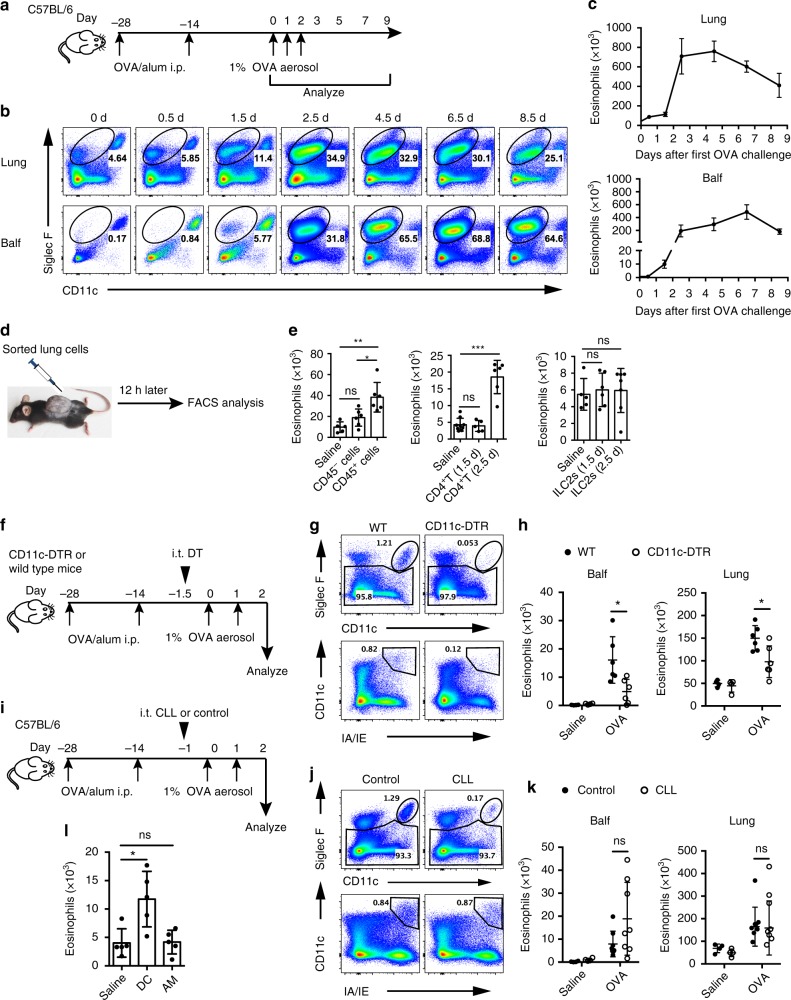
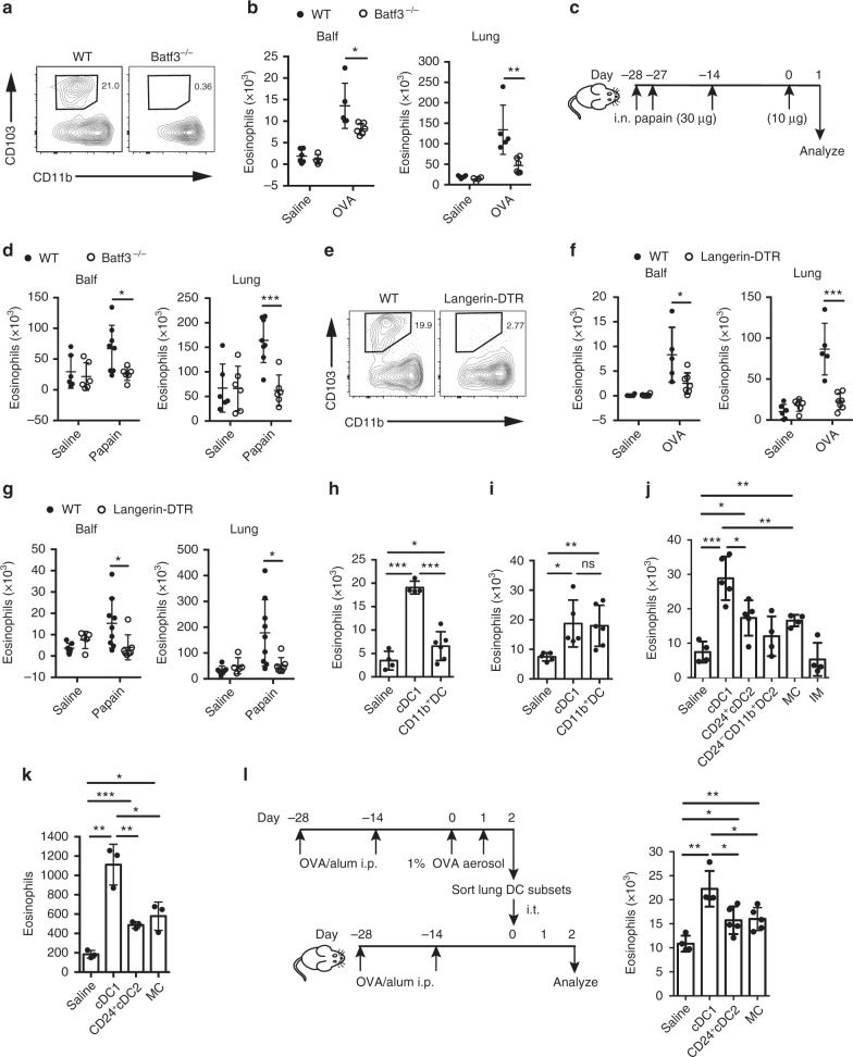
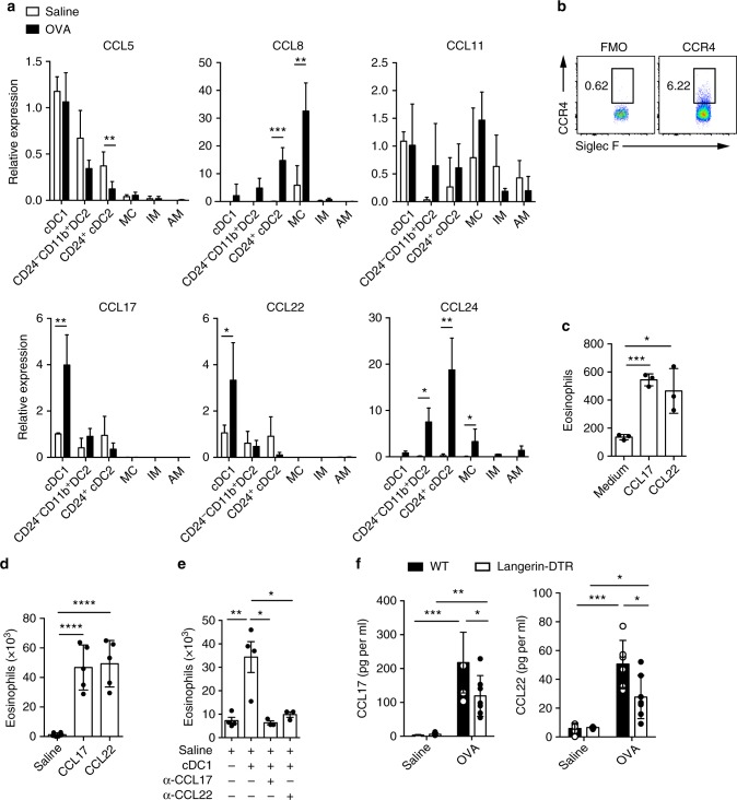
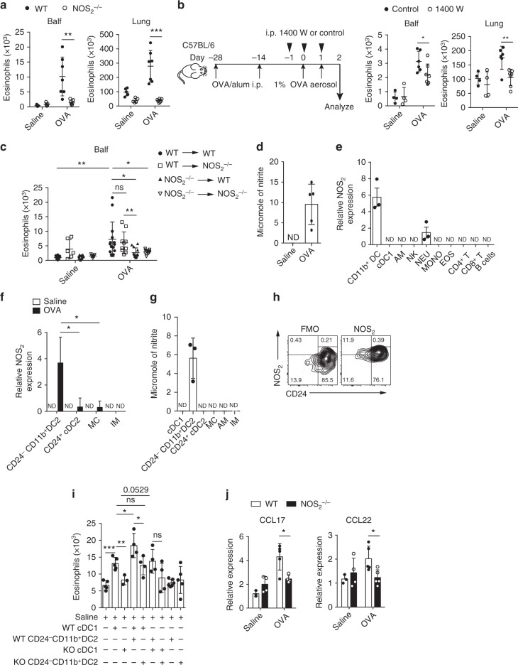
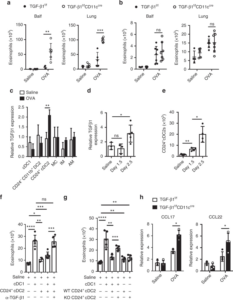
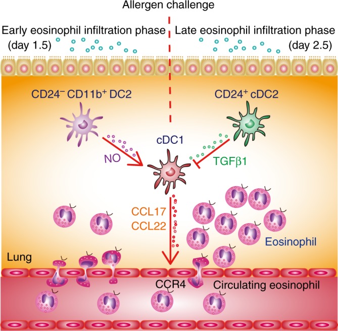
Eosinophil infiltration, a hallmark of allergic asthma, is essential for type 2 immune responses. How the initial eosinophil recruitment is regulated by lung dendritic cell (DC) subsets during the memory stage after allergen challenge is unclear. Here, we show that the initial eosinophil infiltration is dependent on lung cDC1s, which require nitric oxide (NO) produced by inducible NO synthase from lung CD24-CD11b+ DC2s for inducing CCL17 and CCL22 to attract eosinophils. During late phase responses after allergen challenge, lung CD24+ cDC2s inhibit eosinophil recruitment through secretion of TGF-β1, which impairs the expression of CCL17 and CCL22. Our data suggest that different lung antigen-presenting cells modulate lung cDC1-mediated eosinophil recruitment dynamically, through secreting distinct soluble factors during the memory stage of chronic asthma after allergen challenge in the mouse.


Conflict of interest statement

The authors declare no competing interests.

Figures

Fig. 1
Fig. 1
Lung DCs are required for eosinophil recruitment during allergen challenge. a Mice model of kinetics of eosinophil recruitment. b, c FACS (b) and total numbers (c) of eosinophil recruitment in different lung compartments in the murine kinetics model of allergic inflammation shown in a. Upper row, lung tissue homogenates; lower row, Balf. n = 4–8 mice per group. d Protocol for in vivo recruitment assay (air-pouch assay). e Counts of eosinophils recruited into the air pouches of wild-type (WT) mice injected with lung CD45 cells or CD45+ cells (left), lung CD4+ T cells (middle), or ILC2s (right) were analyzed by FACS. n = 5–7 mice per group. fh Eosinophil recruitment of allergic CD11c-DTR Tg (empty circle) or WT (solid circle) mice i.t. injected with DT to delete lung CD11c+ DCs and AMs, where f shows the protocol. g Different deletion efficiencies of DCs and AMs in the lung of CD11c-DTR Tg or WT mice after i.t. instillation with DT. h Total numbers of eosinophils in the lung or Balf. n = 4 mice (Saline) or n = 6 mice (OVA). ik Eosinophil recruitment of allergic C57Bl/6 mice i.t. injected with clodronate liposome (CLL) (empty circle) or control (solid circle) on day −1.5 to delete lung AMs, where i presents the protocol. j 24 h after CLL or empty liposome instillation, lungs were analyzed for the presence of AMs. k The total numbers of eosinophils in the lung or Balf. n = 4 mice (Saline) or n = 8 mice (OVA). l Counts of eosinophils recruited into the air pouches of WT mice injected with pulmonary SiglecFCD11c+IA/IE+ DCs or SiglecF+CD11chigh AMs (5 × 104 cells, 200 μl) from OVA-challenged mice. n = 4–5 mice per group. *P < 0.05, **P < 0.01, ***P < 0.001, unpaired Student’s t test. Means ± SD are shown. Data represent two (ac) and three (di) independent experiments
Fig. 2
Fig. 2
Lung cDC1s are necessary and sufficient for the induction of eosinophilia. a Lung cDC1s from WT and Batf3−/− mice. b Eosinophil counts in the lung or Balf in WT (solid circle) and Batf3−/− (empty circle) mice 1.5 days after the first OVA challenge. n = 4 or 6 mice (Saline) or n = 5–6 mice (OVA). c, d Mice were sensitized and challenged with papain, and eosinophils in the lung or Balf in WT (solid circle) and Batf3−/− (empty circle) mice were analyzed. n = 5–7 mice per group. e Lung cDC1s from DT treatment langerin-DTR or WT mice. f, g OVA/alum-sensitized (f) or papain-sensitized (g) langerin-DTR (empty circle) or WT (solid circle) mice were treated with DT on day −1, and eosinophils in the lung or Balf were assessed after challenge. n = 5–9 mice per group. h, i Eosinophils recruited into the air pouches injected with pulmonary cDC1s (SiglecFCD11c+IA/IE+CD103+CD11b) or CD11b+ DCs (1.5 × 104, 200 μl) sorted from C57BL/6 mice 1.5 days after the first OVA challenge (h) or 1 day after the first papain challenge (i). n = 4–6 mice per group. j Eosinophils recruited into the air pouches injected with pulmonary cDC1s, CD24+ cDC2s, CD24CD11b+ DC2s, MCs, or IMs (1.5 × 104, 200 μl) sorted from C57BL/6 mice 1.5 days after the first OVA challenge. n = 4–6 per group. k The direct chemotactic effect of lung cDC1s, CD24+ cDC2s, or MCs (from C57BL/6 mice 1.5 days after the first OVA challenge) on eosinophils was evaluated in a transwell culture system. n = 3–4 per group. l Protocol (left) and lung eosinophil counts (right) of allergic airway induction by adoptive i.t. injection of lung cDC1s, CD24+ cDC2s, or MCs; see Methods *P < 0.05, **P < 0.01, ***P < 0.001, unpaired Student’s t test. Means ± SD are shown. Data represent two (c, d, g, i, l) and three (a, b, e, f, h, j, k) independent experiments
Fig. 3
Fig. 3
Lung cDC1s directly recruit eosinophils via CCL17 and CCL22. a mRNA expression of CCL5, CCL8, CCL11, CCl17, CCL22, and CCL24 chemokines by lung cDC1s, CD24+ cDC2s, CD24CD11b+ DC2s, MCs, IMs, and AMs isolated from mice 1.5 days after the first OVA challenge. mRNA expression was first normalized against the housekeeping GAPDH genes and then represented as relative expression compared to the cDC1s from saline-treated mice. n = 3–4 mice per group. b FACS analysis of CCR4 expression on lung eosinophils from naive C57Bl/6 mice. c The direct chemotactic effect of CCL17 (100 pg per ml) and CCL22 (100 pg per ml) on eosinophils was evaluated in a transwell culture system. n = 3 per group. d Counts of eosinophils recruited into the air pouches of wild-type mice 5 h after injection of CCL17 (150 pg) or CCL22 (150 pg) with 500 μl of temperature-sensitive surface gel. n = 5–6 mice per group. e Counts of eosinophils recruited into the air pouches of wild-type mice injected with pulmonary cDC1s (SiglecFCD11c+IA/IE+CD103+CD11b) from OVA-challenged C57Bl/6 mice with CCL17 or CCL22 neutralizing antibody or goat IgG isotype control. n = 3–4 mice per group. f ELISA analysis of CCL17 and CCL22 in Balf supernatant from DT-treated langerin-DTR (empty rectangle) or wild-type (solid rectangle) mice 1.5 days after the first OVA challenge. n = 5–7 mice per group. *P < 0.05, **P < 0.01, ***P < 0.001, unpaired Student’s t test. Means ± SD are shown. Data represent two (a, b, d, e, f) and three (c) independent experiments
Fig. 4
Fig. 4
Lung CD24CD11b+ DC2s promote cDC1-mediated eosinophil recruitment. ac Mice were sensitized and challenged with OVA and culled 1.5 days after the first OVA challenge, and eosinophil counts in the lung or Balf were assessed. a WT (solid circle) and NOS2−/− (empty circle) mice. n = 5–7. b 1400 W (empty circle) or control-treated (solid circle) mice, protocol on the left. n = 4–7. c NOS2−/− and WT bone marrow chimeric mice. WT to WT (solid circle), WT to NOS2−/− (empty square), NOS2−/− to WT (solid triangle), and NOS2−/− to NOS2−/− (empty triangle) mice. n = 6–10 (Saline) or n = 10–14 (OVA). df Cells were sorted from mice 1.5 days after the first OVA challenge and used for assay. d NO production measured as nitrite in lung CD45+ cell culture supernatants. n = 4–5 per group. e mRNA expression of NOS2 by different types of lung CD45+ cells, such as CD11b+DCs, cDC1s (SiglecFCD11c+IA/IE+CD103+CD11b), neutrophils (NEU), etc. n = 3 per group. f NOS2 mRNA expression in lung CD24CD11b+ DC2s isolated from OVA (solid rectangle) or saline (empty rectangle) challenged mice. mRNA expression is shown relative to the expression of NEU (e) or CD24+ cDC2s (f). n = 4 per group. g NO production measured in lung CD24CD11b+ DC2 culture supernatants. n = 4 per group. h FACS analysis of NOS2 expression on lung CD11b+ DCs from OVA-challenge mice. i, j Cells were purified from NOS2−/− or WT mice 1.5 days after the first OVA challenge. i Eosinophils recruited into the air pouches injected with pulmonary cDC1s (SiglecFCD11c+IA/IE+CD103+CD11b) (1 × 104 cells), CD24CD11b+ DC2s (1 × 104 cells). n = 4–6 per group. j mRNA expression of CCL17 and CCL22 by lung cDC1s from NOS2−/− (solid rectangle) or WT mice (empty rectangle). n = 3–5 per group. *P < 0.05, **P < 0.01, ***P < 0.001, unpaired Student’s t test. Means ± SD are shown. Data represent two (c, e) and three (a, b, d, fj) independent experiments
Fig. 5
Fig. 5
Lung CD24+ cDC2s inhibit cDC1-mediated eosinophil migration via TGF-β1. a, b Quantification of eosinophils in Balf and lung from TGF-β1fl/flCD11cCre (empty circle) and TGF-β1fl/fl (solid circle) mice 2.5 days (a, n= 5–6 mice per group) or 1.5 days (b, n = 4–5 mice for saline groups and n = 7 mice for OVA groups) after the first OVA challenge. c mRNA expression of TGF-β1 by pulmonary cDC1, CD24+ cDC2, CD24CD11b+ DC2, MC, IM and AM populations separated from C57BL/6 mice 2.5 days after the first OVA (solid rectangle) or saline (empty rectangle) challenge. d mRNA expression of TGF-β1 by pulmonary CD24+ cDC2s separated from C57BL/6 mice 0, 1.5, or 2.5 days after the first OVA challenge. mRNA expression is relative to the expression of cDC1s (c) or CD24CD11b+ DC2s (d) from saline-treated mice. n = 3–5 per group. e Counts of CD24+ cDC2s in lungs from C57BL/6 mice 0, 1.5, or 2.5 days after the first OVA challenge. f Counts of eosinophils recruited into the air pouches injected with pulmonary cDC1s or CD24+ cDC2s (1 × 104 cells, 200 μl) purified from C57BL/6 mice 2.5 days after the first OVA challenge with anti-TGF-β1 (10 μg per ml) antibody or mouse IgG1 isotype control. n = 4–6 mice per group. g Counts of eosinophils recruited into the air pouches injected with pulmonary cDC1s, and CD24+ cDC2s (1 × 104 cells, 200 μl) purified from TGF-β1fl/flCD11cCre (KO) and TGF-β1fl/fl (WT) mice 2.5 days after the first OVA challenge. n = 4–6 mice per group. h mRNA expression of CCL17 and CCL22 chemokines by lung cDC1s (SiglecFCD11c+IA/IE+CD103+CD11b), separated from TGF-β1fl/flCD11cCre (solid rectangle) and TGF-β1fl/fl (empty rectangle) mice 2.5 days after the first OVA challenge. n = 4–5 per group. *P < 0.05, **P < 0.01, ***P < 0.001, unpaired Student’s t test. Means ± SD are shown. Data represent two (c, d) and three (a, b, eh) independent experiments
Fig. 6
Fig. 6
Initial eosinophil recruitment is dynamically regulated by lung DC subsets. After allergen challenge, cDC1s directly recruit eosinophils by secreting CCL17 and CCL22, which are critical for early eosinophil infiltration. This cDC1-mediated eosinophil infiltration is dynamically modulated by other lung DC subsets. After allergen challenge, lung CD24CD11b+ DC2s promoted eosinophil infiltration by producing NO on day 1.5, which promotes CCL17 and CCL22 expression by cDC1s, whereas CD24+ cDC2s inhibit this process by releasing TGF-β1 on day 2.5

References

    1. Lambrecht BN, Hammad H. The immunology of asthma. Nat. Immunol. 2015;16:45–56. doi: 10.1038/ni.3049. - DOI - PubMed
    1. Rothenberg ME. Humanized anti-IL-5 antibody therapy. Cell. 2016;165:509. doi: 10.1016/j.cell.2016.04.020. - DOI - PubMed
    1. Rothenberg ME, Hogan SP. The eosinophil. Annu. Rev. Immunol. 2006;24:147–174. doi: 10.1146/annurev.immunol.24.021605.090720. - DOI - PubMed
    1. Fulkerson PC, Rothenberg ME. Targeting eosinophils in allergy, inflammation and beyond. Nat. Rev. Drug Discov. 2013;12:117–129. doi: 10.1038/nrd3838. - DOI - PMC - PubMed
    1. Lambrecht, B. N., Persson, E. K. & Hammad, H. Myeloid cells in asthma. Microbiol. Spectr.5, doi: 0.1128/microbiolspec.MCHD-0053-2016 (2017). - PMC - PubMed

Publication types

MeSH terms