. 2018 Sep 24;8(1):14289.
doi: 10.1038/s41598-018-32715-5.

Transfer and Integration of Breast Milk Stem Cells to the Brain of Suckling Pups

Affiliations

Transfer and Integration of Breast Milk Stem Cells to the Brain of Suckling Pups

Mehmet Şerif Aydın et al. Sci Rep. .

Abstract

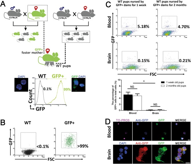
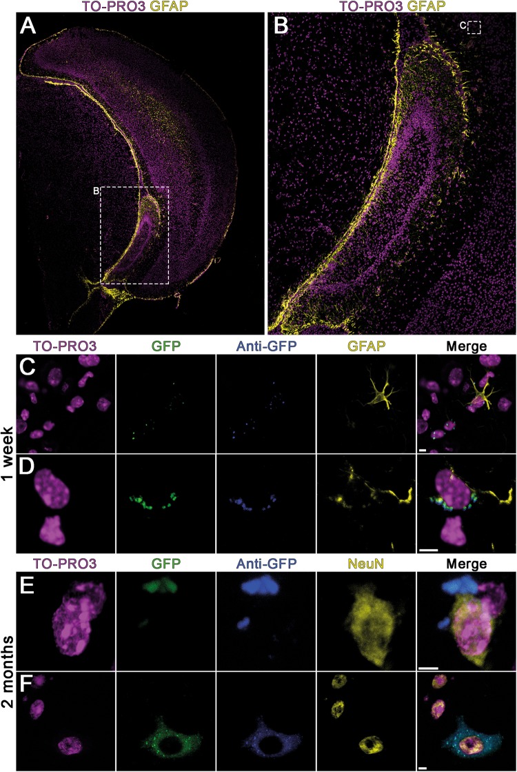
Beside its unique nutritional content breast milk also contains live cells from the mother. Fate of these cells in the offspring has not been adequately described. In this study, we aimed to detect and identify maternal cells in the suckling's blood and the brain. Green fluorescent protein expressing transgenic female mice (GFP+) were used as foster mothers to breastfeed wildtype newborn pups. One week and two months after the birth, blood samples and brains of the sucklings were analyzed to detect presence of GFP+ cells by fluorescence activated cell sorting, polymerase chain reaction and immunohistochemistry on the brain sections and optically cleared brains. The tests confirmed that maternal cells were detectable in the blood and the brain of the pups and that they differentiated into both neuronal and glial cell types in the brain. This phenomenon represents breastfeeding - induced microchimerism in the brain with functional implications remain to be understood.


Conflict of interest statement

The authors declare no competing interests.

Figures

Figure 1
Figure 1
Experimental design and flow cytometric analyses. (A) Breeding and nursing diagram of WT and GFP+ mice. WT newborn pups were immediately delivered to GFP+ foster mothers to be breastfed. Flow cytometry plots present the confirmation of expected GFP expression in the brains of GFP+ and WT mice. (B) Brain samples of WT and GFP+ mice were analyzed with GFP/FSC plot to determine the threshold of GFP signal for further flow cytometry analysis. (C) GFP+ cells were detected in both blood and brain of pups by the end of first week (n = 6) and two months (n = 6) of nursing. Error bars in the percentage of GFP+ cells in total cells graphs are S.D. (D) GFP+ cells detected in the brain by flow cytometry were sorted to verify GFP signal microscopically (scale bars: 10 µm).
Figure 2
Figure 2
Immunohistochemically stained brain sections of sucklings nursed by GFP+ mothers. Breast milk borne maternal cells are randomly distributed in the pup’s brain. (A,B) Representative zoomed images of GFP+ cells in the hippocampal region of the brain. (CF) Glial cell marker (anti-GFAP) and neuronal marker (anti-NeuN) were used to label different cell types, counterstained with nuclear dye TO-PRO3. GFP+ cells differentiated to neuronal and glial cells were detected after one week and two months of nursing (scale bars: 5 µm).
Figure 3
Figure 3
Tissue clearing method and immunohistochemical staining of sucklings brain. iDISCO method was used to determine the specific localization and 3-dimensional morphology of the GFP+ cells in the pup’s brain. GFP+ cells showed a random distribution like in sections. However, the morphology and localization of these cells were much better elucidated in cleared tissue. (AD) GFAP+ and GFP+ cells can be seen in cleared brain section of pups (250 µm z-optical section of 2 mm thick section). (E) Close proximity of a GFP+ maternal cell to a blood vessel is shown. As it also expresses glial marker GFAP, it is likely to be an astrocyte associated with the blood vessel to contribute the blood brain barrier. (F) Orthogonal section of GFP+ cell detected at the top of the blood vessel (scale bars: 15 µm) (See also Movie S1).

References

    1. Walker A. Breast milk as the gold standard for protective nutrients. The Journal of pediatrics. 2010;156:S3–7. doi: 10.1016/j.jpeds.2009.11.021. - DOI - PubMed
    1. Hassiotou F, et al. Maternal and infant infections stimulate a rapid leukocyte response in breastmilk. Clinical & translational immunology. 2013;2:e3. doi: 10.1038/cti.2013.1. - DOI - PMC - PubMed
    1. Hassiotou F, Geddes DT, Hartmann PE. Cells in human milk: state of the science. Journal of human lactation: official journal of International Lactation Consultant Association. 2013;29:171–182. doi: 10.1177/0890334413477242. - DOI - PubMed
    1. Hassiotou F, et al. Breastmilk is a novel source of stem cells with multilineage differentiation potential. Stem cells (Dayton, Ohio) 2012;30:2164–2174. doi: 10.1002/stem.1188. - DOI - PMC - PubMed
    1. Visvader JE. Keeping abreast of the mammary epithelial hierarchy and breast tumorigenesis. Genes & development. 2009;23:2563–2577. doi: 10.1101/gad.1849509. - DOI - PMC - PubMed

Publication types

Substances