. 2018 Dec;63(12):1211-1222.
doi: 10.1038/s10038-018-0515-y. Epub 2018 Sep 25.

Biallelic mutations in FDXR cause neurodegeneration associated with inflammation

Affiliations

Biallelic mutations in FDXR cause neurodegeneration associated with inflammation

Jesse Slone et al. J Hum Genet. 2018 Dec.

Abstract

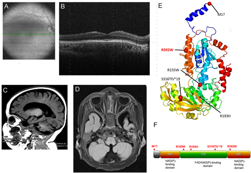
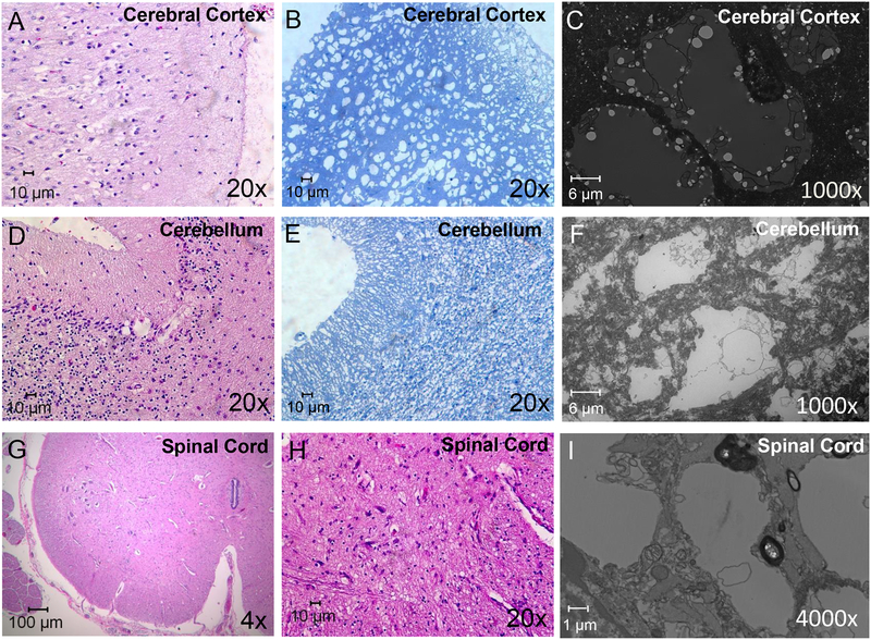
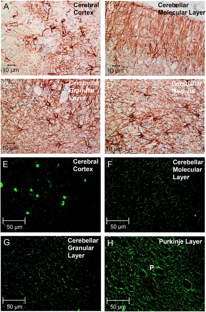
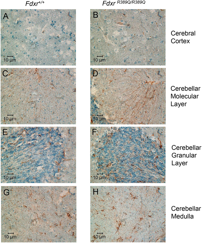
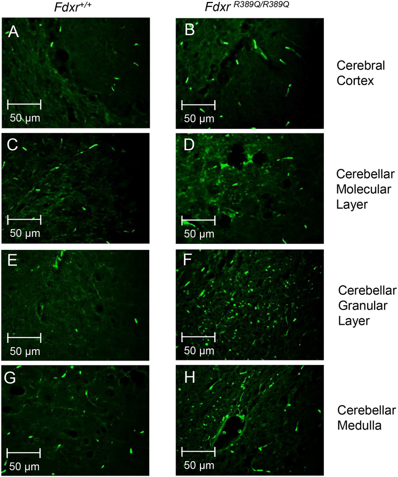
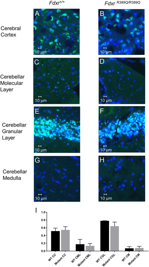
Mitochondrial dysfunction lies behind many neurodegenerative disorders, owing largely to the intense energy requirements of most neurons. Such mitochondrial dysfunction may work through a variety of mechanisms, from direct disruption of the electron transport chain to abnormal mitochondrial biogenesis. Recently, we have identified biallelic mutations in the mitochondrial flavoprotein "ferredoxin reductase" (FDXR) gene as a novel cause of mitochondriopathy, peripheral neuropathy, and optic atrophy. In this report, we expand upon those results by describing two new cases of disease-causing FDXR variants in patients with variable severity of phenotypes, including evidence of an inflammatory response in brain autopsy. To investigate the underlying pathogenesis, we examined neurodegeneration in a mouse model. We found that Fdxr mutant mouse brain tissues share pathological changes similar to those seen in patient autopsy material, including increased astrocytes. Furthermore, we show that these abnormalities are associated with increased levels of markers for both neurodegeneration and gliosis, with the latter implying inflammation as a major factor in the pathology of Fdxr mutations. These data provide further insight into the pathogenic mechanism of FDXR-mediated central neuropathy, and suggest an avenue for mechanistic studies that will ultimately inform treatment.


Conflict of interest statement

Conflict of Interest: The authors declare no conflict of interest. A.C. is a full-time employee of Ambry Genetics.

Figures

Figure 1.
Figure 1.. Optic atrophy and cerebral atrophy in patients carrying biallelic mutations in FDXR.
(A–B) Optical coherence tomography (OCT) of retina from the proband of Family #1. Images were obtained at the age of 35 months. The green line shown in the fundus photograph in panel A indicates the position of the OCT scan shown in panel B. The fovea appears to be well-formed, and the outer retinal layers are normal. The inner retina, however, shows almost complete absence of the ganglion cell layer in both eyes and severe thinning of the nerve fiber layer, consistent with bilateral optic atrophy involving the papillomacular bundle. (C–D) MRI images from the proband of Family #2 also show optic nerve hypoplasia (C) and global cerebral atrophy with marked prominence of sulci and ventricles (D). (E) Patient mutations were mapped to a three-dimensional FDXR protein structure model. The most common variant observed in all patient families (R392W) (10) has been highlighted in red for comparison. (F) Patient mutations were also mapped to a two-dimensional map of the FDXR functional domains. Domain map was generated using the “MyDomains” software provided by PROSITE (34).
Figure 2.
Figure 2.. Atrophy and cell loss in the CNS of the patient autopsy (proband 2) with FDXR mutations.
Histological sectioning of the occipital cerebral cortex with H&E staining (A) indicated that the cerebral cortex lost many of its neurons, and multiple vacuoles were observed in the H&E staining section that formed a spongiform structure. Toluidine staining of the cerebral section (B) also showed the presence of spongiform-like structures in the cerebral cortex, and TEM confirmed this was largely due to the presence of vacuole-like structures (C). The cerebellum of the patient also lost neurons in the molecular layer, Purkinje cell layer, and granular layer (D–F). Most of the Purkinje cells were absent, and multiple vacuoles were observed with H&E staining (D). Toluidine staining (E) and TEM imaging (F) of the cerebellar sections also showed loss of cells and the formation of vacuoles in the cerebellum. H&E staining (G, H) of spinal cord also indicated that the spinal cord lost lower motor neurons in the anterior horn (G), and multiple vacuoles were formed in the spinal cord by H&E staining (H) and TEM (I).
Figure 3.
Figure 3.. Signs of gliosis and neurodegeneration in the autopsy brain from proband 2.
Immunostaining with antibody specific for GFAP revealed extensive gliosis in the cerebral cortex (A), as well as in the molecular layer (B), granular layer (C), and medullary layer (D) of the cerebellum. Astrocytes and their extending branches were observed throughout the cerebrum and cerebellum, and Bergmann glial cells were prominent throughout the molecular layer (B). Immunostaining for Fluoro-Jade C (FJC) also showed signs of neurodegeneration in the cerebral cortex (E), the cerebellar molecular layer (F), the cerebellar granular layer (G), and in the Purkinje layer (H, Purkinje cell indicated by the letter “P”).
Figure 4.
Figure 4.. Astrocyte gliosis in brains of mice with Fdxr gene mutation.
10-month-old mouse mutant (Fdxr R389Q/R389Q) and wildtype control brains were stained with anti-GFAP antibody, and images were compared for the cerebral cortex (A–B), as well as the molecular layer (C––D), granular layer (E–F), and medullary layer (G–H) of the cerebellum. A modestly increased presence of astrocytes and Bergmann glial cells are apparent in the various layers of the cerebellum (C–H) and the cerebral cortex (A–B).
Figure 5.
Figure 5.. Neurodegeneration in Fdxr mutant mouse brain.
Significantly increased immunostaining for Fluoro-Jade C (FJC) was observed in the cerebral cortex (A–B), as well as the molecular layer (C–D), granular layer (E–F), and medullary layer (G–H) of the cerebellum.
Figure 6.
Figure 6.. Modest reduction in the proportion of neurons in Fdxr mutant brains.
IF staining with anti-NeuN antibody was used to determine the proportion of neurons in mutant and wildtype brains. Counterstaining with DAPI was used to show the total number of cells in each image field; cells staining with DAPI alone are largely composed of glial cells, with a small percentage consisting of neurons such as Purkinje cells that do not express NeuN. Staining reveals a similar number of neurons present in the cerebral cortex (A–B), molecular layer (C–D), and medullary layer (G–H) of the cerebellum between Fdxr mutant and wildtype mice. The number of neurons present in the granular layer of mutant cerebellar tissue (E–F) is modestly, but noticeably reduced relative to wildtype control tissues. The proportion of neurons in each brain region and genotype was also determined by dividing the number of NeuN-positive cells by the total number of DAPI-positive cells (I), confirming the modest reduction in neurons seen in the granular layer. Cell counts were generated by manual counting of cells in 3 different fields of view at 40X magnification, and then averaging the result from the 3 counts. A total of 174 to 834 cells were counted for each genotype and brain region / layer, which was determined by the density of neurons in each brain region. Abbreviations are as follows: “CC” = Cerebral Cortex, “CML” = Cerebellar Molecular Layer, “CGL” = Cerebellar Granular Layer, “CM” = Cerebellar Medulla, “WT” = Wildtype.

References

    1. Shoffner JM, Lott MT, Lezza AMS, Seibel P, Ballinger SW, Wallace DC. Myoclonic epilepsy and ragged-red fiber disease (MERRF) is associated with a mitochondrial DNA tRNALys mutation. Cell. 1990;61(6):931–7. - PubMed
    1. Nijtmans LG, Henderson NS, Attardi G, Holt IJ. Impaired ATP synthase assembly associated with a mutation in the human ATP synthase subunit 6 gene. The Journal of biological chemistry. 2001;276(9):6755–62. - PubMed
    1. Goto Y, Nonaka I, Horai S. A mutation in the tRNA(Leu)(UUR) gene associated with the MELAS subgroup of mitochondrial encephalomyopathies. Nature. 1990;348(6302):651–3. - PubMed
    1. Wallace DC. A mitochondrial paradigm of metabolic and degenerative diseases, aging, and cancer: a dawn for evolutionary medicine. Annu Rev Genet. 2005;39:359–407. - PMC - PubMed
    1. Webert H, Freibert SA, Gallo A, Heidenreich T, Linne U, Amlacher S, et al. Functional reconstitution of mitochondrial Fe/S cluster synthesis on Isu1 reveals the involvement of ferredoxin. Nature communications. 2014;5:5013. - PubMed

MeSH terms