Review
. 2018 Oct;21(10):1318-1331.
doi: 10.1038/s41593-018-0234-x. Epub 2018 Sep 24.

The role of brain vasculature in neurodegenerative disorders

Affiliations
Review

The role of brain vasculature in neurodegenerative disorders

Melanie D Sweeney et al. Nat Neurosci. 2018 Oct.

Abstract

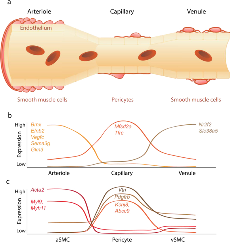
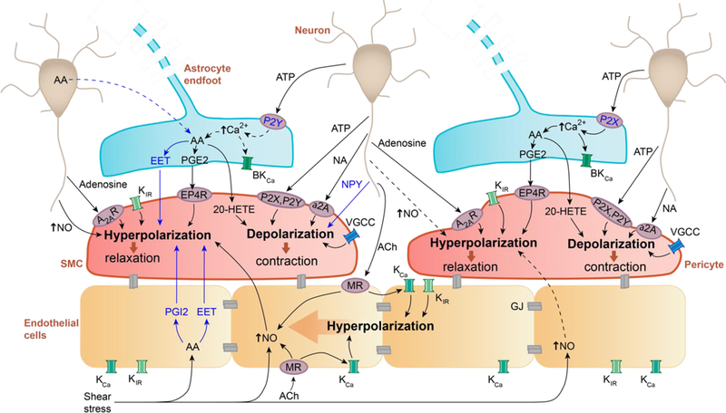
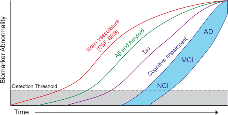
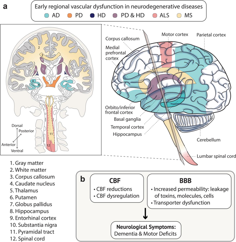
Adequate supply of blood and structural and functional integrity of blood vessels are key to normal brain functioning. On the other hand, cerebral blood flow shortfalls and blood-brain barrier dysfunction are early findings in neurodegenerative disorders in humans and animal models. Here we first examine molecular definition of cerebral blood vessels, as well as pathways regulating cerebral blood flow and blood-brain barrier integrity. Then we examine the role of cerebral blood flow and blood-brain barrier in the pathogenesis of Alzheimer's disease, Parkinson's disease, Huntington's disease, amyotrophic lateral sclerosis, and multiple sclerosis. We focus on Alzheimer's disease as a platform of our analysis because more is known about neurovascular dysfunction in this disease than in other neurodegenerative disorders. Finally, we propose a hypothetical model of Alzheimer's disease biomarkers to include brain vasculature as a factor contributing to the disease onset and progression, and we suggest a common pathway linking brain vascular contributions to neurodegeneration in multiple neurodegenerative disorders.


Figures

Figure 1.
Figure 1.. Molecular definition of the blood-brain barrier and cerebral blood vessels.
(a) The brain vasculature is a continuum from artery to arteriole to capillary to venule to vein. The blood-brain barrier is formed by a continuous endothelium monolayer surrounded by mural cells. Vascular ‘zonation’ refers to the molecular and phenotypic changes along the vascular endothelial continuum. Molecularly, the endothelium is a gradual continuum enriched with cell-specific markers at the arterial/arteriolar, capillary and venule/veins levels. Mural cells also cluster at different vascular segments: smooth muscle cells (SMCs) at arterioles and venules and pericytes at capillaries. (b) Representative curves showing molecular expression patterns of endothelial cells. The arteriole-specific genes include Bmx, Efnb2, Vegfc, Sema3g, and Gkn3, capillary-specific genes include Mfsd2a and Tfrc, and venule-specific genes include Nr2f2 and Slc38a5. (c) Representative curves showing molecular expression patterns of mural cells. Arteriole SMCs enriched genes include Acta2, Myl9, and Myh11. Capillary pericyte enriched genes include Vtn, Pdgfrβ, Kcnj8, and Abcc9. The molecular characterization is informed from recent single cell RNA-sequencing studies in multiple murine models. Arteriole markers represent averaged artery and arteriole expression. See main text for details.
Figure 2.
Figure 2.. Key cellular and molecular pathways regulating cerebral blood flow.
Neuron-mural cells crosstalk. ATP and noradrenaline (NA) released by neurons act on smooth muscle cells (SMCs) and pericytes through adenosine A2A receptors (A2AR) or α2-adrenergic receptors (α2A), respectively, causing cell depolarization and constriction, which reduces blood flow. Adenosine acts via purinergic P2X and P2Y receptors to hyperpolarize SMCs and pericytes, which increases blood flow. Neuropeptide Y (NPY) causes SMCs contraction. In both SMCs and pericytes, nitric oxide (NO) produced by neurons leads to hyperpolarization resulting in blood flow increase. Pericyte response to NO may vary by brain region, indicated by dashed arrows. Extracellular potassium ions (K+) released during neuronal activation can act on K+ (inward rectifier, KIR) and Ca2+ (Voltage-gated, VGCC) channels in SMCs and pericytes to hyperpolarize and relax the cells, or depolarize and contract cells. Astrocyte-mural cells crosstalk. ATP acts on P2X or P2Y receptors on astrocytes, which according to some studies can increase intracellular [Ca2+]. However, the role of arteriolar astrocyte [Ca2+] changes remains debatable (indicated by dashed arrows; see text). [Ca2+] increase triggers production of arachidonic acid (AA) and its metabolites (prostaglandin E2, PGE2, through PGE2 receptor EP4, EP4R; 20-hydroxyeicosatetraenoic acid; 20-HETE; epoxyeicosatetraenoic acids, EETs) that act on SMCs and pericytes to regulate blood flow. Alternatively, neurons may release AA to be further metabolized by astrocytes, indicated by dashed line. Endothelial-mural cells crosstalk. Acetylcholine (ACh) released from neurons or blood-derived ACh act on endothelial muscarinic ACh receptors (MRs) to increase endothelial NO production causing hyperpolarization and relaxation of mural cells, which increases blood flow. Shear stress can also increase NO endothelial production as well as production of AA and metabolites EETs and prostacyclin (PGI2) that hyperpolarize and relax SMCs, increasing arteriolar blood flow. Extracellular [K+] increase or ACh can activate KIR or calcium-activated K+ (KCa) channels on endothelial cells, leading to endothelial hyperpolarization that can propagate via gap junctions (GJs) between endothelial cells in a retrograde direction to increase blood flow. Altogether these findings are informed from various CNS regions and from both in vivo and in vitro studies; see main text for details.
Figure 3.
Figure 3.. Key cellular and molecular pathways regulating blood-brain barrier integrity.
BBB integrity is maintained by tight junction (TJ) and adherens junction (AJ) proteins between endothelial cells and low-level bulk flow transcytosis. Pericyte-endothelial cells crosstalk: Notch ligands-Notch3 receptor signaling promotes pericyte survival. Platelet-derived growth factor-BB (PDGF-BB) binds to PDGFRβ on pericytes causing pericyte survival, proliferation, and migration. Vascular endothelial growth factor-A (VEGFA) binds to endothelial VEGF receptor-2 (VEGFR2) mediating endothelial survival. Pericyte-derived notch ligands bind to endothelial Notch1 receptor which mediates BBB stability, as does endothelial sphingosine-1 phosphate (S1P). Transforming growth factor-β (TGFβ) and TGFβ receptor-2 (TGFβR2) signaling occurs bi-directionally between pericytes and endothelial cells. Pericyte-secreted angiopoietin-1 (Angpt1) binds Tie2 receptor on endothelial cells to promote proliferation. Astrocyte-endothelial cells crosstalk: Astrocyte-secreted APOE2 and APOE3, in contrast to APOE4, suppresses the pro-inflammatory signaling cyclophilin A-NFkB-matrix metalloproteinase-9 (MMP9) pathway in pericytes to maintain BBB stability. Similarly, astrocyte-produced laminin maintains BBB stability. Astrocyte-secreted sonic hedgehog (Shh) interacts with patched-1 (PTCH1) at the endothelium to further promote BBB stability. Smooth muscle cell (SMC)-endothelial cells crosstalk: Ephrin B2 (EphB2) on the endothelium promotes BBB stability. PDGF-BB binds PDGFRβ on SMCs to promote survival and migration. Endothelial-secreted jagged-1 (Jag-1) binds Notch3 to promote SMC maturation and survival. Neuron-endothelial cells crosstalk. Neuron secreted Wnt is a ligand of frizzled (FZD) at the endothelium that promotes endothelial cell differentiation.
Figure 4.
Figure 4.. Hypothetical updated Jack model of Alzheimer’s disease biomarkers to include the role of brain vasculature.
Hypothetical model of Alzheimer’s disease (AD) biomarker changes illustrating that early cerebral blood flow (CBF) and blood-brain barrier (BBB) biomarkers and vascular dysfunction may contribute to initial stages of AD pathophysiological progression from no cognitive impairment (NCI) to mild cognitive impairment (MCI) to AD, which is followed by cerebrospinal fluid and brain changes in Aβ and amyloid, and tau biomarkers. All biomarker curves converge at the top right-hand corner of the plot, that is the point of maximum abnormality. The horizontal axis of disease progression is expressed as time. Cognitive response is illustrated as a zone (blue filled area) with low and high-risk borders. Subjects with high risk of AD-related cognitive impairment are shown with a cognitive response curve that is shifted to the left. In contrast, the cognitive response curve is shifted to the right in subjects with a protective genetic profile, high cognitive reserve and the absence of comorbid brain pathologies.
Figure 5.
Figure 5.. Commonality of an early involvement of brain vasculature in different neurodegenerative disorders.
(a–b) Region-specific brain vascular dysfunction including cerebral blood flow (CBF) shortfalls (reductions and dysregulation) and/or blood-brain barrier (BBB) breakdown (increased vascular permeability and transporter dysfunction) is a common pathway seen early in multiple neurodegenerative disorders, including Alzheimer’s disease (AD), Parkinson’s disease (PD), Huntington’s disease (HD), amyotrophic lateral sclerosis (ALS), and multiple sclerosis (MS), as illustrated schematically in a and b. See Tables 1, 2 and 3 for details. Specifically, some studies suggest that vascular dysfunction (CBF and/or BBB) in the hippocampus, gray matter and entorhinal cortex in AD may precede dementia, brain atrophy and/or detectable Aβ and tau biomarker changes. Similar, vascular dysfunction in the white matter and corpus callosum in MS, basal ganglia (the caudate nucleus, thalamus, putamen, globus pallidus and substantia nigra) in PD and HD, the spinal cord white matter pyramidal tract in MS, and motor cortex and spinal cord in ALS is found by some studies in early stages of these disorders prior to progression of neurological symptoms including motor deficits.

References

    1. Iadecola C The Neurovascular Unit Coming of Age: A Journey through Neurovascular Coupling in Health and Disease. Neuron 96, 17–42 (2017). - PMC - PubMed
    1. Kisler K, Nelson AR, Montagne A & Zlokovic BV Cerebral blood flow regulation and neurovascular dysfunction in Alzheimer disease. Nat. Rev. Neurosci (2017). - PMC - PubMed
    1. Hartmann DA et al. Pericyte structure and distribution in the cerebral cortex revealed by high-resolution imaging of transgenic mice. Neurophotonics 2, 041402 (2015). - PMC - PubMed
    1. Zhao Z, Nelson AR, Betsholtz C & Zlokovic BV Establishment and Dysfunction of the Blood-Brain Barrier. Cell 163, 1064–1078 (2015). - PMC - PubMed
    1. Sweeney MD, Ayyadurai S & Zlokovic BV Pericytes of the neurovascular unit: key functions and signaling pathways. Nat. Neurosci 19, 771–783 (2016). - PMC - PubMed

Publication types