. 2018 Sep 24;9(10):999.
doi: 10.1038/s41419-018-1027-6.

MARVELD1 depletion leads to dysfunction of motor and cognition via regulating glia-dependent neuronal migration during brain development

Affiliations

MARVELD1 depletion leads to dysfunction of motor and cognition via regulating glia-dependent neuronal migration during brain development

Weizhe Liu et al. Cell Death Dis. .

Abstract

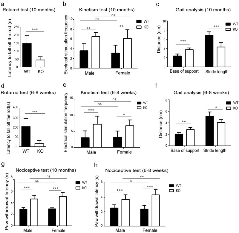
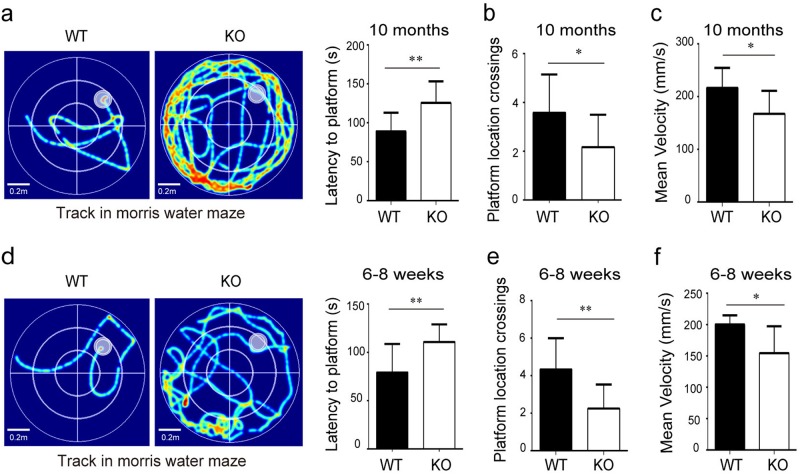
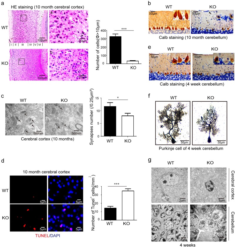
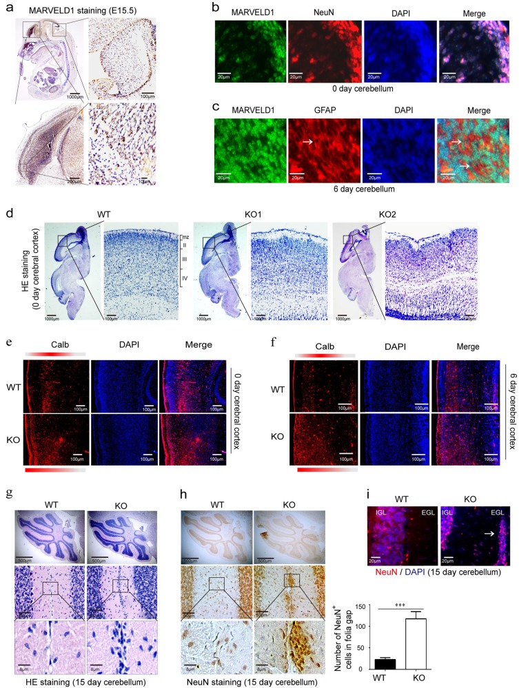
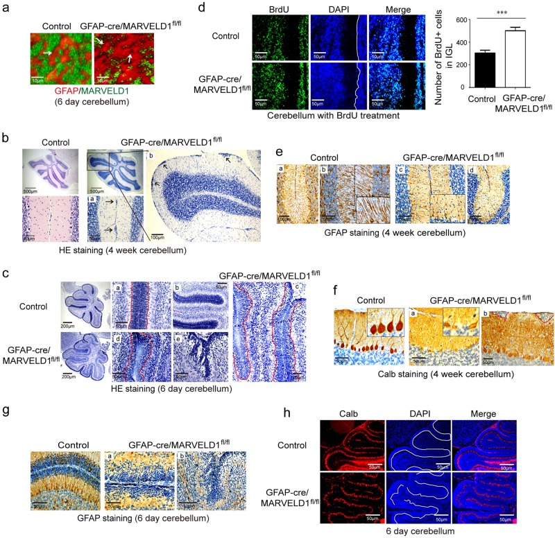
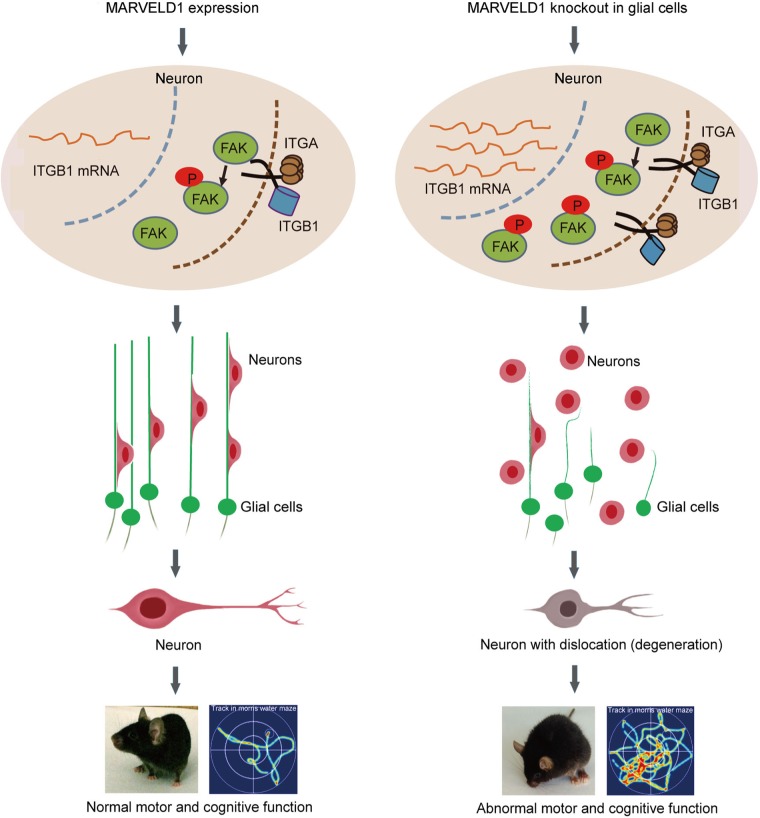
The establishment of functional neuronal connectivity is dependent on the neuronal migration and the accurate positioning of neurons in the developing brain. Abnormal neuronal migration can trigger neuronal maturation defects and apoptosis. However, many genetic bases remain unclear in neuronal migration disorders during brain development. In this study, we reported that MARVELD1-defected mice displayed motor and cognitive dysfunction resulting from aberrant neuronal migration during brain development. The laminar organization of the cerebral cortex and cerebellum in MARVELD1 knockout (KO) mice is disrupted, indicating impaired radial neuronal migration. Furthermore, we used the cerebellum as a model to explore the radial neuronal migration processes, and the results demonstrated that the proper neuronal migration depended on MARVELD1 expression in glial cells of the developing brain. MARVELD1 suppressed the expression of ITGB1 and FAK Tyr397 phosphorylation in glia-dependent manner. The inhibition of the MARVELD1/ITGB1/FAK signalling pathway in MARVELD1 KO mice could reverse the defects in neuronal migration in vitro. Our findings revealed that MARVELD1 regulated neuronal migration by mediating the formation of glial fibres and ITGB1/FAK signalling pathway. The depletion of MARVELD1 during mouse brain development led to the abnormity of motor and cognition functions.


Conflict of interest statement

The authors declare that they have no conflict of interest.

Figures

Fig. 1
Fig. 1. MARVELD1 KO mice displayed motor abnormalities.
a Rotarod test was performed with 10-month-old mice. For WT mice n = 10, and for MARVELD1 KO mice n = 9. b Treadmill test: electrical stimulation frequency was analyzed using 10-month-old mice. For male mice: WT mice n = 10, and MARVELD1 KO mice n = 9; for female mice: WT mice n = 11, and MARVELD1 KO mice n = 9. One-way ANOVA was used in this study. c Gait experiment was tested using 10-month-old mice. Base of support and stride length were analyzed. For WT mice n = 12, and MARVELD1 KO mice n = 10. d Rotarod test was performed with 6–8-week-old mice. For WT and MARVELD1 KO mice, n = 13. e Treadmill test: electrical stimulation frequency was analyzed using 6–8-week-old mice. For male mice: WT and MARVELD1 KO mice n = 10, respectively; for female mice: WT and MARVELD1 KO mice n = 9, respectively. One-way ANOVA was used. f Gait test was tested using 6–8-week-old mice. Base of support and stride length were analyzed. For WT mice n = 11, and MARVELD1 KO mice n = 9. g The nociceptive response was assessed by the radiant heat paw withdrawal test using 10-month-old mice. For male mice: for WT mice n = 11 and for MARVELD1 KO mice n = 9; for female mice: for WT mice n = 10, and for MARVELD1 KO mice n = 9. One-way ANOVA was used. h The nociceptive response was assessed by the radiant heat paw withdrawal test using 6–8-week-old mice. For male mice: for WT mice n = 24 and MARVELD1 KO mice n = 27; for female mice: for WT mice n = 18 and MARVELD1 KO mice n = 23. One-way ANOVA was used. *p < 0.05; **p < 0.01; ***p < 0.001
Fig. 2
Fig. 2. MARVELD1 KO mice displayed cognitive function abnormalities.
a Morris water maze test was performed with 10-month-old mice. Training was conducted during 5 days with training two times per day and latency to the platform is analyzed. The platform was marked by a white pointed circle in the first quadrant. b A probe trial was assessed and the platform location crossing times were analyzed using 10-month-old mice. c The swimming velocity in Morris water maze test was analyzed using 10-month-old mice. The WT group and MARVELD1 KO group n = 11, respectively, in a, b and c. d Morris water maze test with 6–8-week-old mice and latency to the platform was analyzed. e A probe trial was assessed and the platform location crossing times were analyzed using 6–8-week-old mice. f The swimming velocity in Morris water maze test was analyzed using 6–8-week-old mice. For WT mice n = 13 and MARVELD1 KO mice n = 14 in d, e and f, respectively. *p < 0.05; **p < 0.01
Fig. 3
Fig. 3. The depletion of MARVELD1 led to neurodegeneration in mouse brain.
a The cerebral cortex HE staining of 10-month-old mice. Neurons with a diameter greater than 10  μm were counted (per mm2). b Immunohistochemistry staining was performed with Calb antibodies in 10-month-old mice cerebellum. c Transmission electron microscopy: neural synapses were observed in 10-month-old mice cerebral cortex. The arrowheads indicated neural synapses. d The number of TUNEL stained neurons/mm2 were counted in the cerebral cortex of 10-month-old mice. e Immunohistochemistry staining was performed with Calb antibodies in 4-week-old mice cerebella. f Golgi–Cox staining was performed to observe synapses of Purkinje cells in 4-week-old mice cerebellum. g Transmission electron microscopy: apoptotic neurons were observed in 4-week-old mice cerebral cortex and cerebellum. Above all investigations, n = 3 for each genotype. *p < 0.05; ***p < 0.001
Fig. 4
Fig. 4. Mice with MARVELD1 lack showed a disorder of the laminar layers in the cerebral cortex and cerebellum.
a Immunohistochemistry was observed for MARVELD1 protein from WT E15.5 mice. b Immunofluorescence was performed with antibodies of MARVELD1 (green) and a neuronal marker NeuN (red) in 0-day-old mice cerebellum. c Immunofluorescence was observed with antibodies of MARVELD1 (green) and glial cells maker GFAP (red) in 6-day-old mice cerebellum. d Sagittal sections through the cerebral cortex were analyzed by HE staining in 0-day-old mice. ef Immunofluorescence of Calb (red) in 0-day-old and 6-day-old mice cerebral cortex. In control mice, Calb+ cells were present largely in laminaII/III and in MARVELD1 KO mice cerebral cortex they were distributed in superficial lamina or arranged in a disorderly manner. g Sagittal sections of 15-day-old mice cerebellum stained with HE. The whole cerebellum with low magnification shows the overall situation of abnormal cells location in the molecular layer. h Sagittal sections of 15-day-old mice cerebellum stained with immunohistochemistry of NeuN, a granule cell marker in the cerebellum. i Sagittal sections of 15-day-old mice cerebellum stained with immunofluorescence of DAPI and NeuN, a granule cell marker in the cerebellum. di: n = 3 for each genotype. ***p < 0.001
Fig. 5
Fig. 5. MARVELD1 affected granule cell migration but not proliferation.
a Sagittal paraffin-embedded tissue sections of 0- and 6-day-old mice stained with HE. The whole cerebellum with low magnification showed the overall situation of abnormal cells. The arrowheads indicated migrating neurons. b In 6-day-old mice, the width of the EGL and the number of migrating granule cells were analyzed. c Granule cell proliferation and migration were evaluated after a short 1.5-h and a long 30-h chase following BrdU administration in 6-day-old mice in control and MARVELD1 KO animals. The relative cell number was counted. ac: n = 3 for each genotype. **p < 0.01; ***p < 0.001
Fig. 6
Fig. 6. MARVELD1 regulated accurate radial migration by affecting the formation of glial fibres.
a Immunofluorescence staining of MARVELD1 (green) and glial cells marker GFAP (red) in 6-day-old mice cerebellum. The arrowheads indicated glial cells. b HE staining of 4-week-old GFAP-cre/MARVELD1fl/fl mice cerebellum sections. The whole cerebellum with low magnification showed the overall situation of abnormal cells location in the molecular layer. n = 3 for each genotype. a and b were different areas from GFAP-cre/MARVELD1fl/fl cerebellum. c HE staining of 6-day-old control mice and GFAP-cre/MARVELD1fl/fl mice cerebellum. The whole cerebellum with low magnification showed the overall situation of abnormal cells location in the molecular layer. n = 3 for each genotype. a and b were different areas from control cerebellum. c, d and e are different areas from GFAP-cre/MARVELD1fl/fl cerebellum. d Granule cell migration was evaluated by a long 60-h chase following BrdU administration in 6-day-old control mice and GFAP-cre/MARVELD1fl/fl mice. n = 3 for each genotype. e Immunohistochemistry staining with GFAP antibodies in 4-week-old control and GFAP-cre/MARVELD1fl/fl cerebella. n = 3 for each genotype. a and b were different areas from control cerebellum. c and d were different areas from GFAP-cre/MARVELD1fl/fl cerebellum. f Immunohistochemistry staining with Calb antibodies in control and GFAP-cre/MARVELD1fl/fl cerebella in 4-week-old mice. n = 3 for each genotype. a and b are different areas from GFAP-cre/MARVELD1fl/fl cerebellum. g Sagittal sections of 6-day-old mice cerebellum immunostained with anti-GFAP. GFAP-cre/MARVELD1fl/fl mice had no obvious glial fibres. n = 3 for each genotype. a and b were different areas from GFAP-cre/MARVELD1fl/fl cerebellum. h Immunofluorescence of Calb (red) in 6-day-old mice cerebellum. ***p < 0.001
Fig. 7
Fig. 7. MARVELD1/ITGB1/FAK signalling suppressed neuronal cell migration via glia-dependent manner.
a Quantitative analysis indicated increased levels of ITGB1 in 0- and 7-day-old mice. n = 3 for each genotype. One-way ANOVA was used in this study. b ITGB1 and FAK Tyr397 phosphorylation were detected by western blot in 7-day-old WT and MARVELD1 KO cerebellum whole lysates. Quantitative analysis indicates elevated ITGB1 and FAK Tyr397 phosphorylation levels in MARVELD1 KO mice. n = 3 for each genotype. c Immunofluorescence of ITGB1 (red) and NeuN (green) in 6-day-old mice cerebellum. d Immunofluorescence of p397-FAK (red) and NeuN (green) in 6-day-old mice cerebellum. e Neuron migration from 5-day-old mice microexplants of the cerebellum after 30 h was analyzed. DAPI staining revealed that there were more migrating granule cells in MARVELD1 KO mice and there was a reversion after adding an inhibiter (20 μM). The number of granule cells which had migrated to specified distances (zone1: 0–100  μm from the microexplants; zone2: 100  μm beyond) was analyzed. One-way ANOVA was used. f Time-lapse imaging series of migrating granule cells from 5-day-old explants cultured for 30 h before imaging. (Interval time between pictures is 20 min). *p < 0.05; **p < 0.01; ***p < 0.001
Fig. 8
Fig. 8. Schematic representation for MARVELD1-mediated neuronal migration.
MARVELD1 deletion in glial cells induced the abnormality of glial fibres. Meanwhile, the expression of ITGB1 was increased in the pre-mRNA process in neurons, which further activated FAK through increasing its Tyr397 phosphorylation level. This regulation activated FAK-mediated downstream signalling that resulted in neuronal migration. The regulation process of MARVELD1 during brain development was in a glia cell-dependent manner. Furthermore, this process affected the neurodegeneration and behaviour in adult mice

References

    1. Evsyukova I, Plestant C, Anton ES. Integrative mechanisms of oriented neuronal migration in the developing brain. Annu. Rev. Cell Dev. Biol. 2013;29:299–353. doi: 10.1146/annurev-cellbio-101512-122400. - DOI - PMC - PubMed
    1. Riccardo Bocchi., et al. Perturbed Wnt signaling leads to neuronal migration delay, altered interhemispheric connections and impaired social behavior. Nat. Commun. 2017;8:1158. doi: 10.1038/s41467-017-01046-w. - DOI - PMC - PubMed
    1. Sun D, et al. Neogenin, a regulator of adult hippocampal neurogenesis, prevents depressive-like behavior. Cell Death Dis. 2018;9:8. doi: 10.1038/s41419-017-0019-2. - DOI - PMC - PubMed
    1. Jaarsma D, et al. A role for Bicaudal-D2 in radial cerebellar granule cell migration. Nat. Commun. 2014;5:3411. doi: 10.1038/ncomms4411. - DOI - PubMed
    1. Elias LAB, Wang DD, Kriegstein AR. Gap junction adhesion is necessary for radial migration in the neocortex. Nature. 2007;448:901–907. doi: 10.1038/nature06063. - DOI - PubMed

Publication types

MeSH terms