. 2018 Sep 24;8(1):14235.
doi: 10.1038/s41598-018-32427-w.

High resolution 7T and 9.4T-MRI of human cerebral arterial casts enables accurate estimations of the cerebrovascular morphometry

Affiliations

High resolution 7T and 9.4T-MRI of human cerebral arterial casts enables accurate estimations of the cerebrovascular morphometry

Jasper H G Helthuis et al. Sci Rep. .

Abstract

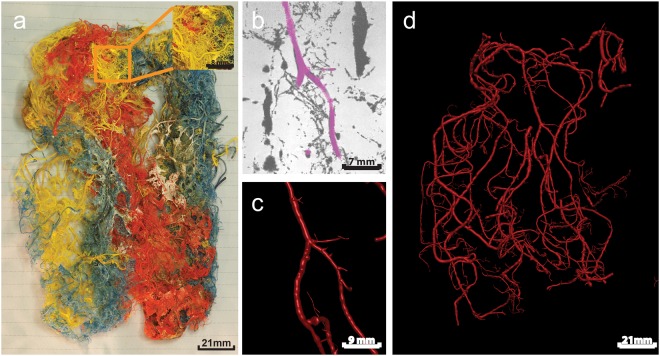
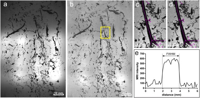
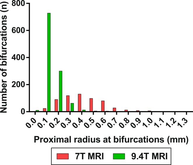
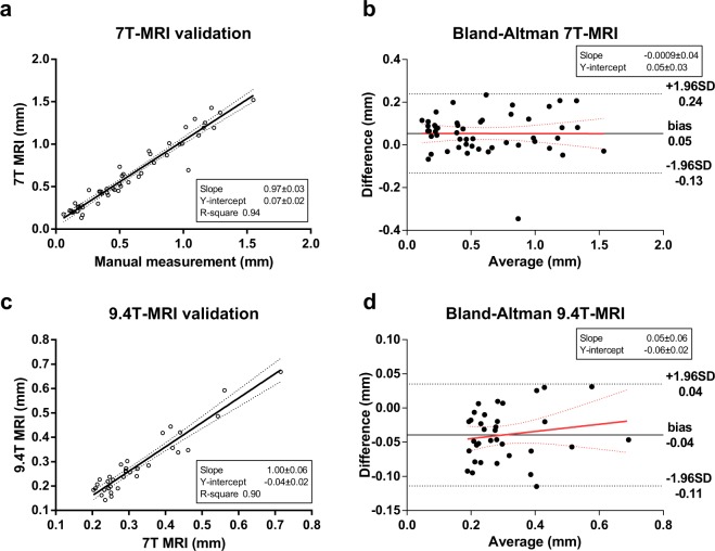
Quantitative data on the morphology of the cerebral arterial tree could aid in modelling and understanding cerebrovascular diseases, but is scarce in the range between 200 micrometres and 1 mm diameter arteries. Traditional manual measurements are difficult and time consuming. 7T-MRI and 9.4T-MRI of human cerebral arterial plastic casts could proof feasible for acquiring detailed morphological data of the cerebral arterial tree in a time efficient method. One cast of the complete human cerebral arterial circulation embedded in gadolinium-containing gelatine gel was scanned at 7T-MRI (0.1 mm isotropic resolution). A small section of another cast was scanned at 9.4T-MRI (30 µm isotropic resolution). Subsequent 3D-reconstruction was performed using a semi-automatic approach. Validation of 7T-MRI was performed by comparing the radius calculated using MRI to manual measurements on the same cast. As manual measurement of the small section was not feasible, 9.4T-MRI was validated by scanning the small section both at 7T-MRI and 9.4T MRI and comparing the diameters of arterial segments. Linear regression slopes were 0.97 (R-squared 0.94) and 1.0 (R-squared 0.90) for 7T-MRI and 9.4T-MRI. This data shows that 7T-MRI and 9.4T-MRI and subsequent 3D reconstruction of plastic casts is feasible, and allows for characterization of human cerebral arterial tree morphology.


Conflict of interest statement

The authors declare no competing interests.

Figures

Figure 1
Figure 1
A human cerebral cast scanned with 7T MRI. (a) Photographic image of the complete cast. The inset shows the enlarged section indicated by the orange square. Different pigments where used for different major cerebral arteries during injection of Araldite F. (b) Detail of a 7T MRI image (100 µm isotropic resolution), with segmented artery as displayed in Simple Neurite Tracer. (c) The segmented artery of panel b in 3D representation. (d) 3D visualisation of all reconstructed arteries. As the reconstruction approach was semi-automatic not all arteries in the casts are segmented, only arteries which were manually selected in Simple Neurite Tracer are reconstructed to 3D.
Figure 2
Figure 2
Small section of the human cerebral cast scanned with 9.4T MRI. (a) The cast section embedded in gel in a small plastic container, (b) Single slice from MRI (30 µm isotropic resolution), (c) Segmented arteries on single slice as displayed in Simple Neurite Tracer. (d) 3D visualisation of all reconstructed arteries.
Figure 3
Figure 3
Post-processing steps. (a) Original 7T MRI single image showing the signal inhomogeneity due to local small receive coils. (b) Normalized image with yellow square marking close-up region of interest. (c) Close-up of non-smoothed centerlines. (d) Close-up of smoothed centerlines. (e) Example of one intensity profile trough the artery on which the estimated radius (obtained with full-width at half-maximum method) of the artery is depicted.
Figure 4
Figure 4
Absolute number of bifurcations for radius (mm) of proximal arteries of all bifurcations as measured on cast. Zero bin contains all values < 0.05 mm.
Figure 5
Figure 5
7T-MRI and 9.4T-MRI validation. (a–b) 7T-MRI validation by comparison of direct manual diameter measurements on cast using digital callipers and measurement performed on 3D reconstructed cast. (c–d) 9.4T-MRI validation by comparison of diameter measurements performed using 7T and 9.4T-MRI on the same segments in a casts. (a) Linear regression (solid line) analysis with 95%-confidence interval (dotted lines) of manual versus 7T MRI measurements. (b) Blant-altman plot of difference versus average for 7T MRI validation (difference: 7T MRI minus manual measurements). (c) Linear regression (solid line) analysis with 95%-confidence interval (dotted lines) of 7T versus 9.4T MRI. (d) Blant-Altman plot of difference versus average for 9.4T MRI validation (difference: 9.4T diameter minus 7T diameter).

References

    1. Hirsch S, Reichold J, Schneider M, Székely G, Weber B. Topology and hemodynamics of the cortical cerebrovascular system. J. Cereb. Blood Flow Metab. 2012;32:952–67. doi: 10.1038/jcbfm.2012.39. - DOI - PMC - PubMed
    1. Baumbach GL, Heistad DD. Regional, segmental, and temporal heterogeneity of cerebral vascular autoregulation. Ann. Biomed. Eng. 1985;13:303–10. doi: 10.1007/BF02584248. - DOI - PubMed
    1. Hall CN, et al. Capillary pericytes regulate cerebral blood flow in health and disease. Nature. 2014;508:55–60. doi: 10.1038/nature13165. - DOI - PMC - PubMed
    1. Cassot F, Lauwers F, Fouard C, Prohaska S, Lauwers-Cances V. A novel three-dimensional computer-assisted method for a quantitative study of microvascular networks of the human cerebral cortex. Microcirculation. 2006;13:1–18. doi: 10.1080/10739680500383407. - DOI - PubMed
    1. Cassot F, et al. Branching patterns for arterioles and venules of the human cerebral cortex. Brain Res. 2010;1313:62–78. doi: 10.1016/j.brainres.2009.12.007. - DOI - PubMed

Publication types

MeSH terms