Sporadic cases of lumpy skin disease among cattle in Sharkia province, Egypt: Genetic characterization of lumpy skin disease virus isolates and pathological findings
- PMID: 30250377
- PMCID: PMC6141277
- DOI: 10.14202/vetworld.2018.1150-1158
Sporadic cases of lumpy skin disease among cattle in Sharkia province, Egypt: Genetic characterization of lumpy skin disease virus isolates and pathological findings
Abstract
Background and aim: Lumpy skin disease (LSD) is a highly infectious viral disease upsetting cattle, caused by LSD virus (LSDV) within the family Poxviridae. Sporadic cases of LSD have been observed in cattle previously vaccinated with the Romanian sheep poxvirus (SPPV) vaccine during the summer of 2016 in Sharkia province, Egypt. The present study was undertaken to perform molecular characterization of LSDV strains which circulated in this period as well as investigate their phylogenetic relatedness with published reference capripoxvirus genome sequences.
Materials and methods: A total of 82 skin nodules, as well as 5 lymph nodes, were collected from suspect LSD cases, and the virus was isolated in embryonated chicken eggs (ECEs). LSD was confirmed by polymerase chain reactions amplification of the partial and full-length sequences of the attachment and G-protein-coupled chemokine receptor (GPCR) genes, respectively, as well as a histopathological examination of the lesions. Molecular characterization of the LSDV isolates was conducted by sequencing the GPCR gene.
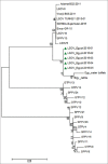
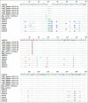
Results: Characteristic skin nodules that covered the whole intact skin, as well as lymphadenopathy, were significant clinical signs in all suspected cases. LSDV isolation in ECEs revealed the characteristic focal white pock lesions dispersed on the chorioallantoic membranes. Histopathologic examination showed characteristic eosinophilic intracytoplasmic inclusion bodies within inflammatory cell infiltration. Phylogenetic analysis revealed that the LSDV isolates were clustered together with other African and European LSDV strains. In addition, the LSDV isolates have a unique signature of LSDVs (A11, T12, T34, S99, and P199).
Conclusion: LSDV infections have been detected in cattle previously vaccinated with Romanian SPPV vaccine during the summer of 2016 and making the evaluation of vaccine efficacy under field conditions necessary.
Keywords: Egypt; Poxviridae; Sharkia province; cattle; lumpy skin disease.
Figures


Similar articles
-
Detection of lumpy skin disease virus in cattle using real-time polymerase chain reaction and serological diagnostic assays in different governorates in Egypt in 2017.Vet World. 2019 Jul;12(7):1093-1100. doi: 10.14202/vetworld.2019.1093-1100. Epub 2019 Jul 24. Vet World. 2019. PMID: 31528038 Free PMC article.
-
Isolation and molecular characterization of lumpy skin disease virus from hard ticks, Rhipicephalus (Boophilus) annulatus in Egypt.BMC Vet Res. 2022 Aug 5;18(1):302. doi: 10.1186/s12917-022-03398-y. BMC Vet Res. 2022. PMID: 35932057 Free PMC article.
-
G-Protein-Coupled Chemokine Receptor Gene in Lumpy Skin Disease Virus Isolates from Cattle and Water Buffalo (Bubalus bubalis) in Egypt.Transbound Emerg Dis. 2016 Dec;63(6):e288-e295. doi: 10.1111/tbed.12344. Epub 2015 Mar 9. Transbound Emerg Dis. 2016. PMID: 25754131
-
Capripoxviruses: Exploring the genetic relatedness between field and vaccine strains from Egypt.Vet World. 2019 Dec;12(12):1924-1930. doi: 10.14202/vetworld.2019.1924-1930. Epub 2019 Dec 11. Vet World. 2019. PMID: 32095042 Free PMC article.
-
Lumpy Skin Disease-An Emerging Cattle Disease in Europe and Asia.Vaccines (Basel). 2023 Mar 2;11(3):578. doi: 10.3390/vaccines11030578. Vaccines (Basel). 2023. PMID: 36992162 Free PMC article. Review.
Cited by
-
Molecular and histopathological characterization of lumpy skin disease in cattle in northern Vietnam during the 2020-2021 outbreaks.Arch Virol. 2022 Nov;167(11):2143-2149. doi: 10.1007/s00705-022-05533-4. Epub 2022 Jul 13. Arch Virol. 2022. PMID: 35831756
-
Oxidative stress, biochemical, and histopathological changes associated with acute lumpy skin disease in cattle.Vet World. 2022 Aug;15(8):1916-1923. doi: 10.14202/vetworld.2022.1916-1923. Epub 2022 Aug 15. Vet World. 2022. PMID: 36313851 Free PMC article.
-
Experimental evaluation of the cross-protection between Sheeppox and bovine Lumpy skin vaccines.Sci Rep. 2020 Jun 1;10(1):8888. doi: 10.1038/s41598-020-65856-7. Sci Rep. 2020. PMID: 32483247 Free PMC article.
-
Molecular diagnosis of three outbreaks during three successive years (2018, 2019, and 2020) of Lumpy skin disease virus in cattle in Sharkia Governorate, Egypt.Open Vet J. 2022 Jul-Aug;12(4):451-462. doi: 10.5455/OVJ.2022.v12.i4.6. Epub 2022 Jul 12. Open Vet J. 2022. PMID: 36118715 Free PMC article.
-
Pathogenicity and virulence of lumpy skin disease virus: A comprehensive update.Virulence. 2025 Dec;16(1):2495108. doi: 10.1080/21505594.2025.2495108. Epub 2025 Apr 27. Virulence. 2025. PMID: 40265421 Free PMC article. Review.
References
-
- Davies F.G. Lumpy skin disease of cattle: A growing problem in Africa and the Near East. Wld. Anim. Rev. 1991;68:37–42.
-
- Ahmed A, Dessouki A. Abattoir-based survey and histopathological findings of lumpy skin disease in cattle at Ismailia Abattoir. Int. J. Biosci. Biochem. Bioinforma. 2013;3:372–375.
-
- El-Neweshy M, El-Shemey T, Youssef S. Pathologic and immunohistochemical findings of natural lumpy skin disease in Egyptian cattle. Pak. Vet. J. 2013;33:60–64.
-
- Buller R.M, Arif B.M, Black D.N, Dumbell K.R, Esposito J.J, Lefkowitz E.J, McFadden G, Moss B, Mercer A.A, Moyer R.W, Skinner M.A, Tripathy D.N. Poxviridae. In: Fauquet C.M, Mayo M.A, Maniloff J, Desselberger U, Ball L.A, editors. Virus Taxonomy: Eighth Report of the International Committee on the Taxonomy of Viruses. Oxford: Elsevier Academic Press; 2005. pp. 117–133.
LinkOut - more resources
Full Text Sources
Other Literature Sources
