

The roles of IL-19 and IL-20 in the inflammation of degenerative lumbar spondylolisthesis
- PMID: 30250404
- PMCID: PMC6145204
- DOI: 10.1186/s12950-018-0195-6
The roles of IL-19 and IL-20 in the inflammation of degenerative lumbar spondylolisthesis
Abstract
Background: Degenerative lumbar spondylolisthesis (DLS) is a major cause of spinal canal stenosis and is often related to lower back pain. IL-20 is emerging as a potent angiogenic, chemotactic, and proinflammatory cytokine related to several chronic inflammatory bone disorders likes intervertebral disc herniation, rheumatoid arthritis (RA), osteoporosis, and bone fracture. IL-19 also acts as a proinflammatory cytokine in RA. The aim of the present study was to investigate whether IL-19 and IL-20 are involved in DLS and compare three different tissues including disc, facet joint, and ligamentum flavum of patients with DLS to verify which tissue is affected more by inflammation.
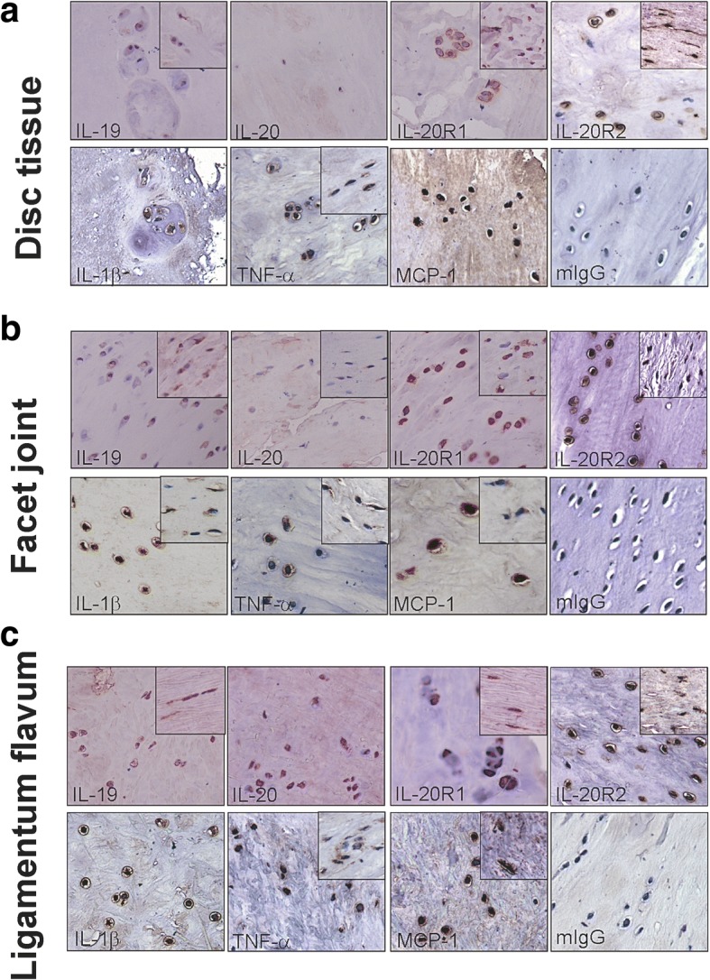
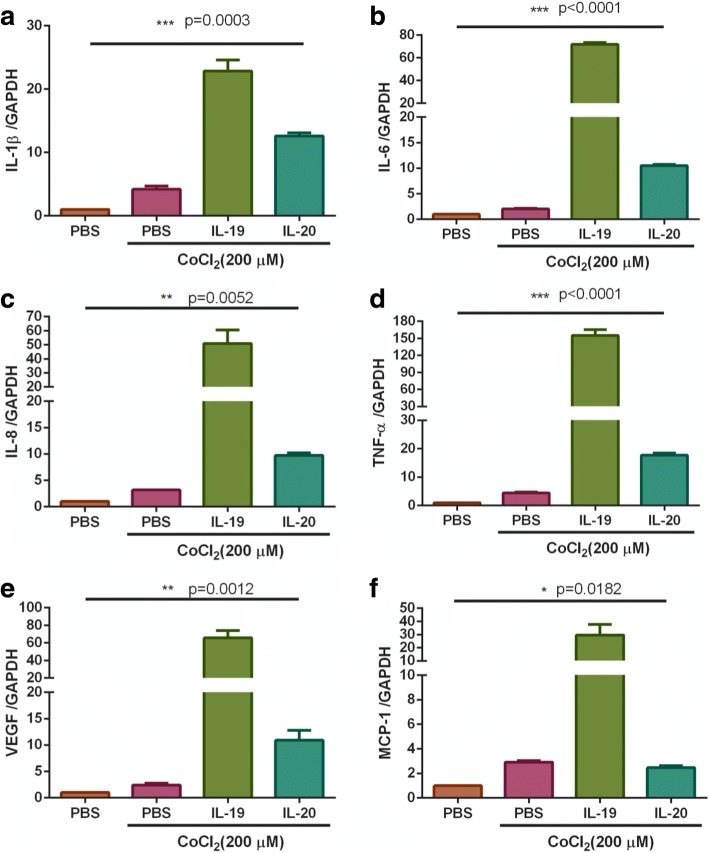
Methods: Disc, facet joint and ligamentum flavum from 13 patients with DLS was retrieved, and the expression pattern of IL-19, IL-20, IL-20R1, IL-20R2, TNF-α, IL-1β, and MCP-1 was evaluated using immunohistochemical staining with specific antibodies. The disc cells were isolated and incubated with IL-19 and IL-20 under CoCl2-mimicked hypoxic conditions to analyze the proinflammatory cytokine expression pattern using real-time quantitative PCR with specific primers.
Results: IL-19 and IL-20 were positively stained and accompanied by abundant expression of TNF-α, IL-1β, and MCP-1 in facet joints of DLS patients. IL-19 and IL-20's receptors (IL-20R1 and IL-20R2) were expressed on chondrocytes and fibrocytes/fibroblasts in facet joint and ligamentum flavum tissues from patients with DLS. There was a significant correlation between the expression of IL-20 and IL-1β in facet joint. In vitro assay, IL-19 and IL-20 upregulated the expression of IL-1β, IL-6, TNF-α, IL-8, VEGF, and MCP-1 in primary cultured DLS disc cells under CoCl2-mimicked hypoxic conditions.
Conclusions: IL-19, IL-20, and their receptors as well as proinflammatory cytokines (TNF-α, IL-1β, and MCP-1) were expressed more in facet joints than the other tissues in patients with DLS; therefore, the etiology of inflammation might be more facet-centric. IL-19 and IL-20 induced proinflammatory cytokine expression in disc cells and might play a role in the pathogenesis of DLS.
Keywords: Degenerative lumbar Spondylolisthesis; IL-19; IL-20; Inflammation.
Conflict of interest statement
The written informed consents for participation in the study were obtained from participants. All the procedures were approved by the Human Experiment and Ethics Committee of National Cheng Kung University Medical Center (IRB approval: ER-96-163.), and were done in accordance with the Guidelines of the Declaration of Helsinki.Not applicable.The authors declare that they have no competing interests.Springer Nature remains neutral with regard to jurisdictional claims in published maps and institutional affiliations.
Figures

Similar articles
-
Inflammatory cytokines released from the facet joint tissue in degenerative lumbar spinal disorders.Spine (Phila Pa 1976). 2004 Oct 1;29(19):2091-5. doi: 10.1097/01.brs.0000141265.55411.30. Spine (Phila Pa 1976). 2004. PMID: 15454697
-
IL-20 may contribute to the pathogenesis of human intervertebral disc herniation.Spine (Phila Pa 1976). 2008 Sep 1;33(19):2034-40. doi: 10.1097/BRS.0b013e31817eb872. Spine (Phila Pa 1976). 2008. PMID: 18758357
-
Correlation between inflammatory cytokines released from the lumbar facet joint tissue and symptoms in degenerative lumbar spinal disorders.J Orthop Sci. 2007 Mar;12(2):154-60. doi: 10.1007/s00776-006-1105-y. Epub 2007 Mar 30. J Orthop Sci. 2007. PMID: 17393271
-
Facet Tropism in Lumbar Spine and Cervical Spine: A Systematic Review and Meta-Analysis.World Neurosurg. 2021 Mar;147:47-65. doi: 10.1016/j.wneu.2020.11.171. Epub 2020 Dec 9. World Neurosurg. 2021. PMID: 33309642
-
The role of IL-1β and TNF-α in intervertebral disc degeneration.Biomed Pharmacother. 2020 Nov;131:110660. doi: 10.1016/j.biopha.2020.110660. Epub 2020 Aug 24. Biomed Pharmacother. 2020. PMID: 32853910 Review.
Cited by
-
The expression of P16 and S100 associated with elastin degradation and fibrosis of the Ligamentum Flavum hypertrophy.BMC Musculoskelet Disord. 2019 Oct 22;20(1):458. doi: 10.1186/s12891-019-2825-4. BMC Musculoskelet Disord. 2019. PMID: 31638980 Free PMC article.
-
DNA Demethylation of Promoter Region Facilitates Atoh-1-Induced Interleukin-19 Expression Activation in Bone Marrow Monocytes of Old Mice.Aging Dis. 2024 Jan 12;16(1):540-51. doi: 10.14336/AD.2024.0108. Online ahead of print. Aging Dis. 2024. PMID: 38300634 Free PMC article.
-
1,25-Dihydroxyvitamin D3 Negatively Regulates the Inflammatory Response to Porcine Epidemic Diarrhea Virus Infection by Inhibiting NF-κB and JAK/STAT Signaling Pathway in IPEC-J2 Porcine Epithelial Cells.Int J Mol Sci. 2022 Sep 13;23(18):10603. doi: 10.3390/ijms231810603. Int J Mol Sci. 2022. PMID: 36142545 Free PMC article.
-
PET/CT imaging of spinal inflammation and microcalcification in patients with low back pain: A pilot study on the quantification by artificial intelligence-based segmentation.Clin Physiol Funct Imaging. 2022 Jul;42(4):225-232. doi: 10.1111/cpf.12751. Epub 2022 Apr 1. Clin Physiol Funct Imaging. 2022. PMID: 35319166 Free PMC article.
-
The Thermosensitive Injectable Celecoxib-Loaded Chitosan Hydrogel for Repairing Postoperative Intervertebral Disc Defect.Front Bioeng Biotechnol. 2022 Jun 28;10:876157. doi: 10.3389/fbioe.2022.876157. eCollection 2022. Front Bioeng Biotechnol. 2022. PMID: 35837544 Free PMC article.
References
LinkOut - more resources
Full Text Sources
Other Literature Sources
Miscellaneous
