

Iron and Alzheimer's Disease: From Pathogenesis to Therapeutic Implications
- PMID: 30250423
- PMCID: PMC6139360
- DOI: 10.3389/fnins.2018.00632
Iron and Alzheimer's Disease: From Pathogenesis to Therapeutic Implications
Abstract
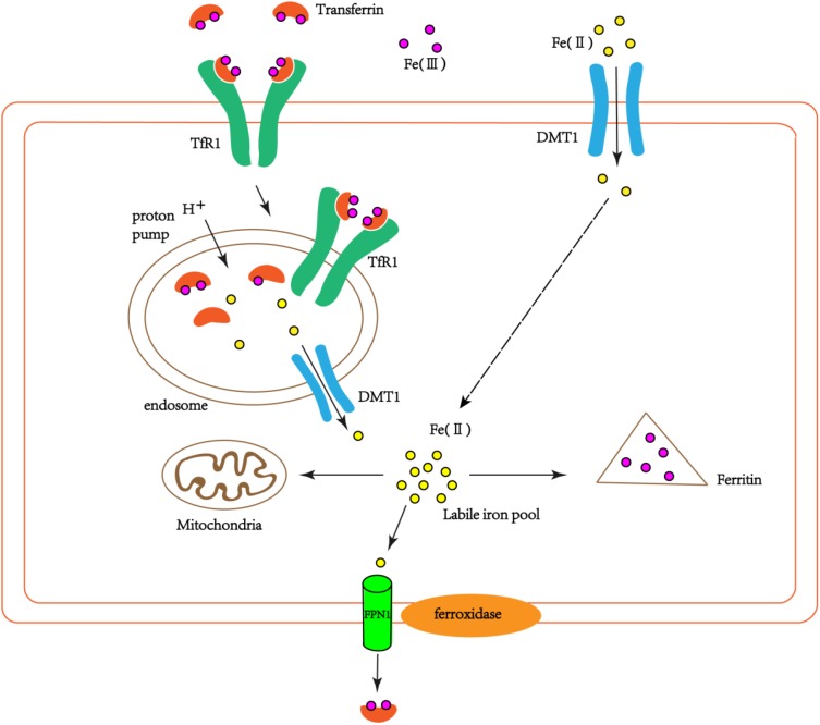
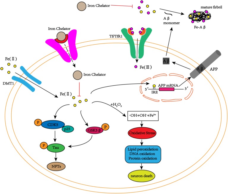
As people age, iron deposits in different areas of the brain may impair normal cognitive function and behavior. Abnormal iron metabolism generates hydroxyl radicals through the Fenton reaction, triggers oxidative stress reactions, damages cell lipids, protein and DNA structure and function, and ultimately leads to cell death. There is an imbalance in iron homeostasis in Alzheimer's disease (AD). Excessive iron contributes to the deposition of β-amyloid and the formation of neurofibrillary tangles, which in turn, promotes the development of AD. Therefore, iron-targeted therapeutic strategies have become a new direction. Iron chelators, such as desferoxamine, deferiprone, deferasirox, and clioquinol, have received a great deal of attention and have obtained good results in scientific experiments and some clinical trials. Given the limitations and side effects of the long-term application of traditional iron chelators, alpha-lipoic acid and lactoferrin, as self-synthesized naturally small molecules, have shown very intriguing biological activities in blocking Aβ-aggregation, tauopathy and neuronal damage. Despite a lack of evidence for any clinical benefits, the conjecture that therapeutic chelation, with a special focus on iron ions, is a valuable approach for treating AD remains widespread.
Keywords: Alzheimer’s disease; alpha-lipoic acid; chelation; iron; lactoferrin.
Figures
Similar articles
-
Iron Pathophysiology in Alzheimer's Diseases.Adv Exp Med Biol. 2019;1173:67-104. doi: 10.1007/978-981-13-9589-5_5. Adv Exp Med Biol. 2019. PMID: 31456206 Review.
-
Porphyromonas Gingivalis May Seek the Alzheimer's Disease Brain to Acquire Iron from Its Surplus.J Alzheimers Dis Rep. 2021 Jan 20;5(1):79-86. doi: 10.3233/ADR-200272. J Alzheimers Dis Rep. 2021. PMID: 33681719 Free PMC article.
-
Effect of metal chelators on the aggregation of beta-amyloid peptides in the presence of copper and iron.Biometals. 2017 Apr;30(2):285-293. doi: 10.1007/s10534-017-0005-2. Epub 2017 Mar 10. Biometals. 2017. PMID: 28281098
-
The Case for Abandoning Therapeutic Chelation of Copper Ions in Alzheimer's Disease.Front Neurosci. 2017 Jun 2;11:317. doi: 10.3389/fnins.2017.00317. eCollection 2017. Front Neurosci. 2017. PMID: 28626387 Free PMC article. Review.
-
Metal Ions in Alzheimer's Disease: A Key Role or Not?Acc Chem Res. 2019 Jul 16;52(7):2026-2035. doi: 10.1021/acs.accounts.9b00248. Epub 2019 Jul 5. Acc Chem Res. 2019. PMID: 31274278
Cited by
-
Biochemistry of mammalian ferritins in the regulation of cellular iron homeostasis and oxidative responses.Sci China Life Sci. 2021 Mar;64(3):352-362. doi: 10.1007/s11427-020-1795-4. Epub 2020 Sep 17. Sci China Life Sci. 2021. PMID: 32974854 Review.
-
A computational protocol for the calculation of the standard reduction potential of iron complexes: application to Fe2+/3+-Aβ model systems relevant to Alzheimer's disease.RSC Adv. 2022 Sep 7;12(37):24077-24087. doi: 10.1039/d2ra03907a. eCollection 2022 Aug 22. RSC Adv. 2022. PMID: 36200023 Free PMC article.
-
Beneficial and Detrimental Roles of Heme Oxygenase-1 in the Neurovascular System.Int J Mol Sci. 2022 Jun 24;23(13):7041. doi: 10.3390/ijms23137041. Int J Mol Sci. 2022. PMID: 35806040 Free PMC article. Review.
-
What Is the Role of Ferroptosis in Neurodegeneration?Neurology. 2023 Aug 15;101(7):312-319. doi: 10.1212/WNL.0000000000207730. Neurology. 2023. PMID: 37580137 Free PMC article. No abstract available.
-
Delivering the Promise of Gene Therapy with Nanomedicines in Treating Central Nervous System Diseases.Adv Sci (Weinh). 2022 Sep;9(26):e2201740. doi: 10.1002/advs.202201740. Epub 2022 Jul 18. Adv Sci (Weinh). 2022. PMID: 35851766 Free PMC article. Review.
References
-
- Alzheimer’s Association (2016). 2016 Alzheimer’s disease facts and figures. Alzheimers Dement. 12 459–509. - PubMed
Publication types
LinkOut - more resources
Full Text Sources
Other Literature Sources
Research Materials