Review
. 2018 Sep 19:13:7.
doi: 10.1186/s13008-018-0040-6. eCollection 2018.

The functional diversity of Aurora kinases: a comprehensive review

Affiliations
Review

The functional diversity of Aurora kinases: a comprehensive review

Estelle Willems et al. Cell Div. .

Abstract

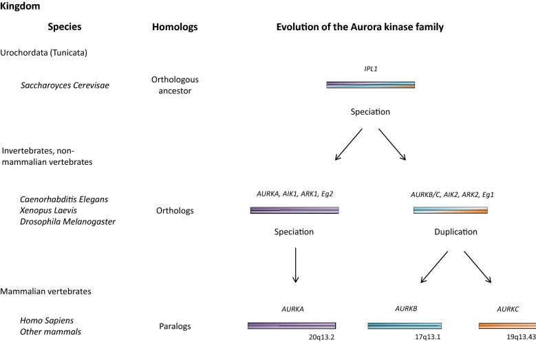
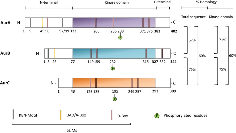
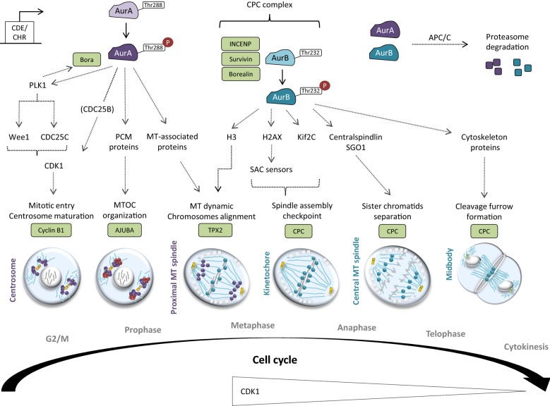
Aurora kinases are serine/threonine kinases essential for the onset and progression of mitosis. Aurora members share a similar protein structure and kinase activity, but exhibit distinct cellular and subcellular localization. AurA favors the G2/M transition by promoting centrosome maturation and mitotic spindle assembly. AurB and AurC are chromosome-passenger complex proteins, crucial for chromosome binding to kinetochores and segregation of chromosomes. Cellular distribution of AurB is ubiquitous, while AurC expression is mainly restricted to meiotically-active germ cells. In human tumors, all Aurora kinase members play oncogenic roles related to their mitotic activity and promote cancer cell survival and proliferation. Furthermore, AurA plays tumor-promoting roles unrelated to mitosis, including tumor stemness, epithelial-to-mesenchymal transition and invasion. In this review, we aim to understand the functional interplay of Aurora kinases in various types of human cells, including tumor cells. The understanding of the functional diversity of Aurora kinases could help to evaluate their relevance as potential therapeutic targets in cancer.

Keywords: Aurora kinase; Cancer; Mitosis.


Figures

Fig. 1
Fig. 1
Evolution of the Aurora kinase family: Aurora members evolved from a single ancestor gene, called Ipl1 and initially identified in Saccharomyces cerevisiae and other Urochordata (renamed Tunicata). In invertebrates and non-mammalian vertebrates (e.g. Caenorhabditis elegans, Xenopus laevis and Drosophila melanogaster), the Aurora family is constituted by two orthologs: the AURKA (also called AIK1, ARK1, Eg2) and the AURKB (also called AIK2, ARK2, Eg1) ancestor genes. In mammals, AURKB/AURKC ancestor gene duplication gave rise to AURKB and AURKC paralogs. In human, AURKA, AURKB and AURKC map on chromosomes 20q13.2, 17p13.1, and 19q13.43 respectively
Fig. 2
Fig. 2
Structure of Aurora kinase domains. Aurora kinases are constituted by (i) an N-terminal domain, believed to control protein localization, (ii) a large and conserved catalytic domain containing the activation T-loop and (iii) a short C-tern domain with a D-box (Destruction Box). AurA and AurB also contain a KEN motif and an A-box that contributes to APC/C-dependent proteolysis. Numbers represent the residue position. Percentages of homology of the total amino acid sequence are evaluated (i) 57% between AurA and AurB, (ii) 75% between AurB and AurC and (iii) 60% between AurA and AurC. Percentages of homology of the catalytic domain are evaluated at (i) 71% between AurA and AurB, (ii) 75% between AurB and AurC and (iii) 60% between AurA and AurC
Fig. 3
Fig. 3
Role of AurA and AurB in mitosis. The cell-cycle dependent transcription of AurA and AurB are under the control of the CDE/CHR elements, which are recognized by the E4TF1 transcription factor. AurA is mainly activated after Thr288 auto-phosphorylation. Bora, a key AurA co-factor, is phosphorylated by AurA and, in return, Bora enhances the kinase activity of AurA. Once activated, AurA phosphorylates and activates CDK1-Cyclin B to allow G2/M checkpoint unlock through various mechanisms, including: (i) PLK1-dependent targeting of Wee1 and CDC25C, (ii) CDC25B-dependent activation of CDK1 and (ii) direct phosphorylation of CDK1. Then, PLK1 mediates Bora degradation to permit mitosis progression. At G2/M, AurA localizes in the centrosome and also contributes to their maturation before mitotic entry. At prophase, AurA—whose activity is maintained by Ajuba- recruits and phosphorylates several PCM proteins (i.e. γ-TuRC, centrosomin, NDEL1, TACC, LATS2 and BRCA1) to organize the MTOC. At metaphase, AurA moves to the proximal MT and targets MT-associated proteins (i.e. Ki2a, TACC3, CKAP5-a) to organize the mitotic spindle. At this time, TPX2 allows the maintenance of the activate state of AurA. AurB binds INCENP, Survivin and Borealin to form the CPC complex and to be activated upon Thr232 auto-phosphorylation. AurB, firstly localized on chromosomes, contributes to their proper alignment at metaphase. Prior anaphase, AurB concentrates to the kinetochore to allow the spindle assembly checkpoint (SAC) crossing through (i) H2AX-dependent activation of SAC sensors and (ii) Kif2C recruitment. Then, AurB moves to the central MT to trigger sister chromatids separation through Centralspindlin and SGO1 recruitment at anaphase. Finally, AurB targets various cytoskeleton regulatory proteins (RhoA, Vimentin, Desmin, GFAP) at the midbody in order to organize the cleavage furrow for cytokinesis. At the end of mitosis, both AurA and AurB undergo ubiquitination and proteasome degradation by APC/C, which happen subsequently to their dephosphorylation by PP2A or PP1

References

    1. National Institute of General Medicine. Mitosis phases image. https://publications.nigms.nih.gov/insidethecell/ch4_phases_allbig.html. 2018. p. 1. https://publications.nigms.nih.gov/order/pubs_gateway.html. Accessed 11 Jun 2018.
    1. Sansregret Laurent, Swanton Charles. The Role of Aneuploidy in Cancer Evolution. Cold Spring Harbor Perspectives in Medicine. 2016;7(1):a028373. doi: 10.1101/cshperspect.a028373. - DOI - PMC - PubMed
    1. Hochegger H, Hégarat N, Pereira-Leal JB. Aurora at the pole and equator: overlapping functions of Aurora kinases in the mitotic spindle. Open Biol. 2013;3(3):120185. doi: 10.1098/rsob.120185. - DOI - PMC - PubMed
    1. Compton DA. Mechanism of aneuploidy. Curr Opin Cell Biol. 2011;23(1):109–113. doi: 10.1016/j.ceb.2010.08.007. - DOI - PMC - PubMed
    1. Manning G. The Protein Kinase Complement of the Human Genome. Science. 2002;298(5600):1912–1934. doi: 10.1126/science.1075762. - DOI - PubMed