

Real-time assessment of platinum sensitivity of primary culture from a patient with ovarian cancer with extensive metastasis and the platinum sensitivity enhancing effect by metformin
- PMID: 30250536
- PMCID: PMC6144930
- DOI: 10.3892/ol.2018.9223
Real-time assessment of platinum sensitivity of primary culture from a patient with ovarian cancer with extensive metastasis and the platinum sensitivity enhancing effect by metformin
Abstract
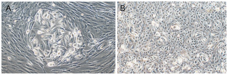
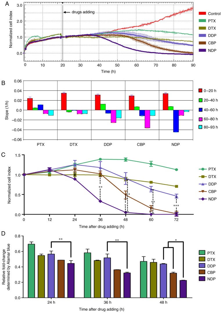
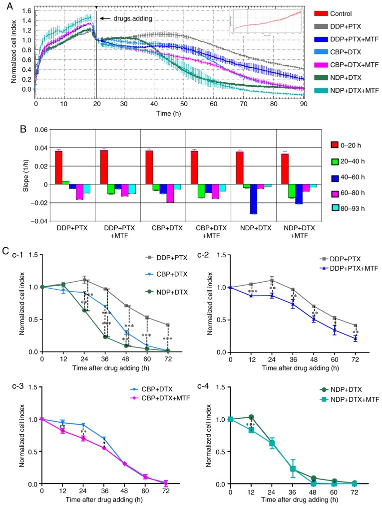
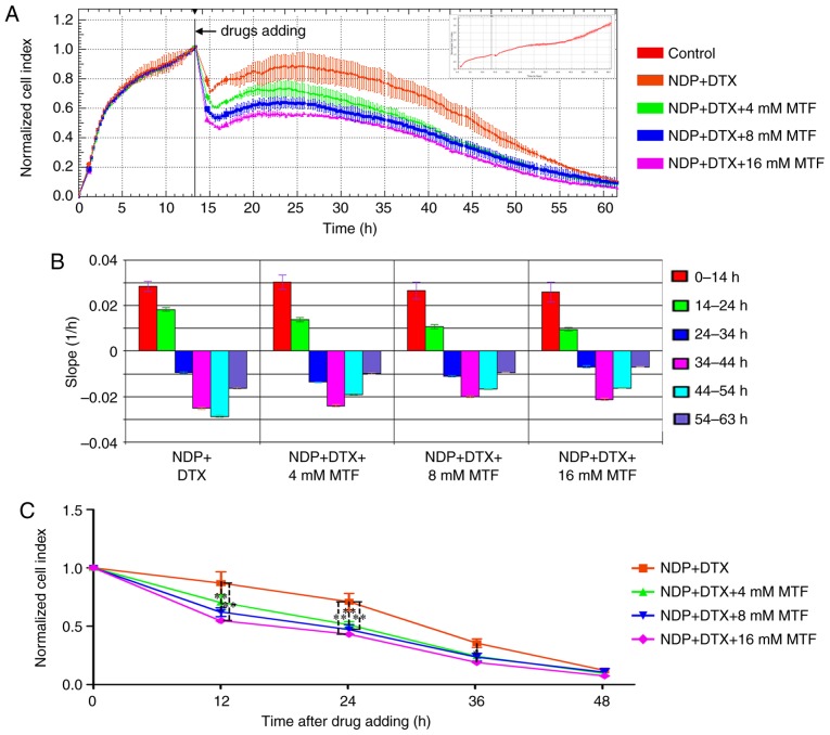
The aim of the present study was to perform a rapid evaluation of the efficiency of commonly used platinum-based chemotherapy regimens for patients with ovarian cancer with extensive metastases using an in vitro method combined with culturing primary cells and real-time monitoring, and to further explore the enhanced effect of metformin on susceptibility of ovarian cancer cells to platinum-based chemotherapy. The primary omental metastatic (OM) cells were isolated from the omentum metastasis of a surgical patient with stage IIIc ovarian carcinoma. Drug sensitivity was evaluated using the xCELLigence system, and screening of the most effective platinum chemotherapy was performed through analysis of cell susceptibility to cisplatin, carboplatin, nedaplatin and paclitaxel or docetaxel alone or in combination. At the same time, this system was used to determine whether metformin was able to increase the sensitivity of cancer cells to platinum chemotherapy. The results revealed that nedaplatin exhibited the most marked cytotoxic effect on the OM cells, followed by those of carboplatin and cisplatin. The addition of docetaxel enhanced the cytotoxic effect, and the combination of platinum and paclitaxel also enhanced the effect. Metformin rapidly increased the sensitivity of cells to platinum-based chemotherapy, and this effect was dose-dependent. The sensitivity of OM cells to different platinum-based regimens was varied. The effect of metformin on chemotherapeutic sensitization of cancer cells is clear in vitro, and the real-time cell analyzer assay has the potential to assist in determining individualized drug regimens for patients with metastatic ovarian cancer.
Keywords: omentum metastases; ovarian cancer; platinum; primary cell culture; real-time cell analyzer system; sensitivity.
Figures
Similar articles
-
Association between in vitro platinum resistance in the EDR assay and clinical outcomes for ovarian cancer patients.Gynecol Oncol. 2002 Oct;87(1):8-16. doi: 10.1006/gyno.2002.6797. Gynecol Oncol. 2002. PMID: 12468336 Clinical Trial.
-
Chemosensitizing effects of metformin on cisplatin- and paclitaxel-resistant ovarian cancer cell lines.Pharmacol Rep. 2018 Jun;70(3):409-417. doi: 10.1016/j.pharep.2017.11.007. Epub 2017 Nov 21. Pharmacol Rep. 2018. PMID: 29627688
-
In vitro phase II comparison of the cytotoxicity of a novel platinum analog, nedaplatin (254-S), with that of cisplatin and carboplatin against fresh, human ovarian cancers.Cancer Chemother Pharmacol. 1997;39(6):493-7. doi: 10.1007/s002800050604. Cancer Chemother Pharmacol. 1997. PMID: 9118460
-
Medical therapy of advanced malignant epithelial tumours of the ovary.Forum (Genova). 2000 Oct-Dec;10(4):323-32. Forum (Genova). 2000. PMID: 11535983 Review.
-
The integration of paclitaxel and new platinum compounds in the treatment of advanced ovarian cancer.Int J Gynecol Cancer. 2001;11 Suppl 1:21-30. doi: 10.1046/j.1525-1438.2001.11(suppl.1)sup1021.x. Int J Gynecol Cancer. 2001. PMID: 11488999 Review.
Cited by
-
Metformin in gynecological disorders: pathogenic insights and therapeutic implications.Front Pharmacol. 2025 Apr 22;16:1526709. doi: 10.3389/fphar.2025.1526709. eCollection 2025. Front Pharmacol. 2025. PMID: 40331195 Free PMC article. Review.
-
Recycling the Purpose of Old Drugs to Treat Ovarian Cancer.Int J Mol Sci. 2020 Oct 20;21(20):7768. doi: 10.3390/ijms21207768. Int J Mol Sci. 2020. PMID: 33092251 Free PMC article. Review.
-
Metformin and ovarian cancer: the evidence.Ann Transl Med. 2020 Dec;8(24):1711. doi: 10.21037/atm-20-1060. Ann Transl Med. 2020. PMID: 33490223 Free PMC article. Review.
-
circ_C20orf11 enhances DDP resistance by inhibiting miR-527/YWHAZ through the promotion of extracellular vesicle-mediated macrophage M2 polarization in ovarian cancer.Cancer Biol Ther. 2021 Sep 2;22(7-9):440-454. doi: 10.1080/15384047.2021.1959792. Epub 2021 Aug 12. Cancer Biol Ther. 2021. PMID: 34382916 Free PMC article.
-
Phase I study of metformin in combination with carboplatin/paclitaxel chemotherapy in patients with advanced epithelial ovarian cancer.Invest New Drugs. 2020 Oct;38(5):1454-1462. doi: 10.1007/s10637-020-00920-7. Epub 2020 Mar 7. Invest New Drugs. 2020. PMID: 32146550 Free PMC article. Clinical Trial.
References
LinkOut - more resources
Full Text Sources
Other Literature Sources