

Increased plasma levels of galectin-1 in pancreatic cancer: potential use as biomarker
- PMID: 30250644
- PMCID: PMC6152472
- DOI: 10.18632/oncotarget.26034
Increased plasma levels of galectin-1 in pancreatic cancer: potential use as biomarker
Abstract
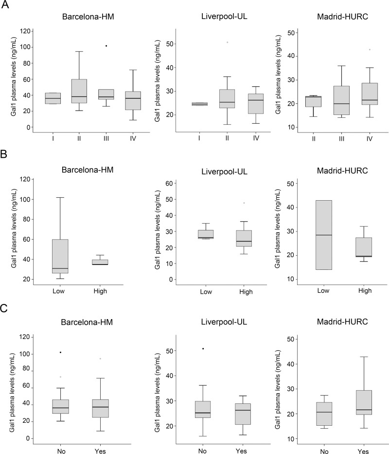
Pancreatic ductal adenocarcinoma (PDA) is the most frequent type of pancreatic cancer and one of the deadliest diseases overall. New biomarkers are urgently needed to allow early diagnosis, one of the only factors that currently improves prognosis. Here we analyzed whether the detection of circulating galectin-1 (Gal-1), a soluble carbohydrate-binding protein overexpressed in PDA tissue samples, can be used as a biomarker for PDA. Gal-1 levels were determined by ELISA in plasma from healthy controls and patients diagnosed with PDA, using three independent cohorts. Patients with chronic pancreatitis (CP) were also included in the study to analyze the potential of Gal-1 to discriminate between cancer and inflammatory process. Plasma Gal-1 levels were significantly increased in patients with PDA as compared to controls in all three cohorts. Gal-1 sensitivity and specificity values were similar to that of the CA19-9 biomarker (the only FDA-approved blood test biomarker for PDA), and the combination of Gal-1 and CA19-9 significantly improved their individual discriminatory powers. Moreover, high levels of Gal-1 were associated with lower survival in patients with non-resected tumors. Collectively, our data indicate a strong potential of using circulating Gal-1 levels as a biomarker for detection and prognostics of patients with PDA.
Keywords: biomarker; chronic pancreatitis; galectin-1; pancreatic cancer.
Conflict of interest statement
CONFLICTS OF INTEREST No conflicts of interest were declared.
Figures


References
-
- Neoptolemos JP, Kleeff J, Michl P, Costello E, Greenhalf W, Palmer DH. Therapeutic developments in pancreatic cancer: current and future perspectives. Nat Rev Gastroenterol Hepatol. 2018;15:333–48. - PubMed
-
- Bardeesy N, DePinho RA. Pancreatic cancer biology and genetics. Nat Rev Cancer. 2002;2:897–909. - PubMed
-
- Hruban RH, Takaori K, Klimstra DS, Adsay NV, Albores-Saavedra J, Biankin AV, Biankin SA, Compton C, Fukushima N, Furukawa T, Goggins M, Kato Y, Kloppel G, et al. An illustrated consensus on the classification of pancreatic intraepithelial neoplasia and intraductal papillary mucinous neoplasms. Am J Surg Pathol. 2004;28:977–87. - PubMed
-
- Lowenfels AB, Maisonneuve P, Cavallini G, Ammann RW, Lankisch PG, Andersen JR, Dimagno EP, Andren-Sandberg A, Domellof L. Pancreatitis and the risk of pancreatic cancer. International Pancreatitis Study Group. N Engl J Med. 1993;328:1433–7. - PubMed
LinkOut - more resources
Full Text Sources
Other Literature Sources
Research Materials
Miscellaneous
