

Case Reports
doi: 10.7759/cureus.3008.
Intra-articular Large Ossicle Associated to Osgood-Schlatter Disease
Affiliations
- PMID: 30250770
- PMCID: PMC6145751
- DOI: 10.7759/cureus.3008
Item in Clipboard
Case Reports
Intra-articular Large Ossicle Associated to Osgood-Schlatter Disease
Cureus.
.
Abstract
Osgood-Schlatter disease (OSD) is known as a self-limiting condition but surgical excision of the ossicles may be required in adults resistant to conservative treatments. The ossicle associated to OSD is generally small and located outside the joint near the tibial tubercle; however, large or intra-articular ossicle has been reported rarely. Here, we report an unusual case of OSD with a separated, large-sized ossicle that protruded into the knee joint and treated by arthroscopy-assisted excision of the ossicle.
Keywords: knee arthroscopy; osgood -schlatter disease; ossicle.
Conflict of interest statement
The authors have declared that no competing interests exist.
Figures
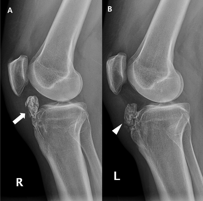
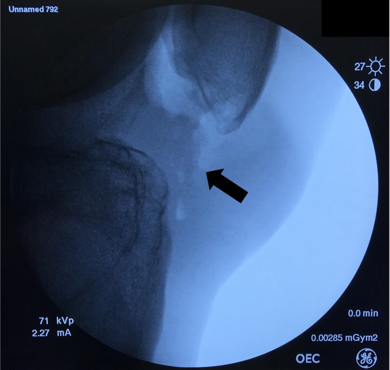
(A) A large osseous lesion (arrow) is seen at the infrapatellar area of the right knee. (B) An osseous body (arrowhead) of equivalent size was partially fused to the hypertrophied tibial tuberosity of the left knee.
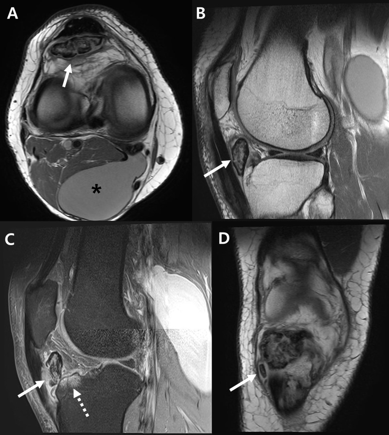
(A) A large ossicle with heterogenous signal intensity (arrow) and a Baker’s cyst (asterisk) are seen on axial image. (B) From T1-weighted sagittal image, the ossicle (arrow) is separately located inside the infrapatellar fat pad. (C) T2-weighted sagittal image shows inflammation of patellar tendon (solid arrow) and bone marrow edematous change of anterior tibia plateau (dotted arrow). (D) Small separated ossicle (arrow) was found inferior to the large ossicle from coronal image.
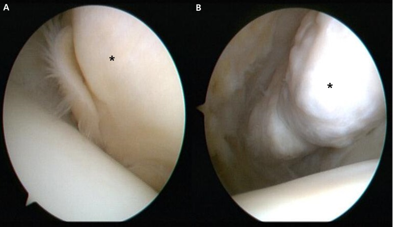
(A) The ossicle was covered by soft tissue (asterisk). (B) After removing the soft tissue by mechanical shaver, the ossicle (asterisk) is exposed.
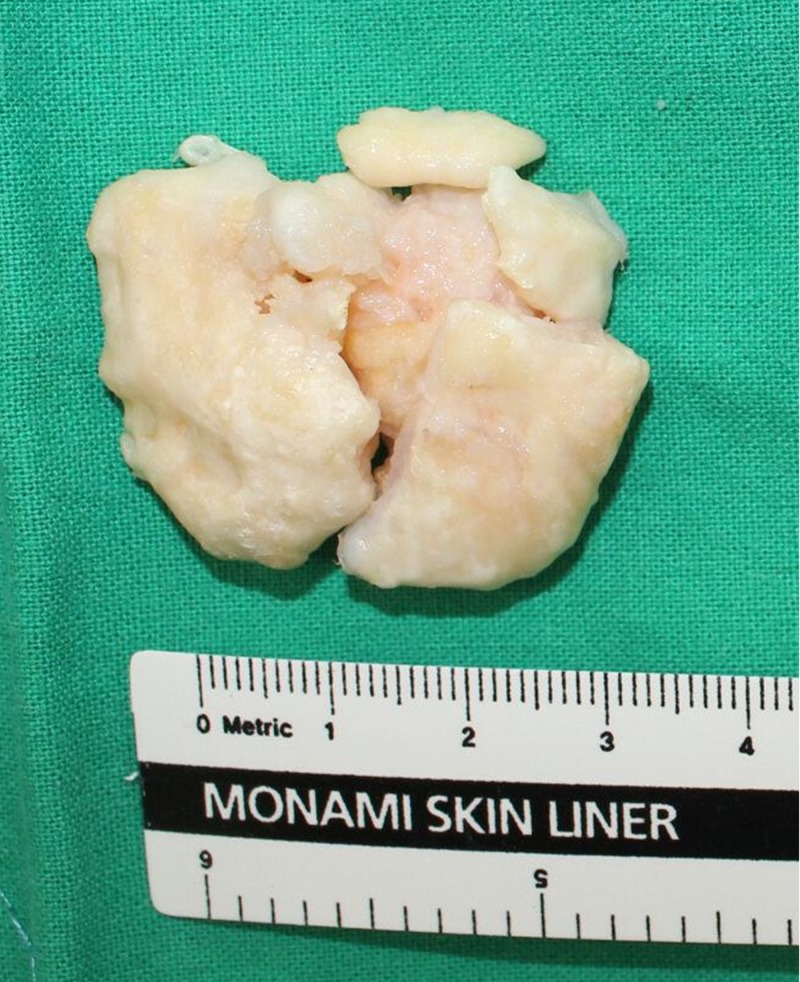
The ossicle was fragmented during surgery.
Complete removal of the ossicle was confirmed (arrow).
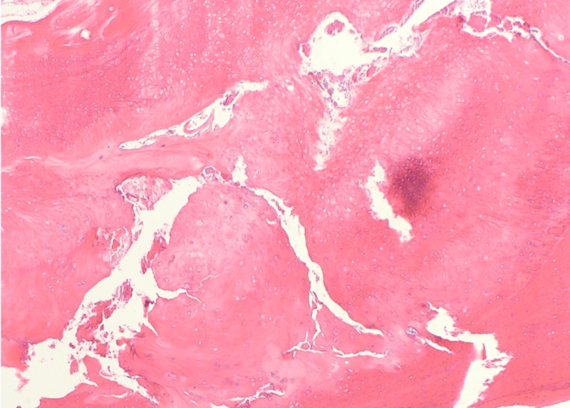
The hematoxylin and eosin staining showed osteocartilaginous tissue without distinct cartilage cap, suggestive of ossicle associated to Osgood-Schlatter disease.
Similar articles
-
Osgood-Schlatter Disease: Ossicle Resection and Patellar Tendon Repair in a Symptomatic Adult.Arthrosc Tech. 2024 Jul 9;13(11):103110. doi: 10.1016/j.eats.2024.103110. eCollection 2024 Nov. Arthrosc Tech. 2024. PMID: 39711897 Free PMC article.
-
Treatment of Osgood-Schlatter disease: review of the literature.Musculoskelet Surg. 2017 Dec;101(3):195-200. doi: 10.1007/s12306-017-0479-7. Epub 2017 Jun 7. Musculoskelet Surg. 2017. PMID: 28593576 Review.
-
Arthroscopic Excision of a Huge Ununited Ossicle Due to Osgood-Schlatter Disease in an Adult Patient.J Orthop Case Rep. 2013 Apr-Jun;3(2):4-7. doi: 10.13107/jocr.2250-0685.092. J Orthop Case Rep. 2013. PMID: 27298897 Free PMC article.
-
Arthroscopic excision of an ununited ossicle due to Osgood-Schlatter disease.Arthroscopy. 2008 Sep;24(9):1081-3. doi: 10.1016/j.arthro.2007.03.010. Epub 2007 May 7. Arthroscopy. 2008. PMID: 18760218
-
Apophysitis of the Tibial Tuberosity (Osgood-Schlatter Disease): A Review.Cureus. 2016 Sep 13;8(9):e780. doi: 10.7759/cureus.780. Cureus. 2016. PMID: 27752406 Free PMC article. Review.
Cited by
-
Not just for boys: a rare case of symptomatic Osgood-Schlatter disease in a skeletally mature woman.BMJ Case Rep. 2019 Mar 26;12(3):e228963. doi: 10.1136/bcr-2018-228963. BMJ Case Rep. 2019. PMID: 30914412 Free PMC article. No abstract available.
-
Osgood-Schlatter Disease: Ossicle Resection and Patellar Tendon Repair in a Symptomatic Adult.Arthrosc Tech. 2024 Jul 9;13(11):103110. doi: 10.1016/j.eats.2024.103110. eCollection 2024 Nov. Arthrosc Tech. 2024. PMID: 39711897 Free PMC article.
References
-
- Arthroscopic excision of an ununited ossicle due to Osgood-Schlatter disease. Beyzadeoglu T, Inan M, Bekler H, Altintas F. Arthroscopy. 2008;24:1081–1083. - PubMed
-
- Results of arthroscopic treatment in unresolved Osgood-Schlatter disease in athletes. Circi E, Beyzadeoglu T. Int Orthop. 2017;41:351–356. - PubMed
-
- Direct bursoscopic ossicle resection in young and active patients with unresolved Osgood-Schlatter disease. Eun SS, Lee SA, Kumar R, Sul EJ, Lee SH, Ahn JH, Chang MJ. Arthroscopy. 2015;31:416–421. - PubMed
-
- Long-term outcome after surgical treatment of unresolved Osgood-Schlatter disease in young men. Pihlajamaki HK, Mattila VM, Parviainen M, Kiuru MJ, Visuri TI. J Bone Joint Surg Am. 2009;91:2350–2358. - PubMed
-
- Treatment of Osgood-Schlatter disease: review of the literature. Circi E, Atalay Y, Beyzadeoglu T. Musculoskelet Surg. 2017;101:195–200. - PubMed
Publication types
LinkOut - more resources
Full Text Sources
Other Literature Sources