

Pre-amyloid stage of Alzheimer's disease in cognitively normal individuals
- PMID: 30250861
- PMCID: PMC6144448
- DOI: 10.1002/acn3.615
Pre-amyloid stage of Alzheimer's disease in cognitively normal individuals
Abstract
Objective: To study risk factors for decreasing aβ1-42 concentrations in cerebrospinal fluid (CSF) in cognitively unimpaired individuals with initially normal amyloid and tau markers, and to investigate whether such aβ1-42 decreases are associated with subsequent decline in cognition and other biomarkers of Alzheimer's disease.
Methods: Cognitively normal subjects (n = 83, 75 ± 5 years, 35(42%) female) with normal CSF aβ1-42 and tau and repeated CSF sampling were selected from ADNI. Subject level slopes of aβ1-42 decreases were estimated with mixed models. We tested associations of baseline APP processing markers (BACE1 activity, aβ1-40, aβ1-38 and sAPP β) and decreasing aβ1-42 levels by including an interaction term between time and APP marker. Associations between decreasing aβ1-42 levels and clinical decline (i.e., progression to mild cognitive impairment or dementia, MMSE, memory functioning) and biological decline (tau, hippocampal volume, glucose processing and amyloid PET) over a time period of 8-10 years were assessed.
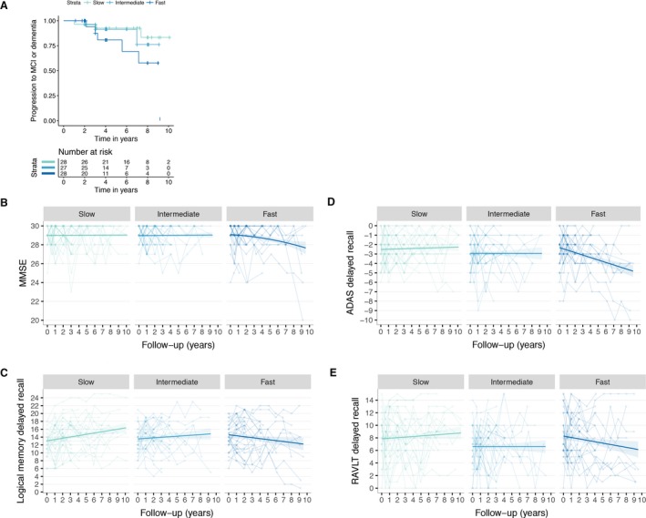
Results: Aβ1-42 levels decreased annually with -4.6 ± 1 pg/mL. Higher baseline BACE1 activity (β(se) = -0.06(0.03), P < 0.05), aβ1-40 (β(se)= -0.11(.03), P < 0.001), and aβ1-38 levels (β(se) = -0.11(0.03), P < 0.001) predicted faster decreasing aβ1-42. The fastest tertile of decreasing aβ1-42 rates was associated with subsequent pathophysiological processes: 11(14%) subjects developed abnormal amyloid levels after 3 ± 1.7 years, showed increased risk for clinical progression (Hazard Ratio[95CI] = 4.8[1.1-21.0]), decreases in MMSE, glucose metabolism and hippocampal volume, and increased CSF tau and amyloid aggregation on PET (all P < 0.05).
Interpretation: Higher APP processing and fast decreasing aβ1-42 could be among the earliest, pre-amyloid, pathological changes in Alzheimer's disease.
Figures

References
-
- Mattsson N, Insel PS, Donohue M, et al. Predicting reduction of cerebrospinal fluid β‐amyloid 42 in cognitively healthy controls. JAMA Neurol 2015;72:554–560. - PubMed
-
- Gomar JJ, Conejero‐Goldberg C, Davies P, et al. Anti‐correlated cerebrospinal fluid biomarker trajectories in preclinical Alzheimer's disease. J Alzheimers Dis 2016;51:1085–1097. - PubMed
Grants and funding
LinkOut - more resources
Full Text Sources
Other Literature Sources
