Review
. 2018 Nov;194(2):166-179.
doi: 10.1111/cei.13163. Epub 2018 Sep 24.

Lymphocytes are a major source of circulating soluble dipeptidyl peptidase 4

Affiliations
Review

Lymphocytes are a major source of circulating soluble dipeptidyl peptidase 4

A Casrouge et al. Clin Exp Immunol. 2018 Nov.

Abstract

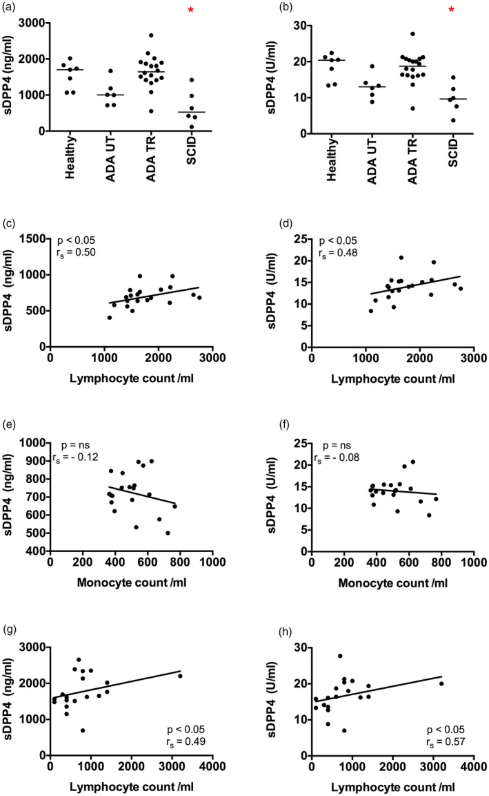
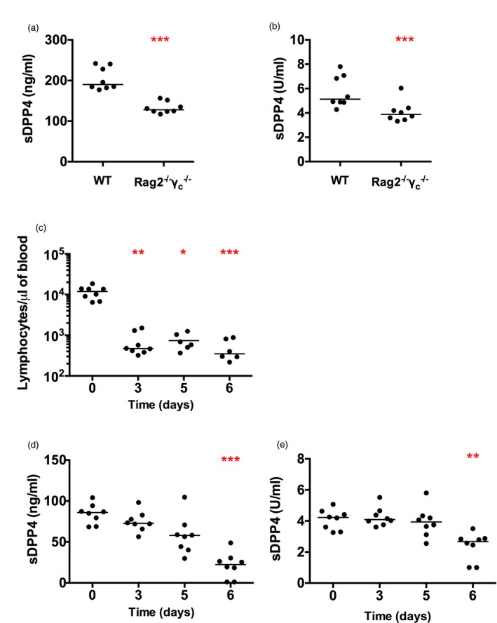
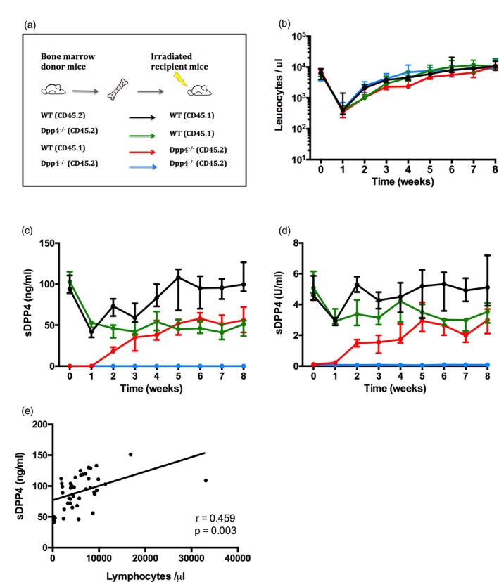
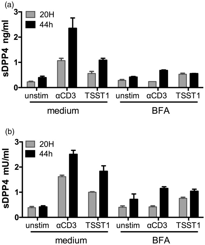
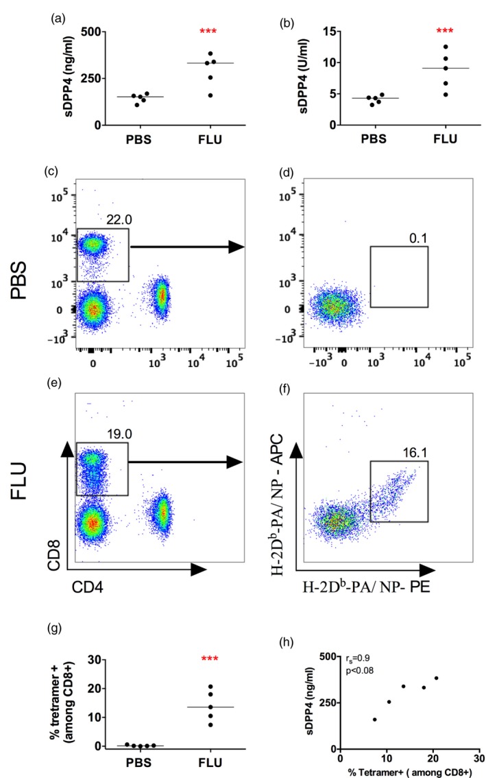
Dipeptidyl peptidase 4 (DPP4, CD26) is a serine protease that is expressed constitutively by many haematopoietic and non-haematopoietic tissues. It exists as a membrane-associated protein, as well as in an active, soluble form (herein called sDPP4), present at high concentrations in bodily fluids. Despite the proposed use of sDPP4 as a biomarker for multiple diseases, its cellular sources are not well defined. Here, we report that individuals with congenital lymphocyte immunodeficiency had markedly lower serum concentrations of sDPP4, which were restored upon successful treatment and restoration of lymphocyte haematopoiesis. Using irradiated lymphopenic mice and wild-type to Dpp4-/- reciprocal bone marrow chimeric animals, we found that haematopoietic cells were a major source of circulating sDPP4. Furthermore, activation of human and mouse T lymphocytes resulted in increased sDPP4, providing a mechanistic link between immune system activation and sDPP4 concentration. Finally, we observed that acute viral infection induced a transient increase in sDPP4, which correlated with the expansion of antigen-specific CD8+ T cell responses. Our study demonstrates that sDPP4 concentrations are determined by the frequency and activation state of lymphocyte populations. Insights from these studies will support the use of sDPP4 concentration as a biomarker for inflammatory and infectious diseases.

Keywords: T cells; chemokines; immunodeficiency diseases.


Figures

Figure 1
Figure 1
Patients with severe combined immunodeficiency have low levels of serum soluble dipeptidyl peptidase 4 (sDPP4). (a,b) The concentration (a) and activity level (b) of sDPP4 was measured in the serum of healthy children (healthy), untreated adenosine deaminase (ADA)‐deficient patients (ADA UT), treated ADA‐deficient patients (ADA TR) and severe combined immunodeficiency (SCID) patients. (c–f) Graphs show circulating lymphocyte or monocyte numbers versus sDPP4 concentration (c,e) or activity (d,f) in the serum of adult healthy donors. (g,h) Graphs show circulating lymphocyte number versus sDPP4 concentration (g) or activity (h) in the serum of treated ADA‐deficient patients. Each dot represents the mean value of one healthy donor or patient measured three times. Horizontal lines represent the median of the group. (a,b) Differences between cohorts were tested using the Kruskal–Wallis test followed by Dunn's multiple comparison test. *P < 0·05. (c–h) Spearman's correlation (r s) and P‐value for each analysis are shown.
Figure 2
Figure 2
Soluble dipeptidyl peptidase 4 (sDPP4) level and activity are decreased in immunodeficient mice and after lymphocyte depletion by irradiation. Graphs show (a) sDPP4 concentration and (b) sDPP4 activity in wild‐type (WT) and Rag –/– γc –/– mice. (c–e) Mice were exposed to a lethal dose of irradiation. Graphs depict (c) the number of circulating lymphocytes, (d) the sDPP4 concentration and (e) sDPP4 activity in irradiated mice over time. Each dot represents one mouse. The experiment was performed twice. Horizontal lines represent the median. (a,b) Differences between groups were tested for statistical significance using the Mann–Whitney test (c–e). Differences among groups were tested for statistical significance using the Kruskal–Wallis test followed by Dunn's multiple comparison test, comparing all time‐points to day 0. *P < 0·05; **P < 0·01; ***P < 0·001.
Figure 3
Figure 3
Bone marrow‐derived cells are a major source of soluble dipeptidyl peptidase 4 (sDPP4) in plasma. (a) Schematic representation of the experimental model. (b–e) Mice were lethally irradiated and reconstituted with bone marrow according to the experimental design as illustrated in (a). (b) Graph shows the number of donor leucocytes in circulation in recipient mice. (c) The graphs depict sDPP4 concentration and (d) sDPP4 activity in the plasma of recipient mice. Lines and dots show the mean for each group, with standard deviations. The experiment was performed twice, with three to five mice per group. One representative experiment is shown. (e) Graph shows circulating lymphocyte number versus sDPP4 concentration in [wild‐type (WT) → WT] group mice during the experimental follow‐up.
Figure 4
Figure 4
Peripheral blood mononuclear cell (PBMC) stimulation by anti‐CD3 and superantigen toxic shock syndrome toxin 1 (TSST1) induces release of soluble dipeptidyl peptidase 4 (sDPP4). PBMCs were isolated from healthy donors and stimulated as indicated and treated or not with brefeldin A (BFA). (a) sDPP4 concentration and (b) activity were measured in supernatants after 20 or 44 h of culture. Bars represent errors of experimental duplicates. A representative experiment of seven independent experiments with different healthy donors is shown.
Figure 5
Figure 5
Induced soluble dipeptidyl peptidase 4 (sDPP4) by influenza virus is correlated with the intensity of tetramer CD8+ T cell‐specific responses. Mice were infected with influenza virus PR8 (influenza). (a,b) Graphs show (a) concentration and (b) activity of sDPP4 in blood of mice 7 days after infection. (c–f) Flow cytometry dot‐plots show combined tetramer (H‐2Db‐PA)‐ and (H‐2Db‐NP)‐specific cells in a representative (c,d) phosphate‐buffered saline (PBS)‐treated and (e,f) influenza‐infected mouse. (g) Graph shows the percentage of tetramer‐positive T cells in a group of five PBS‐treated and five influenza‐infected mice. (h) Graph shows the percentage of circulating tetramer‐positive cells versus sDPP4 levels. A representative experiment of three performed is shown. Spearman's correlation (r s) and P‐value are shown; ***P < 0·001.

References

    1. McCaughan GW, Wickson JE, Creswick PF, Gorrell MD. Identification of the bile canalicular cell‐surface molecule GP110 as the ectopeptidase dipeptidyl peptidase‐IV – an analysis by tissue distribution, purification and N‐terminal amino‐acid‐sequence. Hepatology 1990; 11:534–44. - PubMed
    1. Kameoka J, Tanaka T, Nojima Y, Schlossman SF, Morimoto C. Direct association of adenosine deaminase with a T cell activation antigen, CD26. Science 1993; 261:466–9. - PubMed
    1. Raj VS, Mou H, Smits SL et al Dipeptidyl peptidase 4 is a functional receptor for the emerging human coronavirus – EMC. Nature 2013; 495:251–4. - PMC - PubMed
    1. Chien CH, Huang LH, Chou CY et al One site mutation disrupts dimer formation in human DPP‐IV proteins. J Biol Chem 2004; 279:52338–45. - PubMed
    1. Durinx C, Lambeir AM, Bosmans E et al Molecular characterization of dipeptidyl peptidase activity in serum: soluble CD26/dipeptidyl peptidase IV is responsible for the release of X‐Pro dipeptides. Eur J Biochem 2000; 267:5608–13. - PubMed

Publication types