Review
. 2019;15(1):12-23.
doi: 10.2174/1573403X14666180925095923.

Role of Cardiac Magnetic Resonance Imaging in Patients with Idiopathic Ventricular Arrhythmias

Affiliations
Review

Role of Cardiac Magnetic Resonance Imaging in Patients with Idiopathic Ventricular Arrhythmias

Daniele Muser et al. Curr Cardiol Rev. 2019.

Abstract

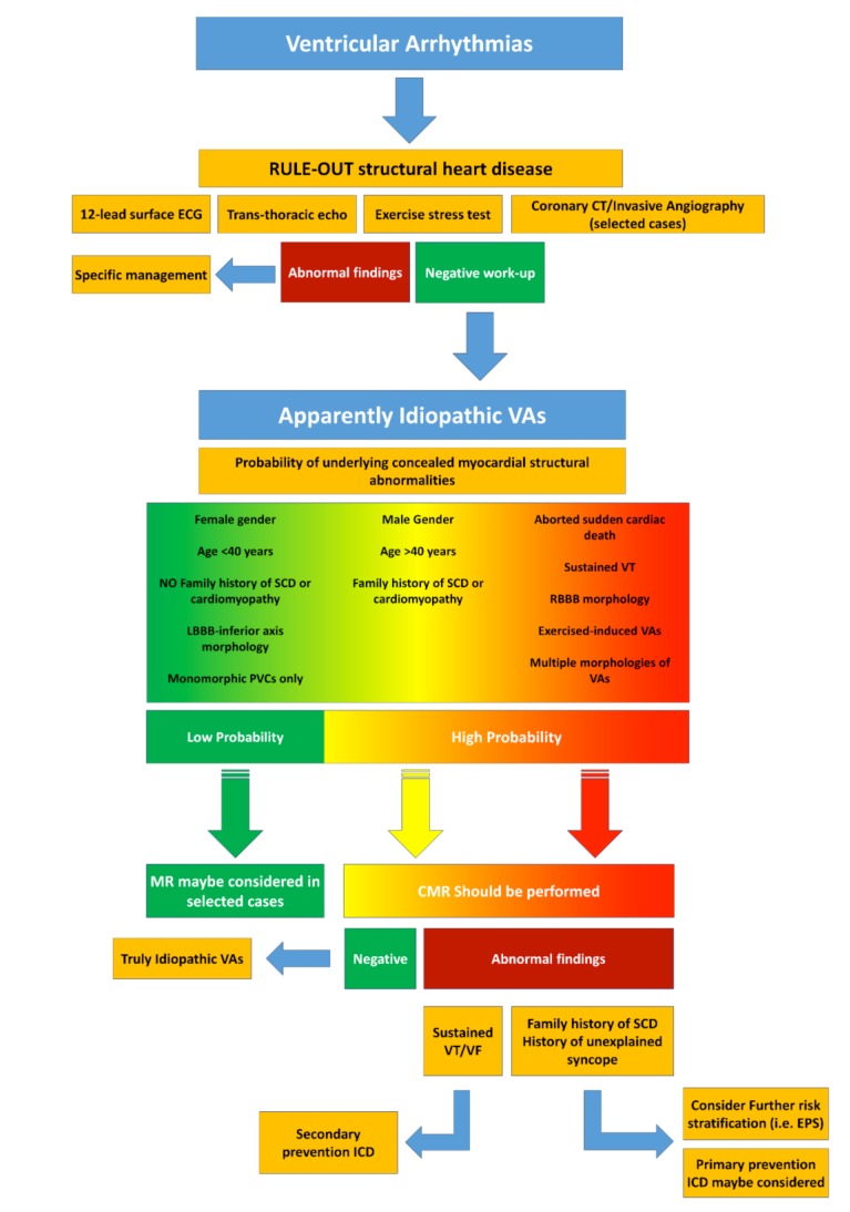
Ventricular Arrhythmias (VAs) may present with a wide spectrum of clinical manifestations ranging from mildly symptomatic frequent premature ventricular contractions to lifethreatening events such as sustained ventricular tachycardia, ventricular fibrillation and sudden cardiac death. Myocardial scar plays a central role in the genesis and maintenance of re-entrant arrhythmias which are commonly associated with Structural Heart Diseases (SHD) such as ischemic heart disease, healed myocarditis and non-ischemic cardiomyopathies. However, the arrhythmogenic substrate may remain unclear in up to 50% of the cases after a routine diagnostic workup, comprehensive of 12-lead surface ECG, transthoracic echocardiography and coronary angiography/ computed tomography. Whenever any abnormality cannot be identified, VAs are referred as to "idiopathic". In the last decade, Cardiac Magnetic Resonance (CMR) imaging has acquired a growing role in the identification and characterization of myocardial arrhythmogenic substrate, not only being able to accurately and reproducibly quantify biventricular function, but, more importantly, providing information about the presence of myocardial structural abnormalities such as myocardial fatty replacement, myocardial oedema, and necrosis/ fibrosis, which may otherwise remain unrecognized. Moreover, CMR has recently demonstrated to be of great value in guiding interventional treatments, such as radiofrequency ablation, by reliably identifying VA sites of origin and improving long-term outcomes. In the present manuscript, we review the available data regarding the utility of CMR in the workup of apparently "idiopathic" VAs with a special focus on its prognostic relevance and its application in planning and guiding interventional treatments.

Keywords: Cardiac magnetic resonance; idiopathic; late gadolinium enhancement; structural heart disease; tissue characterization; ventricular arrhythmias..


Figures

Fig. (1)
Fig. (1)
Schematic representation of a reentry circuit as originally described by Stevenson et al. in 1993. In this “figure of eight” model, two activation wavefronts propagate around two lines of conduction block sharing a common pathway (central isthmus). Areas of dense scar (blue) cannot be excitable during tachycardia. Bystander pathways can be attached to any point in the circuit and represent areas of tissue activated by the wavefront but not playing an active role in the reentrant circuit.
Fig. (2)
Fig. (2)
Ischemic and nonischemic patterns of late Gadolinium enhancement. (a) Ischemic enhancement is subendocardial to transmural in a vascular distribution; (b) Nonischemic enhancement may be midmyocardial, subepicardial, or diffuse subendocardial. Midmyocardial enhancement may be linear, patchy, or at the RV insertion points of the interventricular septum. DCM = dilated cardiomyopathy, HE = hyperenhancement. Reprinted with permission from Rajiah et al. [30].
Fig. (3)
Fig. (3)
Example of a 48-year old man without known cardiovascular risk factors and no previous history of heart disease presenting with sustained ventricular tachycardia of right bundle branch block morphology (A). His 12-Lead ECG obtained after electric cardioversion appeared to be normal (B). Cardiac magnetic resonance late gadolinium enhancement imaging demonstrated non-ischemic myocardial scar involving the basal inferolateral wall, with a subepicardial distribution (C - white arrow); reprinted with permission from Muser et al. [18].
Fig. (4)
Fig. (4)
Example of a 40-year-old man with family history of cardiomyopathy and apparently idiopathic frequent premature ventricular beats with RBBB morphology and superior QRS axis (A). Cardiac magnetic resonance T1-weighted imaging demonstrated signal abnormalities suggestive of myocardial fatty replacement of the lateral left ventricular wall (B). Late gadolinium enhancement with nonischemic pattern of the lateral left ventricular wall was also present (C-D); reprinted with permission from Nucifora et al. [2].
Fig. (5)
Fig. (5)
Example of a 24-years old men with frequent premature ventricular contractions and significant family history for idiopathic dilated cardiomyopathy and sudden cardiac death (SCD) as demonstrated by his family pedigree (A, arrowhead: proband, red: subjects affected by cardiomyopathy, yellow: subjects who died from SCD, blue: subjects affected by breast cancer, males and females are represented by squares and circles, respectively, figures marked by slash: dead patients, number below squares and circles: age of the patients). Proband's 12-lead ECG showed PVC of right bundle branch block morphology and sinus beats within normality (B). Cardiomyopathy-related myocardial morphological changes are shown in histological sections of the explanted heart of the proband's father (C-E). A spectrum of changes is seen: focal degeneration and necrosis of cardiomyocytes with no or minimal cellular response, myocyte loss and replacement fibrosis. Low magnification shows intra-myocardial (C) and sub-epicardial (D) areas of dense fibrotic scar with a patchy distribution (blue with Mallory trichromic stain). Higher magnification reveals foci of myocardial degeneration with vacuolization of cardiomyocytes (E-F, arrows, Hematoxylin and Eosin stain). Necrotic cardiomyocytes exhibit homogeneously brightly eosinophilic sarcoplasm with pyknotic or karyorrhectic nuclei (panels E-F, arrows). Proband's LGE-CMR demonstrated diffuse myocardial scar with a non-ischemic pattern (sub-epicardial and intra-myocardial) involving the interventricular septum and LV lateral wall evident in both three-chamber (G, arrows) and short-axis cross sections (H, arrows). Reprinted with permission from Muser et al. [19].
Fig. (6)
Fig. (6)
Forest plot showing the results of principal studies investigating the risk of major adverse cardiovascular events associated with the presence of myocardial structural abnormalities detected by cardiac magnetic resonance in patients with apparently idiopathic ventricular arrhythmias.
Fig. (7)
Fig. (7)
Example of a patient with frequent premature ventricular contractions and left ventricular non- compaction as seen with cardiac magnetic resonance (A - black arrows). Non- compacted walls do not show evidence of late gadolinium enhancement (B). Endocardial bipolar voltage map did not show any abnormality (C) while a region of low unipolar voltage was present on the inferior and lateral wall in a location consistent with the non-compacted segments (D). Native T1 mapping revealed increased T1 relaxation time (E – red colour indicated by arrows), and post contrast T1 mapping revealed decreased T1 relaxation time (F - white arrows) consistent with interstitial fibrosis. This matched the region of unipolar voltage abnormality (reprinted with permission from Muser et al. [64]).
Fig. (8)
Fig. (8)
Diagram representing the proposed algorithm for the diagnostic work-up and management of patients presenting with apparently idiopathic ventricular arrhythmias. VAs: Ventricular Arrhythmias, VT: Ventricular Tachycardia; SCD: Sudden Cardiac Death; LBBB: Left bundle branch block; RBBB: Right Bundle Branch Block; PVCs: Premature Ventricular Contractions; CMR: Cardiac Magnetic Resonance; VF: Ventricular Fibrillation; ICD: Implantable Cardioverter-defibrillator; EPS: Electrophysiological Study.

Similar articles

Cited by

References

    1. Tan A.Y., Ellenbogen K. Ventricular arrhythmias in apparently normal hearts. Card. Electrophysiol. Clin. 2016;8:613–621. - PubMed
    1. Nucifora G., Muser D., Masci P.G., et al. Prevalence and prognostic value of concealed structural abnormalities in patients with apparently idiopathic ventricular arrhythmias of left versus right ventricular origin A magnetic resonance imaging study. Circ Arrhythm Electrophysiol. 2014;7:456–462. - PubMed
    1. Mahida S., Sacher F., Dubois R., et al. Cardiac imaging in patients with ventricular tachycardia. Circulation. 2017;136:2491–2507. - PubMed
    1. Latif S., Dixit S., Callans D.J. Ventricular arrhythmias in normal hearts. Cardiol. Clin. 2008;26:367–380. - PubMed
    1. Saksena S., Camm A.J. Electrophysiological disorders of the heart: Expert consult. Elsevier Health Sciences; 2011.

MeSH terms