. 2019 Mar 1;11(3):187-199.
doi: 10.1093/jmcb/mjy049.

A single synonymous mutation determines the phosphorylation and stability of the nascent protein

Affiliations

A single synonymous mutation determines the phosphorylation and stability of the nascent protein

Konstantinos Karakostis et al. J Mol Cell Biol. .

Abstract

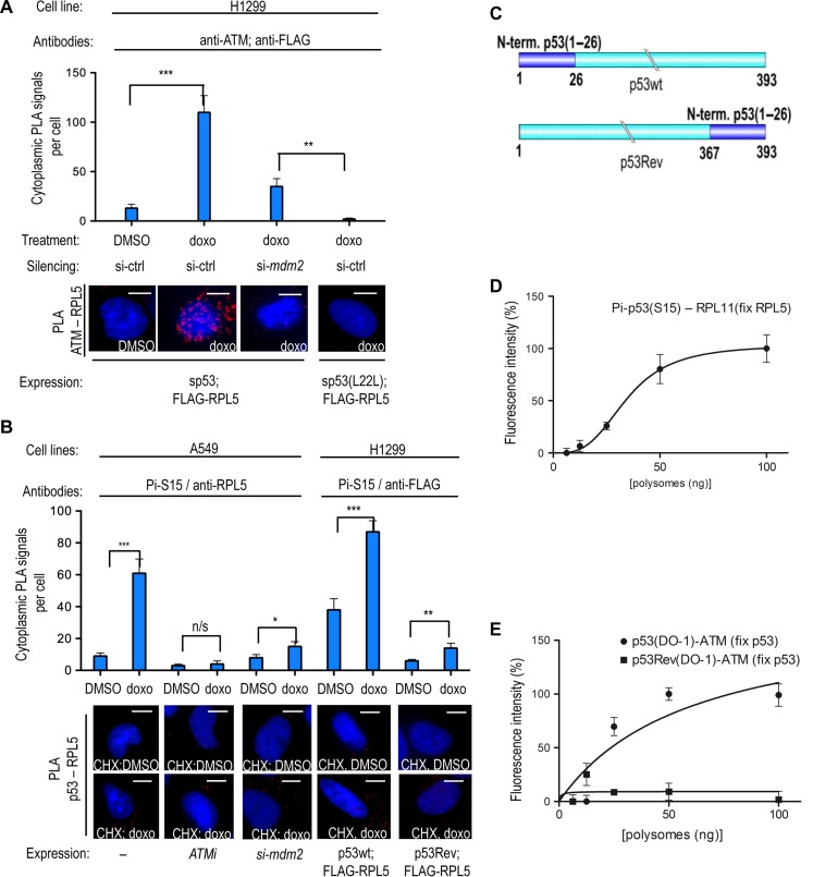
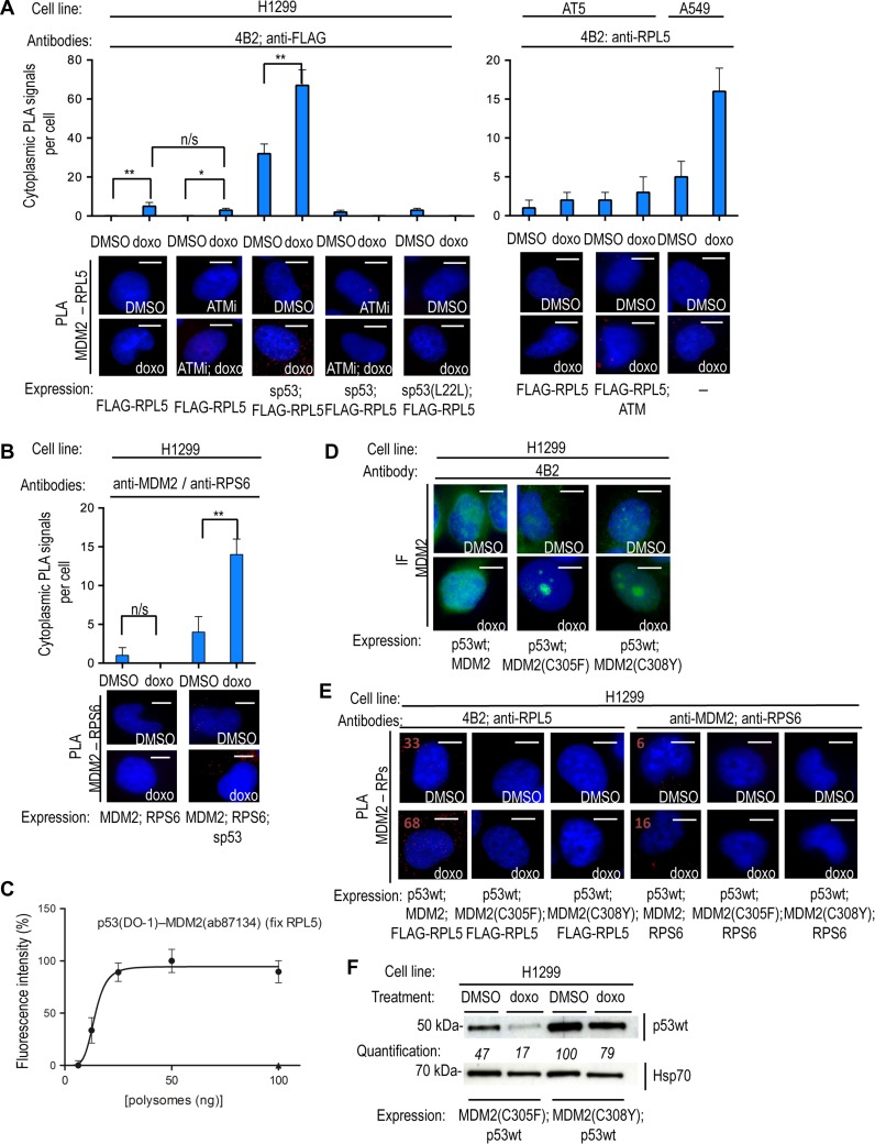
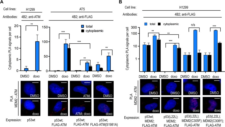
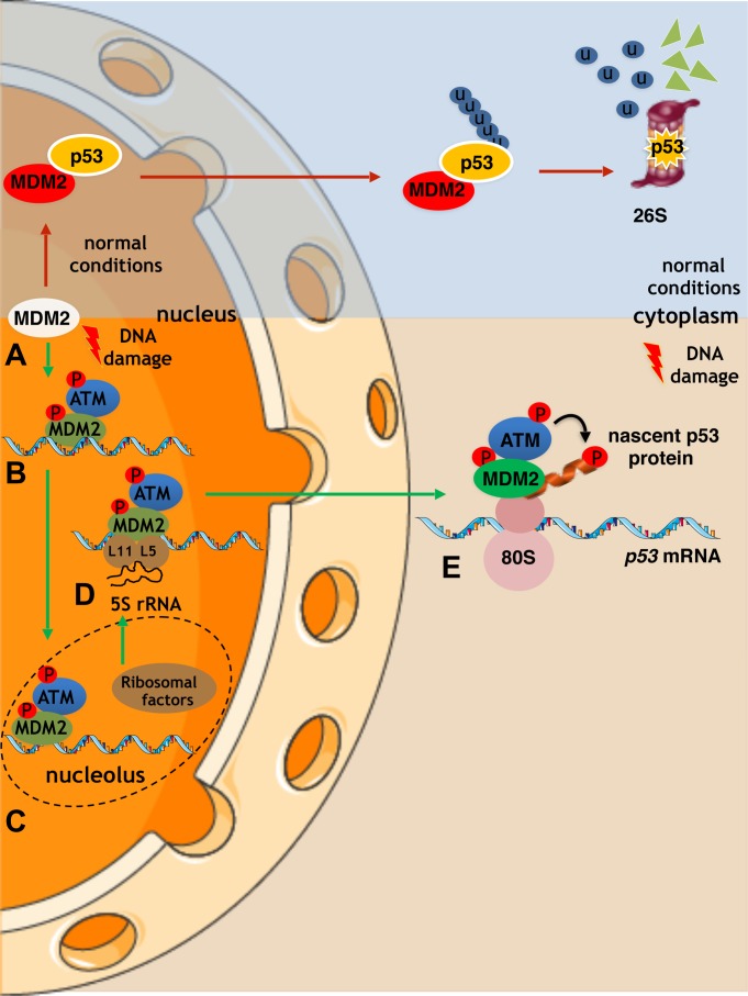
p53 is an intrinsically disordered protein with a large number of post-translational modifications and interacting partners. The hierarchical order and subcellular location of these events are still poorly understood. The activation of p53 during the DNA damage response (DDR) requires a switch in the activity of the E3 ubiquitin ligase MDM2 from a negative to a positive regulator of p53. This is mediated by the ATM kinase that regulates the binding of MDM2 to the p53 mRNA facilitating an increase in p53 synthesis. Here we show that the binding of MDM2 to the p53 mRNA brings ATM to the p53 polysome where it phosphorylates the nascent p53 at serine 15 and prevents MDM2-mediated degradation of p53. A single synonymous mutation in p53 codon 22 (L22L) prevents the phosphorylation of the nascent p53 protein and the stabilization of p53 following genotoxic stress. The ATM trafficking from the nucleus to the p53 polysome is mediated by MDM2, which requires its interaction with the ribosomal proteins RPL5 and RPL11. These results show how the ATM kinase phosphorylates the p53 protein while it is being synthesized and offer a novel mechanism whereby a single synonymous mutation controls the stability and activity of the encoded protein.

Keywords: ATM kinase; MDM2; cell signaling; intrinsically disordered proteins; p53 messenger RNA; synonymous mutations.


Figures

Figure 1
Figure 1
The synonymous p53(L22L) mutation prevents ATM-mediated phosphorylation of the nascent p53 protein and p53 stabilization following genotoxic stress. (A) WB using the anti-p53 DO-1 antibody (N-terminal epitope), monitoring the levels of wild-type p53 protein expressed from the wild-type p53 mRNA or from the L22L mRNA in p53-null H1299 in the absence and presence of MDM2 under normal conditions (DMSO) or DNA damage induced by 0.1 mM doxorubicin (doxo). The p53(L22L) mRNA exhibits a low affinity for MDM2 (Malbert-Colas et al., 2014) and the encoded p53wt protein shows a high rate of degradation in the presence of MDM2 following doxo treatment. The quantification values show the relative density of the bands. (B) The PLA using the phosphospecific Pi-S15 mAb together with the anti-p53 rabbit CM-1 shows the subcellular localization of the phosphorylated p53(S15) in transfected H1299 cells. The graph shows that the number of PLA signals per cell increases following DNA damage. (C) PLA shows the subcellular localization of the MDM2–p53 protein–protein interaction in transfected H1299 (p53-null) and AT5 (ATM-null) cells. The silent p53 (sp53) mRNA carrying mutated AUG codons does not express the p53 protein and was used as a control. The number of MDM2–p53 interactions is reduced following DNA damage induced by 0.1 mM doxorubicin (doxo). This was shown using two different pairs of antibodies (DO-1 for p53 and anti-HA for HA-MDM2 or CM-1 for p53 and 4B2 for MDM2). The phosphospecific anti-p53(S15) mAb gave no PLA signal with anti-MDM2 antibody. In AT5 cells, the MDM2–p53 PLA signal was diminished following doxo treatment only after expression of exogenous ATM. The asterisks represent P-values of three independent experiments: n/s, P > 0.05; *P ≤ 0.05; **P ≤ 0 01; ***P ≤ 0.001. Scale bar, 10 μm. The statistical analyses are based on results obtained by at least 50 cells of each experiment. The antibodies used for the PLA are indicated.
Figure 2
Figure 2
ATM phosphorylates the nascent p53(S15) peptide. (A) PLA showing that ATM associates with the ribosomal RPL5 protein in the presence of sp53 in H1299 cells in the cytoplasm following doxo treatment. Knocking down mdm2 using siRNA prevents the ATM–RPL5 interaction. The sp53(L22L) mRNA has low affinity for MDM2 and does not support the ATM–RPL5 interaction. (B) PLA using the phosphospecific anti-p53(S15) mAb shows the cytoplasmic association of phosphorylated p53(S15) with RPL5 following DNA damage, using endogenous factors, in A549 cells treated with cycloheximide (CHX). This PLA signal is reproduced in H1299 cells expressing p53wt and the FLAG-RPL5, by using the anti-p53(Pi-S15) and anti-FLAG antibodies. Inhibition of ATM or silencing of mdm2 prevents the (Pi-S15)–RPL5 PLA signal in A549 cells. Following doxo and CHX treatment of p53Rev-expressing cells, there is an ~6.5-fold decrease in the PLA signal between RPL5 and p53(Pi-S15) epitope, as compared to p53wt. Following CHX treatment, the Ser15 epitope of the p53Rev is buried within the ribosome on stalled ribosomes. (C) The first 36 aa of p53wt were switched to the C-terminus to generate the p53Rev construct. The cartoon illustrates the p53wt and p53Rev constructs. (D) PLEA showing the association of p53(Pi-S15) epitope with RPL11 on purified polysomes from doxo-treated cell lysates fixed with anti-RPL5 antibodies. (E) PLEA showing the association of p53 with ATM on polysomes treated with doxo and captured with anti-p53 antibodies. The p53Rev shows no association, confirming that the association between the p53 Ser15 epitope and ATM occurs at the p53 polysome. Scale bar, 10 μm. n/s, P > 0.05; *P ≤ 0.05; **P ≤ 0 01; ***P ≤ 0.001. The statistical analyses are based on results obtained by at least 50 cells from three independent experiments.
Figure 3
Figure 3
The p53 mRNA and ribosomal factors govern MDM2’s trafficking to the p53 polysome. (A, left) PLA on H1299 cells transfected with FLAG-RPL5, using the anti-MDM2 mAb 4B2 and anti-FLAG. Following DNA damage, the p53 mRNA and ATM kinase activity are required for MDM2 to associate with RPL5 in the cytoplasm. The p53 mRNA promotes the MDM2–RPL5 interaction in the cytoplasm, which is prevented by inhibition of ATM kinase activity (ATMi) or the sp53(L22L) mutant. (A, right) PLA on AT5 and A549 cells using 4B2 and anti-RPL5 antibodies on transfected and endogenous targets, respectively, confirms that the MDM2–RPL5 interaction requires ATM. (B) PLA on transfected H1299 cells using anti-RPS6 (54D2) and anti-MDM2 (ab87134) antibodies. RPS6 does not bind to MDM2 directly and the p53 mRNA induces the association of MDM2 with RPS6 at the cytoplasm, showing that the p53 mRNA is required for MDM2 to reach ribosomal factors during DDR. (C) PLEA showing the association of nascent p53 with MDM2 on polysomes from H1299 lysates treated with doxo and captured with anti-RPL5, confirming that MDM2 is present at the p53 polysome. (D) IF on H1299 cells comparing the predominately nuclear immuno-staining of MDM2wt with the accumulation of MDM2(C305F) and MDM2(C308Y) mutants in nucleoli following doxo treatment. These mutants do not interact with RPL5 or RPL11. (E) PLA showing that compared to the MDM2wt, the MDM2(C305F) or MDM2(C308Y) mutants do not interact with RPL5 or RPS6, in the presence of p53wt, on H1299 cells under normal conditions or following doxo treatment. PLA signal numbers are noted for the MDM2wt interactions (positive). Also compare with MDM2wt interactions with RPL5 and RPS6 in A and B, respectively. The interaction of MDM2 and ribosomal proteins is required for MDM2 to reach the p53 polysome (see B). (F) WB shows that p53wt is not stabilized in H1299 cells expressing MDM2(C305F) or MDM2(C308Y) following DNA damage. Scale bar, 10 μm. n/s, P > 0.05; *P ≤ 0.05; **P ≤ 0 01; ***P ≤ 0.001. The statistical analyses are based on results obtained by at least 50 cells each from three independent experiments.
Figure 4
Figure 4
The trafficking of MDM2 is required for recruiting ATM to the p53 polysome following DNA damage. (A) PLA on transfected H1299 (left) and AT5 (right) cells using 4B2 and anti-ATM or anti-FLAG antibodies show MDM2 together with ATM predominately in the cytoplasm following doxo treatment. The cytoplasmic MDM2–ATM association is prevented by ATM inhibitors or by inserting the inactive ATM(S1981A) mutation. Black bars represent cytoplasmic PLA signals and blue bars show the total number of PLA signals. (B) The MDM2–ATM association in the cytoplasm requires the interaction of MDM2 with the p53 mRNA and ribosomal factors. Preventing the MDM2–p53 mRNA interaction by using p53(L22L) prevents the induction of the cytoplasmic and total association of MDM2 with ATM following doxo treatment. The MDM2(C305F) mutant shows a strong induction of PLA signal with ATM in the nucleus, but not in the cytoplasm, following doxo treatment. Similarly, the MDM2(C308Y) mutation shows less interaction with ATM in the cytoplasm following doxo treatment. Scale bar, 10 μm. n/s, P > 0.05; *P ≤ 0.05; **P ≤ 0 01; ***P ≤ 0.001. The statistical analyses are based on results obtained by at least 50 cells each of three independent experiments.
Figure 5
Figure 5
A model describes how the p53 mRNA dictates the stability of the encoded protein following DNA damage by guiding MDM2 and ATM to the p53 polysome. (A) Under normal conditions, MDM2 binds to p53 and catalyses its polyubiquitination and the subsequent degradation via the 26S proteasomal pathway. (B) Following DNA damage, ATM phosphorylates MDM2 at Ser395 promoting its binding to the p53 mRNA. (C and D) The ATM–MDM2–p53 mRNA complex is trafficked between the nucleoli and the cytoplasm and associates with the ribosome precursor complex RPN (RPL5–RPL11–5S). This association facilitates the export of the complex to the cytoplasm and the p53 polysome. (E) ATM phosphorylates the nascent p53 peptide at Ser15 and prevents MDM2 from binding to the emerging p53 protein. This model explains how MDM2 can stimulate the translation of p53 without degrading the newly synthesized p53 protein and how a single synonymous mutation can affect the stability of the encoded protein.

Similar articles

Cited by

References

    1. Banin S., Moyal L., Shieh S., et al. . (1998). Enhanced phosphorylation of p53 by ATM in response to DNA damage. Science 281, 1674–1677. - PubMed
    1. Bursac S., Brdovcak M.C., Donati G., et al. . (2014). Activation of the tumor suppressor p53 upon impairment of ribosome biogenesis. Biochim. Biophys. Acta 1842, 817–830. - PubMed
    1. Candeias M.M., Malbert-Colas L., Powell D.J., et al. . (2008). P53 mRNA controls p53 activity by managing Mdm2 functions. Nat. Cell Biol. 10, 1098–1105. - PubMed
    1. Canman C.E., Lim D.S., Cimprich K.A., et al. . (1998). Activation of the ATM kinase by ionizing radiation and phosphorylation of p53. Science 281, 1677–1679. - PubMed
    1. Chen L., Gilkes D.M., Pan Y., et al. . (2005). ATM and Chk2-dependent phosphorylation of MDMX contribute to p53 activation after DNA damage. EMBO J. 24, 3411–3422. - PMC - PubMed

MeSH terms

Substances