

The budding-yeast RWD protein Csm1 scaffolds diverse protein complexes through a conserved structural mechanism
- PMID: 30252178
- PMCID: PMC6237700
- DOI: 10.1002/pro.3515
The budding-yeast RWD protein Csm1 scaffolds diverse protein complexes through a conserved structural mechanism
Abstract
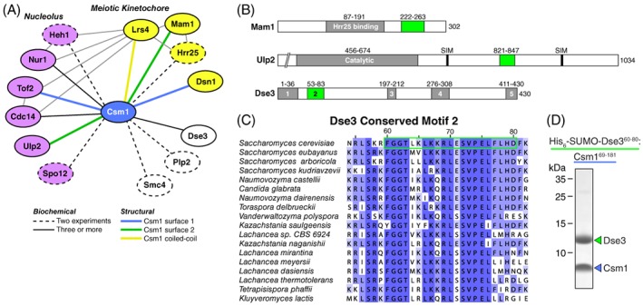
RWD domains mediate protein-protein interactions in a variety of pathways in eukaryotes. In budding yeast, the RWD domain protein Csm1 is particularly versatile, assembling key complexes in the nucleolus and at meiotic kinetochores through multiple protein interaction surfaces. Here, we reveal a third functional context for Csm1 by identifying a new Csm1-interacting protein, Dse3. We show that Dse3 interacts with Csm1 in a structurally equivalent manner to its known binding partners Mam1 and Ulp2, despite these three proteins' lack of overall sequence homology. We theorize that the unique "clamp" structure of Csm1 and the loose sequence requirements for Csm1 binding have led to its incorporation into at least three different structural/signaling pathways in budding yeast.
Keywords: RWD domain; S. cerevisiae; X-ray crystallography; monopolin complex; protein-protein interactions.
© 2018 The Protein Society.
Figures

Similar articles
-
The molecular basis of monopolin recruitment to the kinetochore.Chromosoma. 2019 Sep;128(3):331-354. doi: 10.1007/s00412-019-00700-0. Epub 2019 Apr 30. Chromosoma. 2019. PMID: 31037469 Free PMC article.
-
Binding to small ubiquitin-like modifier and the nucleolar protein Csm1 regulates substrate specificity of the Ulp2 protease.J Biol Chem. 2018 Aug 3;293(31):12105-12119. doi: 10.1074/jbc.RA118.003022. Epub 2018 Jun 14. J Biol Chem. 2018. PMID: 29903909 Free PMC article.
-
Structure of the Saccharomyces cerevisiae Hrr25:Mam1 monopolin subcomplex reveals a novel kinase regulator.EMBO J. 2016 Oct 4;35(19):2139-2151. doi: 10.15252/embj.201694082. Epub 2016 Aug 4. EMBO J. 2016. PMID: 27491543 Free PMC article.
-
Chromosome segregation: clamping down on deviant orientations.Curr Biol. 2003 May 13;13(10):R385-7. doi: 10.1016/s0960-9822(03)00316-6. Curr Biol. 2003. PMID: 12747847 Review.
-
Structures and functions of yeast kinetochore complexes.Annu Rev Biochem. 2007;76:563-91. doi: 10.1146/annurev.biochem.76.052705.160607. Annu Rev Biochem. 2007. PMID: 17362199 Review.
Cited by
-
The molecular basis of monopolin recruitment to the kinetochore.Chromosoma. 2019 Sep;128(3):331-354. doi: 10.1007/s00412-019-00700-0. Epub 2019 Apr 30. Chromosoma. 2019. PMID: 31037469 Free PMC article.
References
-
- Schleiffer A, Maier M, Litos G, Lampert F, Hornung P, Mechtler K, Westermann S (2012) CENP‐T proteins are conserved centromere receptors of the Ndc80 complex. Nat Cell Biol 14:604–613. - PubMed
Publication types
MeSH terms
Substances
Grants and funding
LinkOut - more resources
Full Text Sources
Other Literature Sources
Molecular Biology Databases
