. 2019 Feb;33(2):2314-2326.
doi: 10.1096/fj.201801390R. Epub 2018 Sep 25.

RARβ acts as both an upstream regulator and downstream effector of miR-22, which epigenetically regulates NUR77 to induce apoptosis of colon cancer cells

Affiliations

RARβ acts as both an upstream regulator and downstream effector of miR-22, which epigenetically regulates NUR77 to induce apoptosis of colon cancer cells

Ying Hu et al. FASEB J. 2019 Feb.

Abstract

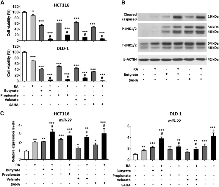
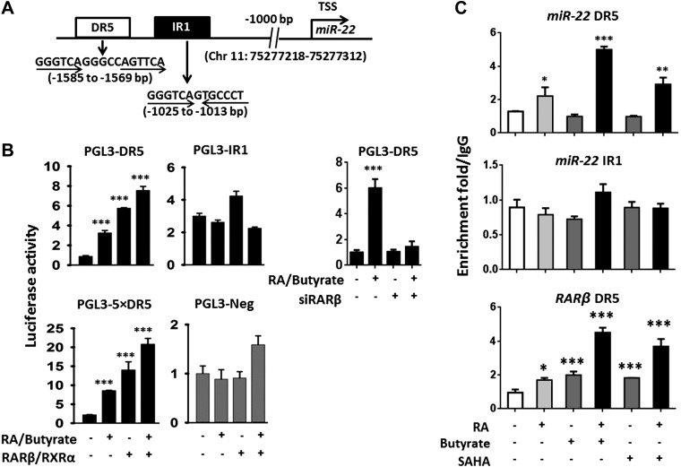
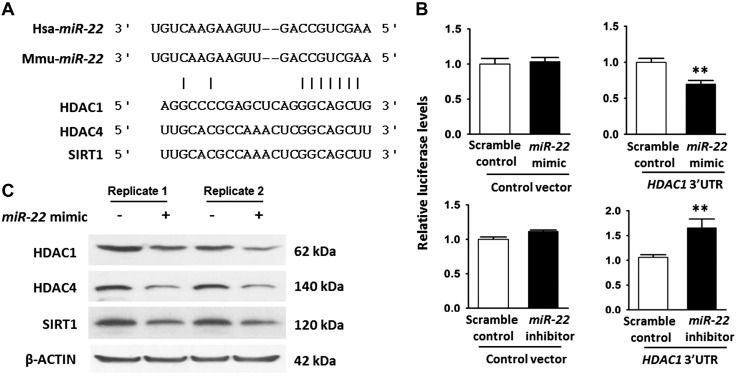
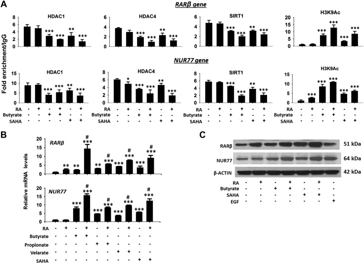
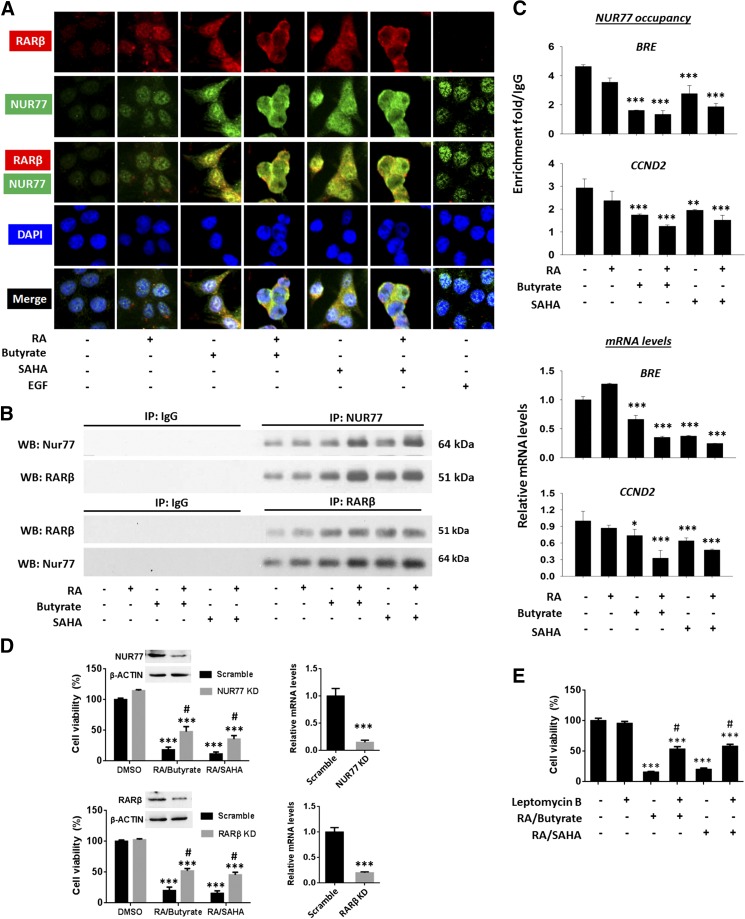
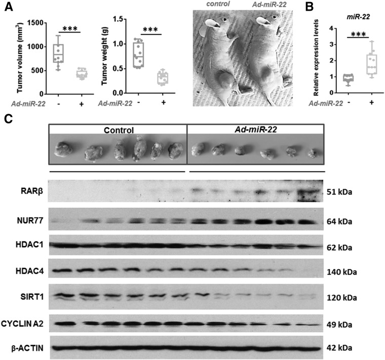
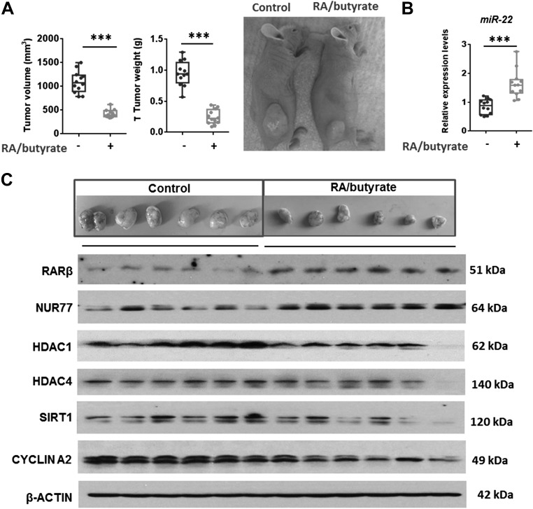
This study investigates the mechanism and consequences of microRNA-22 ( miR-22) induction. Our data revealed for the first time that retinoic acid (RA) and histone deacetylase (HDAC) inhibitors, including short-chain fatty acids and suberanilohydroxamic acid (SAHA), could individually or in combination induce miR-22. This induction was mediated via RA receptor β (RARβ) binding to a direct repeat 5 (DR5) motif. In addition, we uncovered HDAC1 as a novel miR-22 target. In an miR-22-dependent manner, HDAC inhibitors and RA reduced HDAC1, HDAC4, and sirtuin 1 (SIRT1), which were involved in chromatin remodeling of the RARβ and nerve growth factor IB ( NUR77). Thus, HDAC inhibitors and RA-induced miR-22 resulted in simultaneous induction of cytoplasmic RARβ and NUR77, leading to apoptosis of colon cancer cells. In mice, miR-22 and its inducers inhibited the growth of xenograft colon cancer. Moreover, tumor size reduction was accompanied by elevated miR-22, NUR77, and RARβ and by reduced HDACs. In human colon polyps and adenocarcinomas, miR-22 and RARβ were consistently reduced, which was associated with elevated HDAC1, HDAC4, and SIRT1 in colon adenocarcinomas. Results from this study revealed a novel anticancer mechanism of RARβ via miR-22 induction to epigenetically regulate itself and NUR77, providing a promising cancer treatment modality using miR-22 and its inducers.-Hu, Y., French, S. W., Chau, T., Liu, H.-X., Sheng, L., Wei, F., Stondell, J., Garcia, J. C., Du, Y., Bowlus, C. L., Wan, Y.-J. Y. RARβ acts as both an upstream regulator and downstream effector of miR-22, which epigenetically regulates NUR77 to induce apoptosis of colon cancer cells.

Keywords: butyrate; nuclear receptor; propionate; protein deacetylase; short-chain fatty acid.


Conflict of interest statement

The authors thank Dr. Prasant Kumar Jena and Nidhi Nagar (University of California, Davis Health System) for their assistance in preparation of this manuscript. This study was supported by U.S. National Institutes of Health (NIH) National Cancer Institute Grant R01CA222490 (to Y.-J.Y.W.); a Science Translation and Innovative Research (STAIR) Grant (to Y.-J.Y.W.); and National Natural Science Foundation of China Grant 81772572 (to Y.D.). The authors declare no conflicts of interest.

Figures

Figure 1
Figure 1
HDAC inhibitors and RA reduce cell viability and induce miR-22 in human colon cancer HCT116 and DLD-1 cells. A) Cell viability of HCT116 and DLD-1 cells treated with butyrate (5 mM), propionate (10 mM), valerate (10 mM), SAHA (5 μM), and/or RA (10 μM) for 48 h. B) Protein levels of cleaved caspase 3, phosphorylated (P)-JNK1/2, and total (T)-JNK1/2 in HCT116 cells in response to RA, butyrate, or SAHA treatment. C) miR-22 level in HCT116 and DLD-1 cells treated with and without RA and/or HDAC inhibitors for 24 h. Data are expressed as means ± sd. *P < 0.05, **P < 0.01, ***P < 0.001 between treated and DMSO control; #P < 0.05 between combination and single treatment (n = 3).
Figure 2
Figure 2
HDAC inhibitors and RA induce miR-22 through RARβ. A) DR5 and IR1 found within 2 kb upstream from the transcriptional start site (TSS) of miR-22. B) Relative luciferase activities in HCT116 cells transfected with RARβ and RXRα expression plasmids followed by the combination of RA and butyrate treatment. DR5 and IR1 motifs were each cloned into a pGL3 vector. HCT116 cells were cotransfected with RARβ and RXRα expression plasmids or siRARβ along with indicated pGL3 reporter constructs. Six hours after transfection, cells were treated with indicated chemicals for 24 h. pGL3-Neg and pGL3-5 × DR5 were used as negative and positive controls, respectively. C) ChIP-qPCR data revealed the combination effect of butyrate (5 mM) or SAHA (5 µM) plus RA (10 µM) on the recruitment of RARβ to the studied motifs. Data are expressed as means ± sd. *P < 0.05, **P < 0.01, ***P < 0.001 between treated and control groups (n = 3).
Figure 3
Figure 3
miR-22 directly targets HDAC1. A) miR-22 sequence is conserved in humans and mice; miR-22 partially pairs with the 3′UTR of the HDAC1, HDAC4, and SIRT1 genes. B) psiCHECK2-HDAC1 containing the 3′UTR of HDAC1 was cotransfected with either miR-22 mimics or miR-22 inhibitors into HCT116 cells. Scramble constructs were used as negative controls. Data are expressed as means ± sd. **P < 0.01. C) HDAC1, HDAC4, and SIRT1 protein levels using lysates of HCT116 cells transfected with miR-22 mimics or scramble controls.
Figure 4
Figure 4
RA and butyrate-reduced deacetylases is miR-22 dependent. A) Levels of indicated proteins in HCT116 cells treated with butyrate (5 mM), SAHA (5 μM), and/or RA (10 μM) for 24 h. B) Levels of indicated proteins in HCT116 cells treated with RA plus butyrate 48 h after transfection with miR-22 inhibitors or scramble controls. C) Basal mRNA levels of HDACs in HCT116 and DLD-1 cells.
Figure 5
Figure 5
Histone modification of the NUR77 and RARβ genes in response to RA and HDAC inhibitor treatment. A) HCT116 cells were treated with RA (10 µM), butyrate (5 mM), or SAHA (5 µM) for 24 h. ChIP was performed using the indicated antibodies followed by qPCR to amplify NUR77 and RARβ. Binding is expressed relative to binding of IgG antibody. B) mRNA levels of NUR77 and RARβ in HCT116 cells treated with RA and HDAC inhibitors for 48 h. Data are expressed as means ± sd (n = 3). *P < 0.05, **P < 0.01, ***P < 0.001 between treated and DMSO control; #P < 0.05 between combined and single treatment. C) Protein levels of NUR77 and RARβ in HCT116 cells in response to RA, butyrate, SAHA, or EGF treatment.
Figure 6
Figure 6
HDAC inhibitors plus RA promote apoptosis via cytosolic RARβ and NUR77 induction in HCT116 cells. A) Induction and localization of NUR77 and RARβ in HCT116 cells treated by RA, HDAC inhibitors, or EGF. Treated cells were immunostained with anti-NUR77 and anti-RARβ antibodies followed by Alexa Fluor secondary antibodies. B) NUR77 and RARβ interaction in HCT116 cells in response to RA, butyrate, and SAHA treatment. Protein extracts were immunoprecipitated using anti-NUR77, anti-RARβ, or IgG antibody followed by Western blot with anti-RARβ and/or anti-NUR77 antibody. C) ChIP-qPCR data of NUR77 binding to the BRE and CCND2; BRE and CCND2 mRNA level in response to RA and/or HDAC treatments. D) NUR77 KD and RARβ KD increased cell viability in RA/butyrate or RA/SAHA-treated HCT116 cells. HCT116 cells were transfected with control (scramble), Nur77 shRNA plasmid, or siRARβ (transfection efficiency >80%) and treated with DMSO, RA/Butyrate, or RA/SAHA for 48 h. RT-PCR and Western blot of NUR77 and RARβ were performed to assess KD efficiency. Cell viability was measured by MTT assay. E) Inhibition of NUR77 nuclear export by leptomycin B (2 ng/ml) increased the cell viability in RA/butyrate- or RA/SAHA-treated HCT116 cells. HCT116 cells were treated with DMSO, RA/Butyrate, or RA/SAHA in the absence or presence of leptomycin B. Data are expressed as means ± sd (n = 3). *P < 0.05, **P < 0.01, ***P < 0.001 between treated and DMSO control; #P < 0.001 between KD and scramble control or with and without leptomycin B pretreatment.
Figure 7
Figure 7
miR-22 reduces tumor size and silences protein deacetylases in a mouse xenograft model. A) Volume and weight of HCT116 cell–generated tumors in mice receiving either Ad-control or Ad-miR-22. B, C) miR-22 (B) and indicated protein (C) levels in response to miR-22 treatment (n = 12). ***P < 0.001 between treated and control groups.
Figure 8
Figure 8
Butyrate plus RA reduce tumor size and increase miR-22–silenced protein deacetylases in a mouse xenograft model. Volume and weight of HCT116-generated tumors (A), miR-22 level (B), and indicated protein levels (C) in response to butyrate plus RA treatment (n = 12). ***P < 0.001 between treated and control groups.
Figure 9
Figure 9
Human colon polyps and adenocarcinomas have reduced miR-22 and RA signaling. A) Expression levels of miR-22, RARβ, and CYCLINA2 in human colon polyps (P, n = 20) and adenocarcinomas (T) (n = 20) and their paired adjacent normal tissues (N). B) Levels of indicated proteins in human colon adenocarcinomas (T) and their adjacent normal tissues (N) (n = 4).
Figure 10
Figure 10
The mechanism by which RA and HDAC inhibitors induce miR-22, which reduces protein deacetylases, thereby leading to increased cytoplasmic RARβ/NUR77 and apoptosis.

Similar articles

Cited by

References

    1. Tsuchiya, N., Izumiya, M., Ogata-Kawata, H., Okamoto, K., Fujiwara, Y., Nakai, M., Okabe, A., Schetter, A. J., Bowman, E. D., Midorikawa, Y., Sugiyama, Y., Aburatani, H., Harris, C. C., Nakagama, H. (2011) Tumor suppressor miR-22 determines p53-dependent cellular fate through post-transcriptional regulation of p21. Cancer Res. 71, 4628–4639 - PMC - PubMed
    1. Yang, F., Hu, Y., Liu, H. X., Wan, Y. J. (2015) MiR-22-silenced cyclin A expression in colon and liver cancer cells is regulated by bile acid receptor. J. Biol. Chem. 290, 6507–6515 - PMC - PubMed
    1. Zhang, J., Yang, Y., Yang, T., Liu, Y., Li, A., Fu, S., Wu, M., Pan, Z., Zhou, W. (2010) microRNA-22, downregulated in hepatocellular carcinoma and correlated with prognosis, suppresses cell proliferation and tumourigenicity. Br. J. Cancer 103, 1215–1220 - PMC - PubMed
    1. Xiong, J., Du, Q., Liang, Z. (2010) Tumor-suppressive microRNA-22 inhibits the transcription of E-box-containing c-Myc target genes by silencing c-Myc binding protein. Oncogene 29, 4980–4988 - PubMed
    1. Yamakuchi, M., Yagi, S., Ito, T., Lowenstein, C. J. (2011) MicroRNA-22 regulates hypoxia signaling in colon cancer cells. PLoS One 6, e20291 - PMC - PubMed

Publication types

MeSH terms

LinkOut - more resources