. 2018 Sep 25;13(9):e0204567.
doi: 10.1371/journal.pone.0204567. eCollection 2018.

Colonic levels of vasoactive intestinal peptide decrease during infection and exogenous VIP protects epithelial mitochondria against the negative effects of IFNγ and TNFα induced during Citrobacter rodentium infection

Affiliations

Colonic levels of vasoactive intestinal peptide decrease during infection and exogenous VIP protects epithelial mitochondria against the negative effects of IFNγ and TNFα induced during Citrobacter rodentium infection

Arpan K Maiti et al. PLoS One. .

Abstract

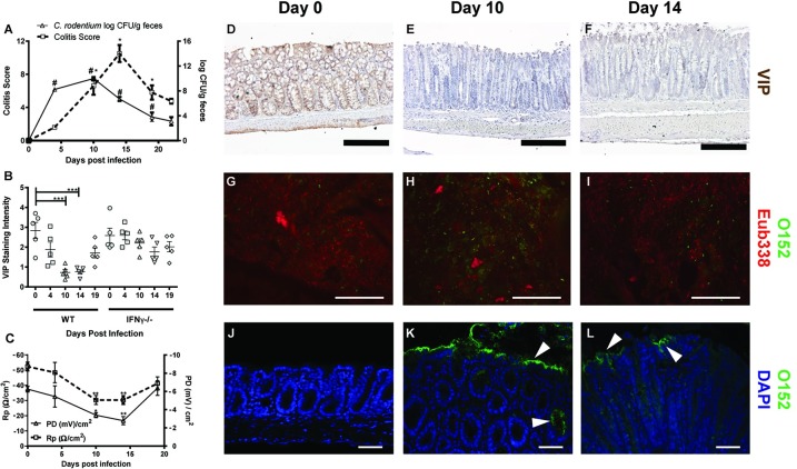
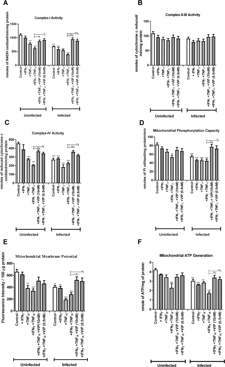
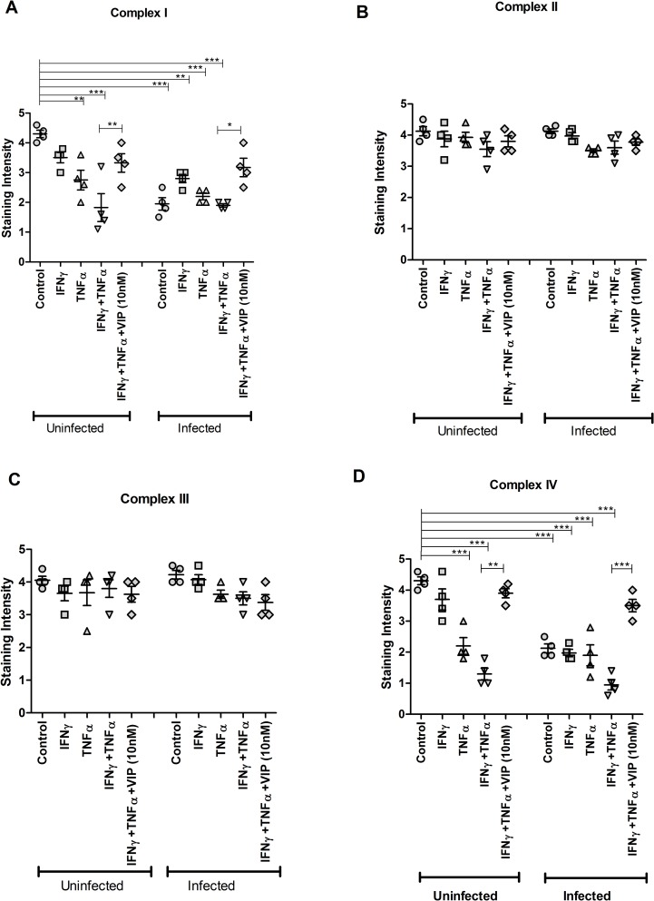
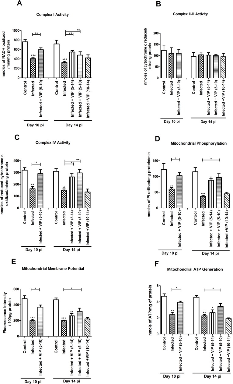
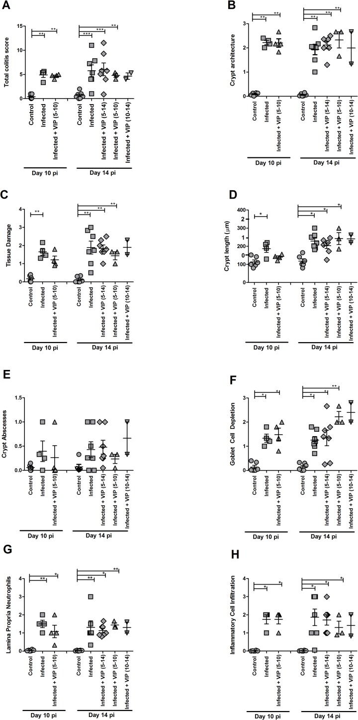
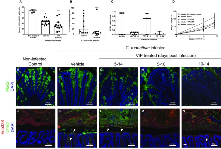
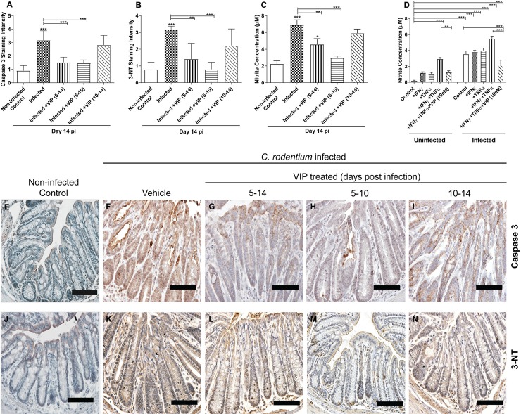
Citrobacter rodentium infection is a model for infection with attaching and effacing pathogens, such as enteropathogenic Escherichia coli. The vasoactive intestinal peptide (VIP) has emerged as an anti-inflammatory agent, documented to inhibit Th1 immune responses and successfully treat animal models of inflammation. VIP is also a mucus secretagogue. Here, we found that colonic levels of VIP decrease during murine C. rodentium infection with a similar time dependency as measurements reflecting mitochondrial function and epithelial integrity. The decrease in VIP appears mainly driven by changes in the cytokine environment, as no changes in VIP levels were detected in infected mice lacking interferon gamma (IFNγ). VIP supplementation alleviated the reduction of activity and levels of mitochondrial respiratory complexes I and IV, mitochondrial phosphorylation capacity, transmembrane potential and ATP generation caused by IFNγ, TNFα and C. rodentium infection, in an in vitro mucosal surface. Similarly, VIP treatment regimens that included the day 5-10 post infection period alleviated decreases in enzyme complexes I and IV, phosphorylation capacity, mitochondrial transmembrane potential and ATP generation as well as increased apoptosis levels during murine infection with C. rodentium. However, VIP treatment failed to alleviate colitis, although there was a tendency to decreased pathogen density in contact with the epithelium and in the spleen. Both in vivo and in vitro, NO generation increased during C. rodentium infection, which was alleviated by VIP. Thus, therapeutic VIP administration to restore the decreased levels during infection had beneficial effects on epithelial cells and their mitochondria, but not on the overall infection outcome.


Conflict of interest statement

The authors have declared that no competing interests exist.

Figures

Fig 1
Fig 1. In vitro mucosal model.
HT29 MTX-E12 cells cultured for 28 days post confluency under semi-wet interface with mechanical stimulation treated with DAPT for the first six days. The membranes were fixed in Methanolic Carnoy’s and stained with PAS/Alcian blue. The image was taken at 400x and the scale bar represent 20 μm.
Fig 2
Fig 2. C. rodentium density, colonic epithelial health status and VIP staining intensity in the murine distal colon during infection.
A: Fecal C. rodentium density and total colitis score: C. rodentium CFU in fecal pellets from individual mice. The colitis score is presented as the sum of the scores from tissue damage, crypt architecture, crypt length, crypt abscesses, goblet cell depletion, inflammatory cell infiltration and neutrophils in lamina propria. (B) Semi-quantification (visually scored 0–5) of the VIP staining intensity in the murine distal colon of wild type (WT) and IFN-γ-/- mice. (C) Colonic epithelial health status measuring Rp (Ω/cm2) and PD (mV/cm2) of mice following day 4, 10, 14 and 19 post-infection in wild type mice infected with C. rodentium. (D-F) Representative images of distal colonic specimens immunohistochemically stained for VIP (brown). Scale bar 200 μm, magnification x200. (G-L) Colonic tissue sections co-stained with a general eubacterial probe (Eub338, red) and an antibody against E. coli lipopolysaccharide antigen O152 (green), which also stains C. rodentium. (G-I) Representative images of feces (scale bar 50 μm, magnification x400) and (J-L) epithelial surface (scale bar 50 μm, magnification x200). Statistics: data are presented as mean ± S.E.M. and analyzed using ANOVA with Student Newman-Keuls Multiple Comparison post hoc test: * P<0.05, ** P<0.01*** P <0.001 vs. control. (n = 5 mice/group). The experiments were performed twice.
Fig 3
Fig 3. In vitro effects of VIP on mitochondrial dysfunction caused by C. rodentium infection and cytokines.
An in vivo like in vitro intestinal mucosal model was treated with cytokines and infected in the presence and absence of VIP, and then six aspects of mitochondrial function were analyzed: (A) complex-I activity (B) complex-II-III activity (C) complex-IV activity (D) mitochondrial phosphorylation capacity (E) mitochondrial membrane potential and (F) mitochondrial ATP generation. Statistics: data are presented as mean ± S.E.M. (n = 4, biological replicates, results were pooled from two separate experiments) and analyzed by ANOVA with Student Newman-Keuls Multiple Comparison post hoc test: * P<0.05, ** P<0.01, *** P<0.001.
Fig 4
Fig 4. Effects of VIP on restoring the decreased levels of mitochondrial respiratory enzyme complexes caused by cytokines and C. rodentium infection in vitro.
An in vivo like in vitro intestinal mucosal model was treated with cytokines and infected in the presence and absence of VIP, stained immunohistochemically and the intensity of the stain was scored. (A) Semi-quantification for complex I (MTND6 antibody), (B) complex-II (SDHA antibody) (C) complex-III (CYC1 antibody) and (D) complex-IV (CCO-VIc antibody). Statistics: data are presented as mean ± S.E.M. (n = 4, biological replicates, results were pooled from two separate experiments) and analyzed by ANOVA with Student Newman-Keuls Multiple Comparison post hoc test: * P<0.05, ** P<0.01, *** P<0.001.
Fig 5
Fig 5. Mitochondrial function in the murine distal colon during C. rodentium infection with and without VIP administration.
Mice were harvested at day 10 and 14 post infection. VIP was administered daily in three treatment groups of mice: mice treated with VIP for day 5–10 were harvested both at day 10 and 14, and mice treated from day 5 to 14 and from day 10 to 14 post-infection were harvested on day 14. (A) complex I activity (B) complex II-III activity (C) complex IV activity (D) mitochondrial phosphorylation capacity (E) mitochondrial membrane potential (F) mitochondrial ATP generation. Statistics: data are presented as mean ± S.E.M. and analyzed by ANOVA with Student Newman-Keuls Multiple Comparison post hoc test: * P <0.05, ** P <0.01, *** P <0.001 vs. control. (n = 2–7 mice/group). Of the mice harvested day 14 post infection, one mouse died in the group that was administered VIP day 5–10 and two in the group that were administered VIP day 10–14. Data about dead animals is not included in the graphs, and the group with only two mice remaining (VIP day 10–14) was omitted from the statistical analysis.
Fig 6
Fig 6. Effect of VIP on colitis during C. rodentium infection.
A. The total colitis score is the sum of the scores present in A, B, C, D, E, G and H: The goblet cell depletion score (F) was not included, due to the fact that VIP is a mucus secretagogue. (B) crypt architecture, (C) tissue damage, (D) crypt length (the crypt lengths were translated into scores for incorporation into the total colitis score according to the numbers 1–3 on the y-axes), (E) crypt abscesses, (F) goblet cell depletion, (G) neutrophils in lamina propria and (H) infiltration of inflammatory cells. Statistics: data are presented as mean ± S.E.M. (n = 2–7 mice, as indicated by the individual symbols in the graph) and analyzed using ANOVA with Student Newman-Keuls Multiple Comparison post hoc test: * P<0.05, ** P<0.01, *** P<0.001 vs. control. Of the mice harvested day 14 post infection, one mouse died in the group that was administered VIP day 5–10 and two in the group that were administered VIP day 10–14. Data about dead animals is not included in the graphs, and the group with only two mice remaining (VIP day 10–14) was omitted from the statistical analysis.
Fig 7
Fig 7. C. rodentium density and localization during VIP treatment at day 14 post-infection.
Colonic tissue sections were stained with anti-Muc2 or co-stained with a general eubacterial probe and an antibody against E. coli lipopolysaccharide antigen O152, which also stains C. rodentium. (A) The number of engorged/Muc2 filled goblet cells per 100 goblet cells in the upper third of the epithelium, scores were pooled for the three VIP regimens. Statistics: Kruskal Wallis test. (B) Percentage of the epithelial surface covered by O152 positive bacteria, scores were pooled from the three VIP regimens. Statistics: Mann-Whitney U-test. (C) The amount of C. rodentium (CFU/g) in the spleen in VIP treated and non-treated infected mice. Each data point represents an individual mouse, and bars represent median and interquartile range (panels A-C). Of the mice harvested day 14 post infection, one mouse died in the group that was administered VIP day 5–10 and two in the group that were administered VIP day 10–14. (D) Change in body weights presented as mean±S.E.M. (E-N) Representative images (images were taken from sections that obtained the median score for each group) of colonic tissue sections stained for Muc2 (green) (E-I) or co-stained using the eubacterial probe (Eub338, red) and anti-O152 (green) (J-N) of non-infected control (E and J), infected vehicle treated (F and K), and infected VIP treated mice for day 5–14 (G and L), day 5–10 (H and M) and day 10–14 (I and N) (magnification x 200).
Fig 8
Fig 8. Caspase-3, 3-Nitrotyrosine (3-NT) staining scores and nitrite measurements at 14 day post-infection.
(A) Semi-quantification of immunohistochemical Caspase-3 staining in distal colon from C. rodentium infected mice with and without VIP treatment (B) Semi-quantification of immunohistochemical 3-NT in distal colon from infected mice with and without VIP treatment. (C) Nitrite concentration in the murine distal colon of infected and VIP treated mice (n = 2–7 mice/group). Of the mice harvested day 14 post infection, one mouse died in the group that was administered VIP day 5–10 and two in the group that were administered VIP day 10–14. Data about dead animals is not included in the graphs, and the group with only two mice remaining (VIP day 10–14) was omitted from the statistical analysis. (D) Nitrite concentration in the in vitro mucosal intestinal model infected with C. rodentium and treated with cytokines in the presence and absence of VIP. (E-I) Representative photos showing caspase-3 tissue localization in distal colon from C. rodentium infected mice with and without VIP treatment. Note that although many cells in the tissue from infected mice stains pale brown, a study that performed both Caspase-3 and Terminal deoxynucleotidyl transferase dUTP Nick-End Labeling (TUNEL) assays in parallel to detect DNA degradation, demonstrated that the proportion of cells that are in late stage apoptosis correspond to the strongly stained cells, not the light brown cells [58]. (J-N) Representative photos showing 3-NT tissue localization in distal colon from C. rodentium infected mice with and without VIP treatment. Scale bar 200 μm, magnification x400. Statistics: data are presented as mean ± S.E.M. and analysed by ANOVA with Student Newman-Keuls Multiple Comparison post hoc test: * P<0.05, ** P<0.01, *** P<0.001.

Similar articles

Cited by

References

    1. Barnett Foster D, Abul-Milh M, Huesca M, Lingwood CA. Enterohemorrhagic Escherichia coli induces apoptosis which augments bacterial binding and phosphatidylethanolamine exposure on the plasma membrane outer leaflet. Infect Immun. 2000; 68(6):3108–15. - PMC - PubMed
    1. Deng W, Li Y, Vallance BA, Finlay BB. Locus of enterocyte effacement from Citrobacter rodentium: sequence analysis and evidence for horizontal transfer among attaching and effacing pathogens. Infect Immun. 2001; 69(10):6323–35. 10.1128/IAI.69.10.6323-6335.2001 - DOI - PMC - PubMed
    1. Frankel G, Phillips AD, Novakova M, Field H, Candy DC, Schauer DB, et al. Intimin from enteropathogenic Escherichia coli restores murine virulence to a Citrobacter rodentium eaeA mutant: induction of an immunoglobulin A response to intimin and EspB. Infect Immun. 1996; 64:5315–25. - PMC - PubMed
    1. Johnson E, Barthold SW. The ultrastructure of transmissible murine colonic hyperplasia. Am J Pathol. 1979; 97:291–313. - PMC - PubMed
    1. Barthold SW. The microbiology of transmissible murine colonic hyperplasia. Lab Anim Sci. 1980; 30: 167–173. - PubMed

Publication types

MeSH terms

Substances