. 2019 Feb;124(1):e63.
doi: 10.1002/cpim.63. Epub 2018 Sep 25.

Mouse Model for Human Vitiligo

Affiliations

Mouse Model for Human Vitiligo

Rebecca L Riding et al. Curr Protoc Immunol. 2019 Feb.

Abstract

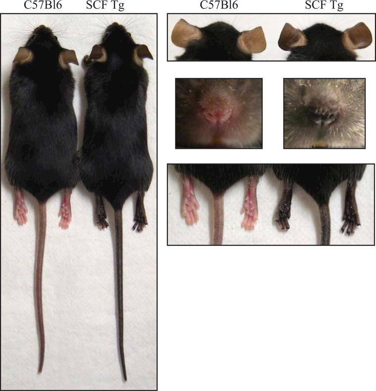
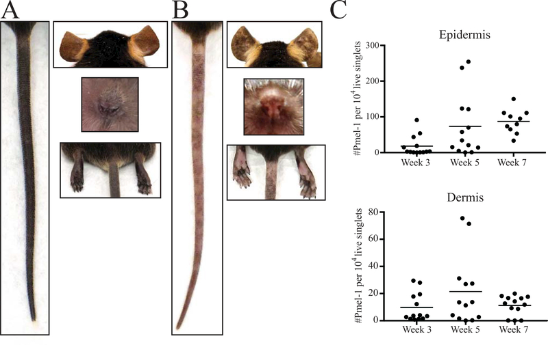
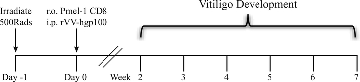
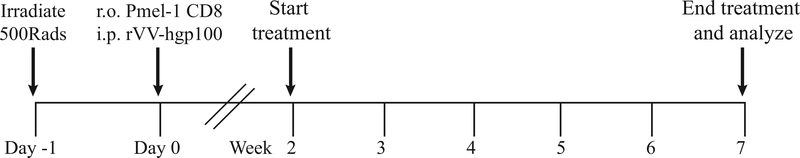
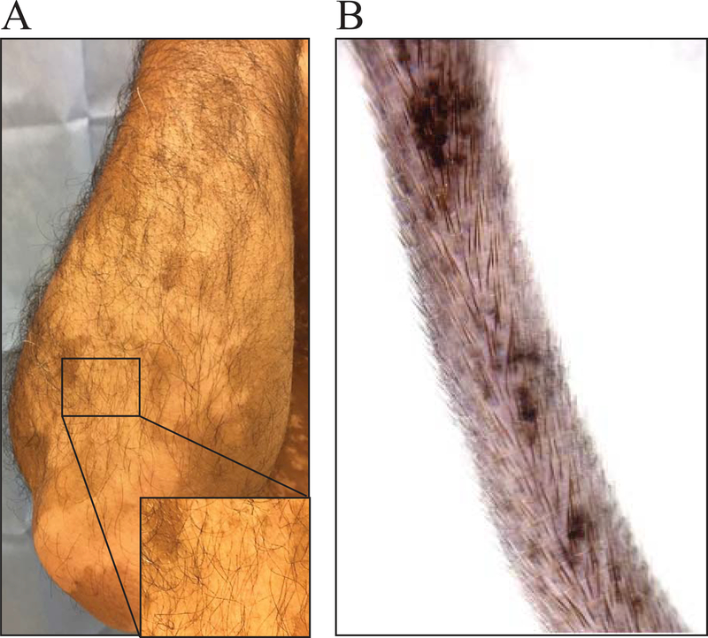
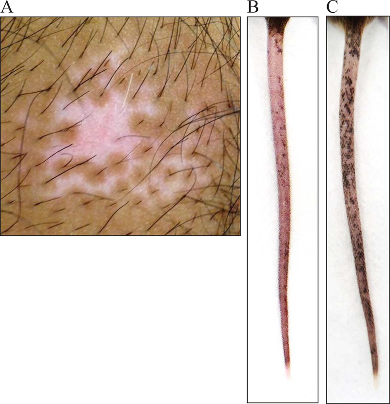
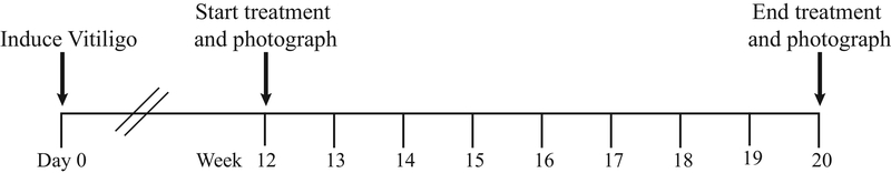
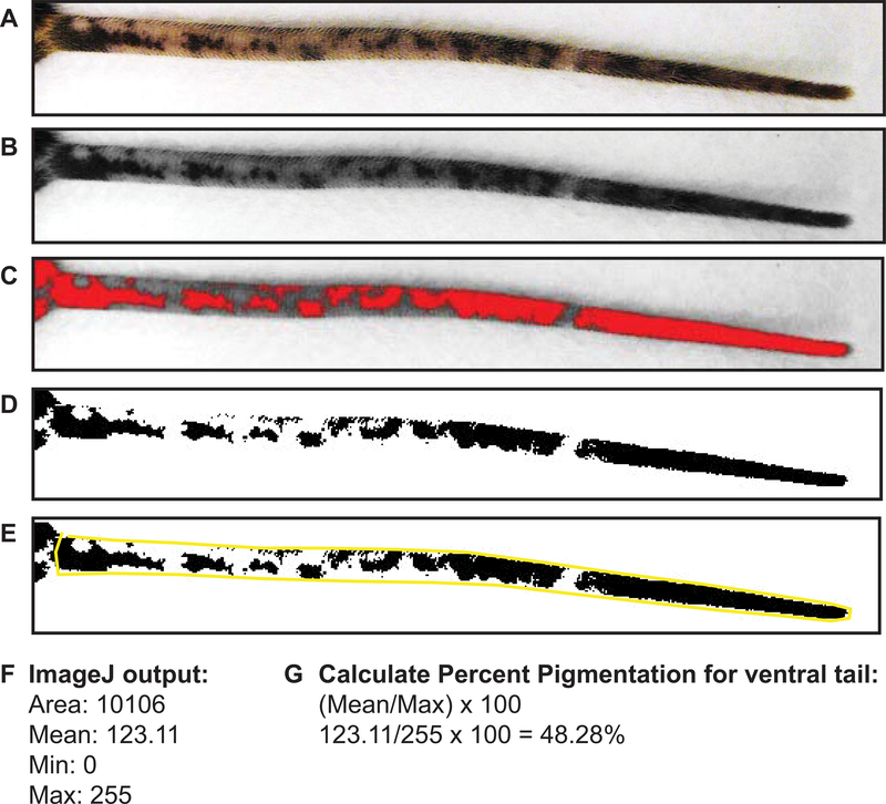
Vitiligo is an autoimmune skin disease in which the pigment-producing melanocytes are destroyed by autoreactive CD8+ T cells. As a result, patients develop disfiguring white spots on the skin. This article discusses the first mouse model of vitiligo that develops epidermal depigmentation, similar to disease in human patients. To achieve epidermal depigmentation, mice are genetically engineered to retain melanocytes in the skin epidermis. Induction of disease occurs by adoptive transfer of melanocyte-specific CD8+ T cells into recipient mice and the subsequent activation of these T cells using a viral vector. Depigmentation of the epidermis occurs within 5 to 7 weeks in a patchy pattern similar to patients with vitiligo. This article describes the methods of vitiligo induction, quantification of lesion progression and regression, processing of the skin for detailed analysis, and how to use this model to inform clinical studies. © 2018 by John Wiley & Sons, Inc.

Keywords: CD8 T cells; autoimmunity; melanocyte; mice; vitiligo.


Figures

Figure 1:
Figure 1:
KRT14-kitl*4XTG2Bjl (SCF) mice have black skin due to melanocyte retention in the epidermis. Images contrast normal (no vitiligo) C57Bl6 and SCF Tg mouse tails, ears, nose, and rear footpads.
Figure 2:
Figure 2:
Vitiligo development in SCF mice. A) Images of the tail, ears, nose and rear footpad of an SCF mouse without vitiligo. B) Images revealing spots of depigmentation on the tail, ears, nose and rear footpads of an SCF mouse 7 weeks post vitiligo induction. C) The number of infiltrating Pmel-1 (normalized to live singlets) in the epidermis and dermis during vitiligo progression.
Figure 3:
Figure 3:
Summary of vitiligo induction in mice. Induction of vitiligo in SCF mice includes sublethal irradiation, transfer of Pmel-1 CD8+ T cells, and infection with recombinant vaccinia virus that expresses their cognate antigen gp100. SCF mice develop epidermal depigmentation over the course of 7 weeks.
Figure 4:
Figure 4:
Prevention Protocol. For preventative studies in SCF vitiligo mice, induce vitiligo as in Basic Protocol 1. Start treatment of mice at 2 weeks post vitiligo induction when wild-type SCF mice have cleared vaccinia virus. Treat mice for 5 weeks and score mice for disease quantification.
Figure 5:
Figure 5:
Hair follicle melanocytes are spared in human vitiligo and in SCF vitiligo mice. A) Human vitiligo lesion with hair follicle pigment maintained. B) SCF vitiligo mouse, 12 weeks post vitiligo induction with pigmented hair follicles within lesions.
Figure 6:
Figure 6:
SCF vitiligo mice show perifollicular repigmentation following successful treatment, similar to in human vitiligo. A) Human vitiligo lesion responding to therapy with repigmentation emerging from hair follicles. B) 12 week old SCF vitiligo mouse with severe depigmentation on the tail. C) Tail of SCF vitiligo mouse 8 weeks after CD8 T cell depletion with perifollicular repigmentation.
Figure 7:
Figure 7:
Repigmentation Protocol. For pre-clinical studies in mice, induce vitiligo as described in Basic Protocol 1. Allow SCF vitiligo mice to develop vitiligo until disease is stabilized, 10–12 weeks post induction. Start treatment by 12 weeks post vitiligo induction and treat for 8 weeks. After treatment is stopped, quantify the percent repigmentation using ImageJ.
Figure 8:
Figure 8:
Quantification of repigmentation using ImageJ analysis. A) Imported ventral tail image. B) Ventral tail image converted to 8-bit black and white. C) Ventral tail image with adjusted brightness threshold to detect areas of pigmentation (red) and depigmentation (grey). D) End result of adjusted ventral tail. E) Ventral tail outlined using the polygon tool. ImageJ will analyze the area outlined to determine the mean area that is pigmented. F) The output from ImageJ after measuring mean pigmentation. G) Formula to calculate percent pigmentation.
Figure 9:
Figure 9:
Visualization of Pmel-1 and chemokine expression in the skin by confocal imaging. A) Sample image from a vitiligo mouse at week 7 depicting GFP+ Pmel-1 CD8+ T cells in the tail skin. Pmel-1 are rounded punctate cells (arrows). Bar = 50 μm. Hairs are visible due to autofluorescence. B) Tail skin of REX3 SCF vitiligo mouse showing clouds of CXCL10-BFP and CXCL9-RFP chemokine expression (10× z-stack).

Similar articles

Cited by

References

    1. Agarwal P, Rashighi M, Essien KI, Richmond JM, Randall L, Pazoki-Toroudi H, et al. (2015). Simvastatin Prevents and Reverses Depigmentation in a Mouse Model of Vitiligo. Journal of Investigative Dermatology, 135(4), 1080–1088. 10.1038/jid.2014.529 - DOI - PMC - PubMed
    1. Alikhan A, Felsten LM, Daly M, & Petronic-Rosic V (2011). Vitiligo: A comprehensive overview. Journal of American Dermatology, 65(3), 473–491. 10.1016/j.jaad.2010.11.061 - DOI - PubMed
    1. Alkhateeb A, Fain PR, Thody A, Bennett DC, & Spritz RA (2003). Epidemiology of vitiligo and associated autoimmune diseases in Caucasian probands and their families. Pigment Cell Research, 16(3), 208–214. - PubMed
    1. Autengruber A, Gereke M, Hansen G, Hennig C, & Bruder D (2012). Impact of enzymatic tissue disintegration on the level of surface molecule expression and immune cell function. European Journal of Microbiology & Immunology, 2(2), 112–120. 10.1556/EuJMI.2.2012.2.3 - DOI - PMC - PubMed
    1. Birlea SA, Costin G-E, Roop DR, & Norris DA (2017). Trends in Regenerative Medicine: Repigmentation in Vitiligo Through Melanocyte Stem Cell Mobilization. Medicinal Research Reviews, 37(4), 907–935. 10.1002/med.21426 - DOI - PMC - PubMed

Publication types

Substances