. 2018 Jan 1:17:1533033818802305.
doi: 10.1177/1533033818802305.

Moderate Heat Application Enhances the Efficacy of Nanosecond Pulse Stimulation for the Treatment of Squamous Cell Carcinoma

Affiliations

Moderate Heat Application Enhances the Efficacy of Nanosecond Pulse Stimulation for the Treatment of Squamous Cell Carcinoma

Chelsea M Edelblute et al. Technol Cancer Res Treat. .

Abstract

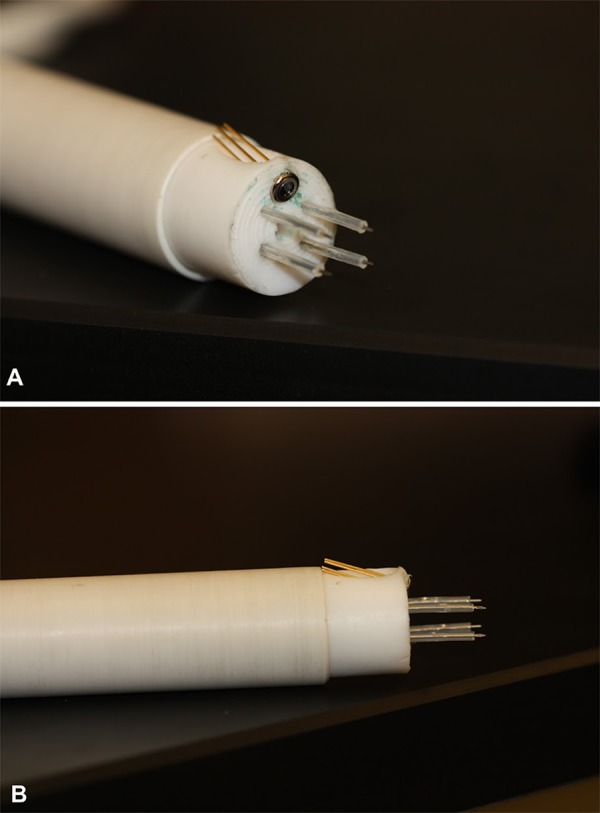
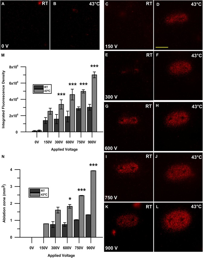
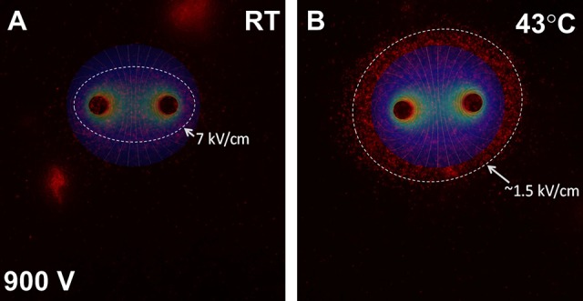
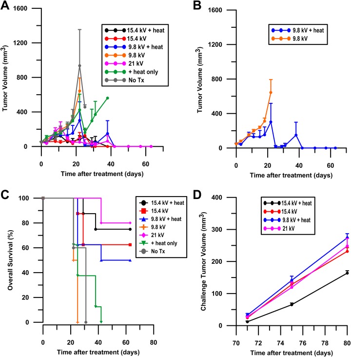
Nanosecond pulse stimulation as a tumor ablation therapy has been studied for the treatment of various carcinomas in animal models and has shown a significant survival benefit. In the current study, we found that moderate heating at 43°C for 2 minutes significantly enhanced in vitro nanosecond pulse stimulation-induced cell death of KLN205 murine squamous cell carcinoma cells by 2.43-fold at 600 V and by 2.32-fold at 900 V, as evidenced by propidium iodide uptake. Furthermore, the ablation zone in KLN205 cells placed in a 3-dimensional cell-culture model and pulsed at a voltage of 900 V at 43°C was 3 times larger than in cells exposed to nanosecond pulse stimulation at room temperature. Application of moderate heating alone did not cause cell death. A nanosecond pulse stimulation electrode with integrated controllable laser heating was developed to treat murine ectopic squamous cell carcinoma. With this innovative system, we were able to quickly heat and maintain the temperature of the target tumor at 43°C during nanosecond pulse stimulation. Nanosecond pulse stimulation with moderate heating was shown to significantly extend overall survival, delay tumor growth, and achieve a high rate of complete tumor regression. Moderate heating extended survival nearly 3-fold where median overall survival was 22 days for 9.8 kV without moderate heating and over 63 days for tumors pulsed with 600, 100 ns pulses at 5 Hz, at voltage of 9.8 kV with moderate heating. Median overall survival in the control groups was 24 and 31 days for mice with untreated tumors and tumors receiving moderate heat alone, respectively. Nearly 69% (11 of 16) of tumor-bearing mice treated with nanosecond pulse stimulation with moderate heating were tumor free at the completion of the study, whereas complete tumor regression was not observed in the control groups and in 9.8 kV without moderate heating. These results suggest moderate heating can reduce the necessary applied voltage for tumor ablation with nanosecond pulse stimulation.

Keywords: ablation; electroporation; irreversible electroporation; nanosecond pulse electric field; nanosecond pulse stimulation.


Conflict of interest statement

Declaration of Conflicting Interests: The author(s) declared the following potential conflicts of interest with respect to the research, authorship, and/or publication of this article: With respect to duality of interest and financial disclosures, Drs K. Schoenbach and R. Heller are inventors on patents which cover the technology that was used in the work reported in this manuscript. In addition, Drs K. Schoenbach and R. Heller own stock in Pulse Biosciences, Inc.

Figures

Figure 1.
Figure 1.
Electrode array for MH-NPS. Four-needle high-voltage electrodes are arranged in a 5 mm × 7 mm array with center port for an infrared laser fiber. Thermopile for surface temperature monitoring is angled toward target (A). Ethylene tetrafluoroethylene tubing sheaths high-voltage needle electrodes leaving 3 mm exposed to insert into target tumor (B). MH-NPS indicates nanosecond pulse stimulation with moderate heating.
Figure 2.
Figure 2.
Propidium iodide uptake after nanosecond pulse stimulation with and without the application of moderate heat. A 2-needle electrode with a 1 mm gap was used to pulse KLN205 cells with 200, 300 ns pulses at 50 Hz with voltages of 150, 300, 600, 750, and 900 V at either room temperature (RT; C, E, G, I, K) or 43°C (D, F, H, J, L). A sham control (0 V) without pulses at RT (A) and 43°C (B) was also recorded. Micrographs are representative of 3 to 5 replicates per treatment group. Scale bar = 1 mm. Quantification of cell death after nanosecond pulse stimulation is represented as the integrated fluorescence density of each individual sample using ImageJ software to draw a region of interest around the area of fluorescence signal. Results shown are the mean of 3 to 5 replicates per group (standard deviation). ***P < .001 (M). The area of the ellipse-shaped ablation zone (mm2) was calculated by multiplying the semi-major and semi-minor axes indicative of propidium iodide uptake by π wherein A = πab. The calculated area is shown as mm2. Results shown are the mean of 3 to 5 replicates per group (± standard deviation). *P < .05, *P < .01, ***P < .001 (N).
Figure 3.
Figure 3.
Two-dimensional electrostatic model of NPS with and without MH. Fluorescence micrograph indicating propidium iodide uptake after NPS with 200, 300 ns pulses at 50 Hz with 900 V at RT (A) and MH-NPS at 43°C (B) and overlaid electric field contour. Highest level of propidium iodide uptake was observed at an electric field strength of 7 kV/cm (A) and 1.5 kV/cm (B) for cells pulsed at RT and preheated to 43°C, respectively. MH-NPS indicates nanosecond pulse stimulation with moderate heating; RT, room temperature.
Figure 4.
Figure 4.
Tumor regression after MH-NPS. KLN205 ectopic murine squamous cell carcinoma tumors were given a single MH, NPS, or MH-NPS treatment on day 0 at an approximate size of 50 mm3. Tumor volume was recorded in surviving animals until day 63 (A). Groups include an untreated no treatment control (no Tx), tumors treated with heat alone, tumors pulsed at a voltage of 9.8 kV or 15.4 kV with and without the application of heat, and tumors pulsed at a voltage 21 kV. A total of 50 mice were used in this experiment. Results of 9.8 kV with and without heat are displayed separately to observe difference between tumors heated and not heated at that voltage (B). Results shown are the mean of 5 to 8 replicates per group (± standard error of the mean) at day 0. Sample size is lower at later time points due to euthanasia by clinical criteria. Kaplan-Meier survival curves are reported until the completion of the study at day 63 (C). A total of 50 mice were used for this experiment. All groups included 8 mice per treatment with the exception of the no treatment control (no Tx) and 21 kV groups, which each contained 5 animals. Mice tumor free at 63 days where challenged on the opposite flank with 5 × 10⁵ KLN2015 cells. Challenge tumor volumes were recorded. Results shown are the mean of 4 to 6 tumor-free replicates per group (± standard error of the mean), as not all mice were tumor free by day 63 (D). MH-NPS indicates nanosecond pulse stimulation with moderate heating.

Similar articles

Cited by

References

    1. Que SKT, Zwald FO, Schmults CD. Cutaneous squamous cell carcinoma: incidence, risk factors, diagnosis, and staging. J Am Acad Dermatol. 2018;78(2):237–247. - PubMed
    1. van Meerbeeck JP, Fennell DA, De Ruysscher DK. Small-cell lung cancer. Lancet. 2011;378(9804):1741–1755. - PubMed
    1. Scheffer HJ, Nielsen K, de Jong MC, et al. Irreversible electroporation for nonthermal tumor ablation in the clinical setting: a systematic review of safety and efficacy. J Vasc Interv Radiol. 2014;25(7):997–1011; quiz 1011. - PubMed
    1. Jiang C, Davalos RV, Bischof JC. A review of basic to clinical studies of irreversible electroporation therapy. IEEE Trans Biomed Eng. 2015;62(1):4–20. - PubMed
    1. Paiella S, Butturini G, Frigerio I, et al. Safety and feasibility of irreversible electroporation (IRE) in patients with locally advanced pancreatic cancer: results of a prospective study. Digest Surg. 2015;32(2):90–97. - PubMed

Publication types