

Anatomic double-bundle medial patellofemoral ligament reconstruction with aperture fixation using an adjustable-length loop device: a 2-year follow-up study
- PMID: 30253770
- PMCID: PMC6156865
- DOI: 10.1186/s12891-018-2261-x
Anatomic double-bundle medial patellofemoral ligament reconstruction with aperture fixation using an adjustable-length loop device: a 2-year follow-up study
Abstract
Background: To assess the clinical availability of an adjustable-length loop device for use in the double-bundle technique with aperture fixation at the patella and femur during anatomic double-bundle medial patellofemoral ligament reconstruction (DB-MPFLR) for recurrent patellar dislocation.
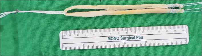
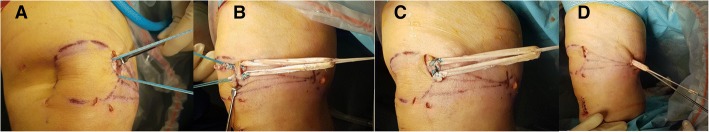
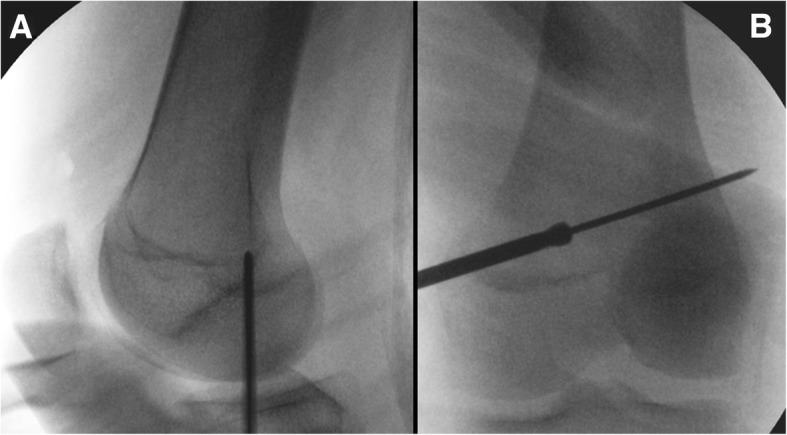
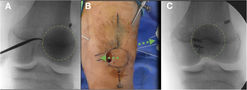
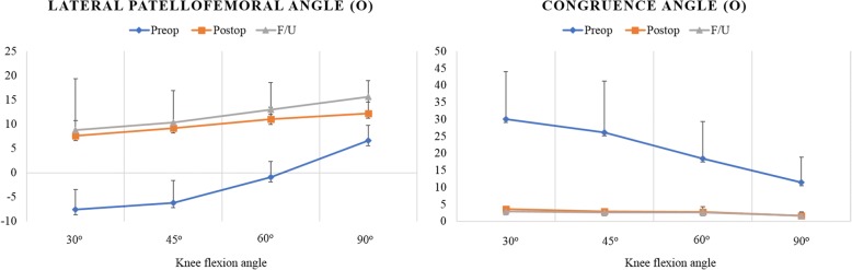
Methods: We retrospectively investigated 11 patients (12 knees) with recurrent patellar dislocation who underwent anatomic DB-MPFLR with an ipsilateral semitendinosus tendon autograft. The graft was folded in half, and its central portion was hanged using the adjustable-length loop device. Both free ends of the graft were fixed at the proximal and distal ends of the medial edge of the patella by using suture anchors, and the hanged graft loop was pulled into the femoral tunnel while maintaining equal tension on both bundles. Manual traction of the suture loops was applied to fix the graft appropriately in full range of motion (ROM) of the knee joint under arthroscopic guidance. Clinical outcomes such as re-dislocation, ROM, clinical scores (Kujala score, Lysholm score, and visual analogue scale score for anterior knee pain), and complications were assessed preoperatively and at 2 years postoperatively. Radiographic parameters indicating patellar position, including congruence angle and lateral patellofemoral angle, were measured at 4 different angles of knee flexion (30°, 45°, 60°, and 90°).
Results: At 4 different flexion angles of the knee joint, the preoperative congruence angle decreased significantly and the lateral patellofemoral angle increased significantly at the final follow-up (P < 0.001). Notably, the improvements in these angles were maintained with no significant differences at the 4 different flexion angles. None of the patients experienced subluxation or re-dislocation after surgery. The patellar instability symptoms improved, as confirmed on the basis of radiographic and other clinical outcomes.
Conclusion: New DB technique with aperture fixation at the patella and femur by using an adjustable-length loop device offers high stability with full ROM of the knee joint, can be considered as a feasible procedure and technique for recurrent patellar dislocation.
Keywords: Adjustable-length loop device; Aperture fixation; Double bundle; Medial patellofemoral ligament; Patella; Recurrent patellar dislocation.
Conflict of interest statement
Ethics approval and consent to participate
All procedures performed in studies involving human participants were in accordance with the ethical standards of the institutional and/or national research committee and with the 1964 Helsinki Declaration and its later amendments or comparable ethical standards. The current study obtained Institutional Review Board approval from our institution (Gachon University of Medicine and Science, GA IRB 2017–236) before study onset, and our protocol was also approved. Informed consent was obtained from all patients. All patients provided written informed consent for participation. For adolescent participants, written informed consent was obtained from their parents/guardians.
Consent for publication
Not applicable.
Competing interests
The authors declare that they have no competing interests.
Publisher’s Note
Springer Nature remains neutral with regard to jurisdictional claims in published maps and institutional affiliations.
Figures
References
-
- White BJ, Sherman OH. Patellofemoral instability. Bull NYU Hosp Jt Dis. 2009;67:22–29. - PubMed
-
- Dejour H, Walch G, Neyret P, Adeleine P: [dysplasia of the femoral trochlea]. Rev Chir Orthop Reparatrice Appar Mot 1990, 76:45–54. - PubMed
-
- Goutallier D, Bernageau J, Lecudonnec B. the measurement of the tibial tuberosity. Patella groove distanced technique and results (author's transl) Rev Chir Orthop Reparatrice Appar Mot. 1978;64:423–428. - PubMed
MeSH terms
LinkOut - more resources
Full Text Sources
Other Literature Sources
Medical
Miscellaneous
