. 2018 Nov 16;293(46):17716-17730.
doi: 10.1074/jbc.RA118.004803. Epub 2018 Sep 25.

Insertion sequence 1 from calpain-3 is functional in calpain-2 as an internal propeptide

Affiliations

Insertion sequence 1 from calpain-3 is functional in calpain-2 as an internal propeptide

Christian-Scott E McCartney et al. J Biol Chem. .

Abstract

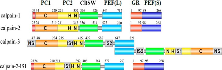
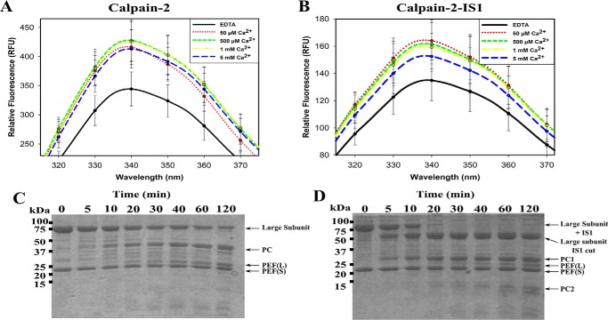
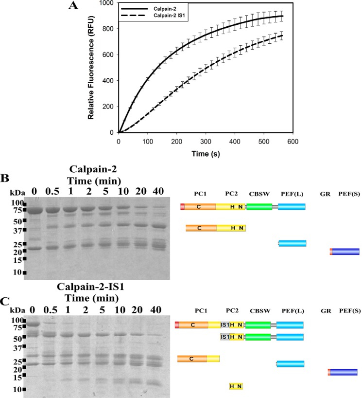
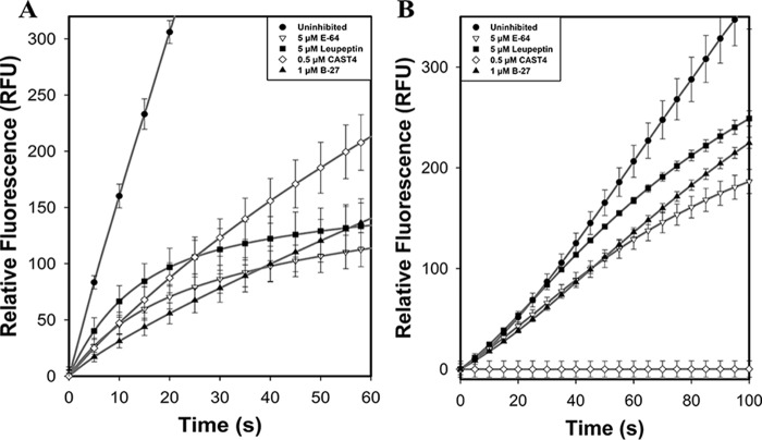
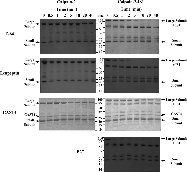
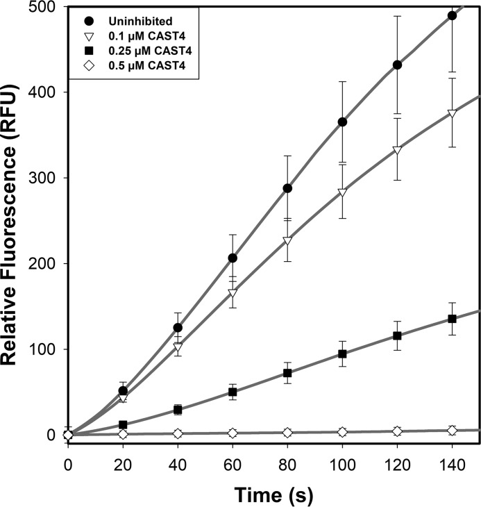
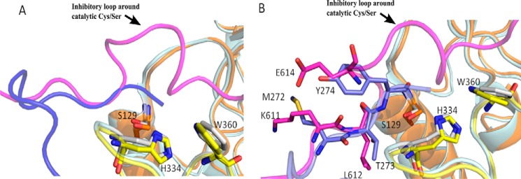
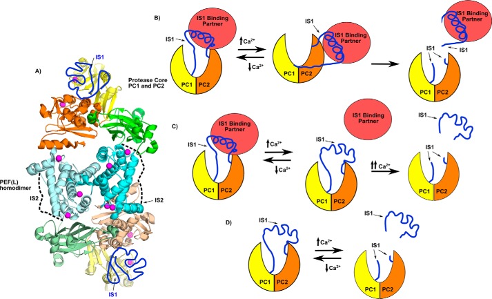
Calpains are intracellular, calcium-activated cysteine proteases. Calpain-3 is abundant in skeletal muscle, where its mutation-induced loss of function causes limb-girdle muscular dystrophy type 2A. Unlike the small subunit-containing calpain-1 and -2, the calpain-3 isoform homodimerizes through pairing of its C-terminal penta-EF-hand domain. It also has two unique insertion sequences (ISs) not found in the other calpains: IS1 within calpain-3's protease core and IS2 just prior to the penta-EF-hand domain. Production of either native or recombinant full-length calpain-3 to characterize the function of these ISs is challenging. Therefore, here we used recombinant rat calpain-2 as a stable surrogate and inserted IS1 into its equivalent position in the protease core. As it does in calpain-3, IS1 occupied the catalytic cleft and restricted the enzyme's access to substrate and inhibitors. Following activation by Ca2+, IS1 was rapidly cleaved by intramolecular autolysis, permitting the enzyme to freely accept substrate and inhibitors. The surrogate remained functional until extensive intermolecular autoproteolysis inactivated the enzyme, as is typical of calpain-2. Although the small-molecule inhibitors E-64 and leupeptin limited intermolecular autolysis of the surrogate, they did not block the initial intramolecular cleavage of IS1, establishing its role as a propeptide. Surprisingly, the large-molecule calpain inhibitor, calpastatin, completely blocked enzyme activity, even with IS1 intact. We suggest that calpastatin is large enough to oust IS1 from the catalytic cleft and take its place. We propose an explanation for why calpastatin can inhibit calpain-2 bearing the IS1 insertion but cannot inhibit WT calpain-3.

Keywords: calpain; calpain inhibitor; calpain-2; calpain-3/p94; calpastatin; cysteine protease; insertion sequence 1 (IS1); limb-girdle muscular dystrophy; muscular dystrophy; p94; propeptide; protease; protease inhibitor; protein engineering; structure-function.


Conflict of interest statement

The authors declare that they have no conflicts of interest with the contents of this article

Figures

Figure 1.
Figure 1.
Domain architecture of calpain-1, -2, and -3 and calpain-2–IS1. The domain nomenclature is based on the structures of calpain-1 and -2, where PC1 (orange) and PC2 (yellow) represent the protease core, CBSW (green) represents the calpain-type β-sandwich, and PEF (blue) represents the large (L) and small (S) subunit penta-EF hand domains. The catalytic triad residues of the cysteine protease core are indicated by C, H, and N. The unique insertion sequences of calpain-3 are shown in gray. The 48-residue IS1 was inserted in calpain-2 after Tyr250.
Figure 2.
Figure 2.
Intrinsic tryptophan fluorescence and tryptic digestion of Ca2+-activated calpain-2 and calpain-2–IS1. A and B, the intrinsic tryptophan fluorescence emission spectra of 50 μm inactive C105S calpain-2 (A) and calpain-2–IS1 (B) titrated with 50 μm to 5 mm Ca2+. Measurements were made at an excitation wavelength of 280 nm and were carried out in 50 mm Tris-HCl (pH 7.4), 150 mm NaCl, and 0.1% β-mercaptoethanol. Error bars, S.D (n = 3). C and D, SDS-PAGE analysis of the tryptic digestion of 5 μm Ca2+-activated inactive C105S calpain-2 (C) and 5 μm inactive C105S calpain-2–IS1 (D). Reactions were carried out in 50 mm HEPES-NaOH (pH 7.4), 5 mm CaCl2, and 0.1% β-mercaptoethanol. Reactions were initiated by adding 0.1 μm trypsin and were stopped at each time point by the addition of SDS-PAGE loading buffer and heating at 95 °C.
Figure 3.
Figure 3.
The N-terminal portion of IS1 modeled into the calpain-2 catalytic cleft produces interactions similar to those in the active site of the calpain-3 protease core. A, model of the N-terminal portion of IS1 (green) from Asn271* to Gly275* (calpain-3 numbering) in the active site of calpain-2 C105S protease core (PDB code 3BOW). B, X-ray crystal structure of the calpain-3 protease core C129S (PDB code 6BDT) (13). Residues belonging to the PC1 domain are shown in orange, and those belonging to the PC2 domain are shown in yellow. Dotted lines represent hydrogen bonding between IS1 and the calpain-2 and/or -3 catalytic cleft. Key binding residues are represented as sticks. Oxygen atoms are colored red, and nitrogen atoms are colored blue.
Figure 4.
Figure 4.
The proteolytic and autoproteolytic behavior of calpain-2 and calpain-2–IS1. A, hydrolysis of 10 μm small molecule FRET substrate (EDANS)-EPLFAERK-(DABCYL) by 250 nm calpain-2 (solid black line) and calpain-2–IS1 (dotted black line). Substrate fluorescence was excited at 335 nm and emission was measured at 500 nm. Reactions were initiated by adding 5 mm CaCl2 and were carried out in 50 mm HEPES-NaOH (pH 7.4) and 0.1% β-mercaptoethanol at room temperature. Error bars, S.D. (n = 3). B and C, SDS-PAGE analysis of the autoproteolytic cleavage of 5 μm Ca2+-activated calpain-2 (B) and calpain-2–IS1 (C). Reactions were carried out under the same conditions as above and were stopped at each time point by the addition of SDS-PAGE loading buffer and heating at 95 °C. The resulting fragments were identified by in-gel tryptic digestion followed by LC-MS. The domain compositions of the autolytic fragments are shown in diagrammatic form on the right.
Figure 5.
Figure 5.
Calpain-2 and calpain-2–IS1 FRET substrate hydrolysis assay in the presences of inhibitors. A, the hydrolysis of 10 μm FRET substrate (EDANS)-EPLFAERK-(DABCYL) by 250 nm calpain-2 (black circles) in the presence of 5 μm E-64 (white triangles), 5 μm leupeptin (black squares), 500 nm CAST4 (white diamonds), and 1 μm B-27 (black triangles). Substrate fluorescence was excited 335 nm, and emission was measured at 500 nm. Reactions were initiated by adding 5 mm CaCl2 and were done in 50 mm HEPES-NaOH (pH 7.4) and 0.1% β-mercaptoethanol at room temperature. B, hydrolysis of (EDANS)-EPLFAERK-(DABCYL) by 250 nm calpain-2–IS1 in the presence of E-64 (white triangles), leupeptin (black squares), CAST4 (white diamonds), and B-27 (black triangles). Reactions were carried out under the same conditions as above. Error bars, S.D. (n = 3).
Figure 6.
Figure 6.
Calpain-2 and calpain-2–IS1 autoproteolysis in the presence of inhibitors E-64, leupeptin, CAST4, and B-27. Left and right, SDS-PAGE analysis of the autoproteolytic cleavage of 5 μm Ca2+-activated calpain-2 (left) and calpain-2–IS1 (right) in the presence of 50 μm E-64, 50 μm leupeptin, 10 μm calpastatin CAST4, and 10 μm calpastatin B-27. Reactions were initiated by adding 5 mm CaCl2 and were carried out in 50 mm HEPES-NaOH (pH 7.4) and 0.1% β-mercaptoethanol at room temperature. Reactions were stopped at each time point by the addition of SDS-PAGE loading buffer and heating at 95 °C. Major bands in the digest are identified to the right of the gels. SDS-PAGE analysis of the uninhibited autolytic digestion of calpain-2 and calpain-2–IS1 is shown in Fig. 4.
Figure 7.
Figure 7.
Titration of calpain-2–IS1 with calpastatin CAST4. The hydrolysis of 10 μm FRET substrate (EDANS)-EPLFAERK-(DABCYL) by 250 nm calpain-2–IS1 (black line) in the presence of 0.1 μm (dotted line), 0.25 μm (dashed line), and 0.5 μm (dashed and dotted line) calpastatin CAST4 is shown. Substrate fluorescence was excited at 335 nm, and emission was measured at 500 nm. Reactions were initiated by adding 5 mm CaCl2 and were carried out in 50 mm HEPES-NaOH (pH 7.4) and 0.1% β-mercaptoethanol at room temperature. Error bars, S.D. (n = 3).
Figure 8.
Figure 8.
GFP-CAST is captured by His-tagged calpain. Pulldown experiments were performed by binding N-terminally GFP-tagged calpastatin CAST4 (GFP-CAST) (A), inactive C105S His6-tagged calpain-2 (B), or inactive C105S His6-tagged calpain-2–IS1 (C) to Ni-NTA–agarose resin. A 1 mg/ml aliquot of GFP-CAST was mixed with bound calpain-2 in the presence of 50 mm MgCl2 (D) or 50 mm CaCl2 (E). GFP-CAST was also mixed with bound calpain-2–IS1 in the presence of 50 mm MgCl2 (F) or 50 mm CaCl2 (G). Protein binding was carried out in, and washed with, buffer containing 50 mm Tris-HCl (pH 7.5), 150 mm NaCl, 2% (v/v) glycerol, and 5 mm imidazole. A, immobilized calpain–CAST-GFP complex (50 μl) was diluted to 100 μl with wash buffer, and its fluorescence was measured with excitation at 488 nm and emission at 510 nm in triplicate in a 96-well plate. Error bars, S.D. (n = 3). B, SDS-PAGE analysis of the calpain–CAST-GFP complex. The black arrow indicates the GFP-CAST band pulled down by calpain binding.
Figure 9.
Figure 9.
The N-terminal portion of IS1 shows partial overlap with part of the B-peptide of calpastatin in the active site of the calpain-3 protease core. Superposition of structures of calpain-3 protease core C129S (13) (PDB code 6BDT) and calpain-2 C129S in complex with calpastatin (PDB code 3BOW) shows that the backbone of the N-terminal portion of IS1 (blue) and the backbone of the B-peptide of calpastatin (magenta) partially overlap in the active site. The partial overlap of the backbones of IS1 and calpastatin is represented as a cartoon (A) and as sticks (B).
Figure 10.
Figure 10.
Calpain-3 structural model and calpain-3 activation schemes. A, the calpain-3 whole-enzyme model based on the calpain-3 homodimer PEF(L) structure (PDB code 4OKH) (19) (cyan), calpain-3 protease core (PDB code 6BDT) (13), and calpain-2 (20). This model places a protease core at both ends of the protein, with the protease core domains PC1 and PC2 colored yellow and orange, respectively. The CBSW domain is colored green. Hypothetical positions of IS1 (dark blue) and IS2 (dotted line) are indicated. B, activation mechanism scheme where a binding partner (red) pulls IS1 away from the active site to allow for access of substrates and/or inhibitors, where IS1 remains intact. C, scheme where the binding partner (red) pulls on IS1 to draw it away from the active-site Cys, protecting it from autolytic cleavage. Following the conformation change required for activation, the partner releases IS1, so that it may be cleaved. D, the current in vitro model, were IS1 cleavage is required before substrate can access the active-site cleft.

References

    1. Ono Y., Saido T. C., and Sorimachi H. (2016) Calpain research for drug discovery: challenges and potential. Nat. Rev. Drug. Discov. 15, 854–876 10.1038/nrd.2016.212 - DOI - PubMed
    1. Campbell R. L., and Davies P. L. (2012) Structure-function relationships in calpains. Biochem. J. 447, 335–351 10.1042/BJ20120921 - DOI - PubMed
    1. Croall D. E., and Ersfeld K. (2007) The calpains: modular designs and functional diversity. Genome Biol. 8, 218 10.1186/gb-2007-8-6-218 - DOI - PMC - PubMed
    1. Goll D. E., Thompson V. F., Li H., Wei W., and Cong J. (2003) The calpain system. Physiol. Rev. 83, 731–801 10.1152/physrev.00029.2002 - DOI - PubMed
    1. Ono Y., and Sorimachi H. (2012) Calpains: an elaborate proteolytic system. Biochim. Biophys. Acta 1824, 224–236 10.1016/j.bbapap.2011.08.005 - DOI - PubMed

Publication types

MeSH terms

Grants and funding