. 2018 Sep 25;9(5):e01376-18.
doi: 10.1128/mBio.01376-18.

CdrA Interactions within the Pseudomonas aeruginosa Biofilm Matrix Safeguard It from Proteolysis and Promote Cellular Packing

Affiliations

CdrA Interactions within the Pseudomonas aeruginosa Biofilm Matrix Safeguard It from Proteolysis and Promote Cellular Packing

Courtney Reichhardt et al. mBio. .

Abstract

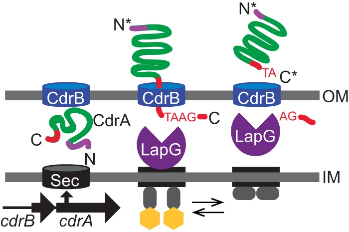
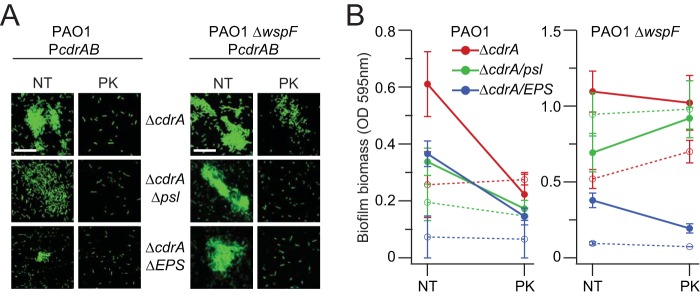
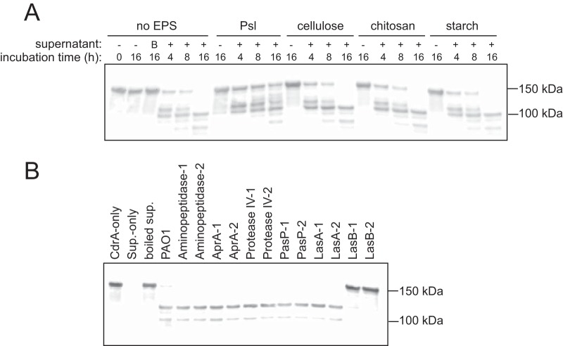
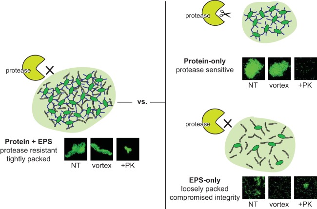
Biofilms are robust multicellular aggregates of bacteria that are encased in an extracellular matrix. Different bacterial species have been shown to use a range of biopolymers to build their matrices. Pseudomonas aeruginosa is a model organism for the laboratory study of biofilms, and past work has suggested that exopolysaccharides are a required matrix component. However, we found that expression of the matrix protein CdrA, in the absence of biofilm exopolysaccharides, allowed biofilm formation through the production of a CdrA-rich proteinaceous matrix. This represents a novel function for CdrA. Similar observations have been made for other species such as Escherichia coli and Staphylococcus aureus, which can utilize protein-dominant biofilm matrices. However, we found that these CdrA-containing matrices were susceptible to both exogenous and self-produced proteases. We previously reported that CdrA directly binds the biofilm matrix exopolysaccharide Psl. Now we have found that when CdrA bound to Psl, it was protected from proteolysis. Together, these results support the idea of the importance of multibiomolecular components in matrix stability and led us to propose a model in which CdrA-CdrA interactions can enhance cell-cell packing in an aggregate that is resistant to physical shear, while Psl-CdrA interactions enhance aggregate integrity in the presence of self-produced and exogenous proteases.IMPORTANCEPseudomonas aeruginosa forms multicellular aggregates or biofilms using both exopolysaccharides and the CdrA matrix adhesin. We showed for the first time that P. aeruginosa can use CdrA to build biofilms that do not require known matrix exopolysaccharides. It is appreciated that biofilm growth is protective against environmental assaults. However, little is known about how the interactions between individual matrix components aid in this protection. We found that interactions between CdrA and the exopolysaccharide Psl fortify the matrix by preventing CdrA proteolysis. When both components-CdrA and Psl-are part of the matrix, robust aggregates form that are tightly packed and protease resistant. These findings provide insight into how biofilms persist in protease-rich host environments.

Keywords: CdrA; Pseudomonas aeruginosa; Psl; biofilm; elastase; exopolysaccharides.


Figures

FIG 1
FIG 1
CdrA is the cargo of the two-partner secretion system encoded by the cdrAB operon. CdrA is found in both cell-associated and secreted fractions. Periplasmic protease LapG can cleave CdrA near its C terminus, which liberates CdrA from the bacterial cell surface. (Adapted from reference with permission of the publisher.)
FIG 2
FIG 2
CdrA can mediate bacterial aggregation in the absence of Psl or other EPS. (A) Aggregation of wild-type PAO1 and mutant strains that no longer produce Psl and/or other EPS (Pel and alginate) was evaluated after induction of PcdrAB with arabinose. Relative aggregation levels were determined by calculating the difference in OD600 between the PcdrAB strain and its corresponding vector control strain, dividing by the OD600 of the vector control strain, and then multiplying by 100%. Data represent the means of results from three replicates, and error bars indicate standard deviations. An asterisk indicates a significant difference in the levels of aggregation of Psl and EPS mutants compared to their parent strains (either strain PAO1 ΔcdrA PcdrAB or strain PAO1 ΔwspF ΔcdrA PcdrAB) (Student’s t test; P < 0.05). (B) Aggregates of bacteria constitutively expressing GFP were imaged using confocal laser scanning microscopy. Representative images of each strain are shown and were obtained from microscopy of at least three biological replicates. Scale bars represent 25 μm, and “Δpsl pel algD” is abbreviated as “ΔEPS.”
FIG 3
FIG 3
CdrA can mediate static biofilm formation in the absence of Psl or EPS. Static biofilm formation of cdrAB overexpression strains was measured by crystal violet staining. Green bars indicate control treatments without arabinose induction, and red bars indicate arabinose induction treatment. Data represent the means of results from six replicates, and error bars indicate standard deviations. An asterisk indicates a significant difference in biofilm biomass compared to the uninduced control (Student’s t test; P < 0.0005); n.s., not statistically significant compared to the uninduced control. “Δpsl pel algD” is abbreviated as “ΔEPS.”
FIG 4
FIG 4
CdrA-CdrA interactions are likely responsible for EPS-independent aggregation. (A and B) Microscopy of aggregates formed by strain PAO1 ΔwspF ΔcdrA ΔEPS transformed with either PcdrAB or the empty vector control. For this experiment, mixed-culture aggregates were grown from a 1:1 inoculum of each strain. (A) The mixed culture of PcdrAB (GFP+) and PcdrAB (mCherry+) showed intermixing of the two strains. (B) The mixed culture of the empty vector control (GFP+) and PcdrAB (mCherry+) did not show mixing of the two strains. Representative images of each condition are shown and were obtained from microscopy of at least three biological replicates. (C) Light microscopy showed that beads aggregated when they were adsorbed with CdrA. Beads adsorbed with BSA or treated only with PBS buffer did not aggregate. Red arrows indicate some of the aggregates that were observed when beads were adsorbed with CdrA. The experiment was repeated three times, and representative images for each condition are shown.
FIG 5
FIG 5
Proteinase K diminishes the amount of CdrA-dependent aggregation and static biofilm formation. (A) Aggregates of bacteria constitutively expressing GFP were imaged using confocal laser scanning microscopy with proteinase K (PK) treatment or no treatment (NT). Representative images of each strain and condition are shown and were obtained from microscopy of at least three biological replicates. (B) Static biofilm formation of cdrAB overexpression strains (solid lines) and isogenic strains carrying the empty vector control (dashed lines) was measured by crystal violet staining with PK treatment or NT. Data represent the means of results from 3 to 6 replicates, and error bars indicate standard deviations. Scale bars represent 25 μm, and “Δpsl pel algD” is abbreviated as “ΔEPS.”
FIG 6
FIG 6
CdrA is susceptible to P. aeruginosa proteases, and CdrA-Psl interactions are protective. (A) Anti-CdrA Western blot analysis showed that Psl, but not cellulose, chitosan, or starch, protected CdrA from degradation by P. aeruginosa supernatant proteases. Intact secreted CdrA that had not been treated with supernatant was detected at 150 kDa. Treating CdrA with boiled supernatant (indicated as “B” above lane 3) did not result in CdrA proteolysis. (B) Anti-CdrA Western blot analysis showed that LasB proteolyzed CdrA. Purified CdrA was treated with cell-free stationary-phase supernatant collected from a panel of protease mutants from the P. aeruginosa PAO1 transposon mutant library. Six extracellular proteases (aminopeptidase, AprA, protease IV, PasP, LasA, and LasB) were surveyed, and two mutants were tested for each protease.
FIG 7
FIG 7
P. aeruginosa can assemble aggregates using both EPS and CdrA. When both components are part of the matrix, robust aggregates that are tightly packed and protease resistant are formed. In contrast, when only CdrA is present, the aggregates are highly susceptible to proteolytic degradation. While EPS-only aggregates are protease resistant, they assemble as loosely packed aggregates whose structural integrity is easily disrupted.

Similar articles

Cited by

References

    1. Hall-Stoodley L, Costerton JW, Stoodley P. 2004. Bacterial biofilms: from the natural environment to infectious diseases. Nat Rev Microbiol 2:95–108. doi:10.1038/nrmicro821. - DOI - PubMed
    1. Lebeaux D, Chauhan A, Rendueles O, Beloin C. 2013. From in vitro to in vivo models of bacterial biofilm-related infections. Pathogens 2:288–356. doi:10.3390/pathogens2020288. - DOI - PMC - PubMed
    1. Römling U, Balsalobre C. 2012. Biofilm infections, their resilience to therapy and innovative treatment strategies. J Intern Med 272:541–561. doi:10.1111/joim.12004. - DOI - PubMed
    1. Costerton JW, Stewart PS, Greenberg EP. 1999. Bacterial biofilms: a common cause of persistent infections. Science 284:1318–1322. doi:10.1126/science.284.5418.1318. - DOI - PubMed
    1. Bjarnsholt T, Alhede M, Alhede M, Eickhardt-Sørensen SR, Moser C, Kühl M, Jensen PØ, Høiby N. 2013. The in vivo biofilm. Trends Microbiol 21:466–474. doi:10.1016/j.tim.2013.06.002. - DOI - PubMed

Publication types

MeSH terms