. 2018 Dec;24(12):1856-1870.
doi: 10.1261/rna.068221.118. Epub 2018 Sep 25.

Early developmental arrest and impaired gastrointestinal homeostasis in U12-dependent splicing-defective Rnpc3-deficient mice

Affiliations

Early developmental arrest and impaired gastrointestinal homeostasis in U12-dependent splicing-defective Rnpc3-deficient mice

Karen Doggett et al. RNA. 2018 Dec.

Abstract

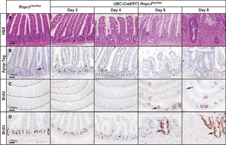
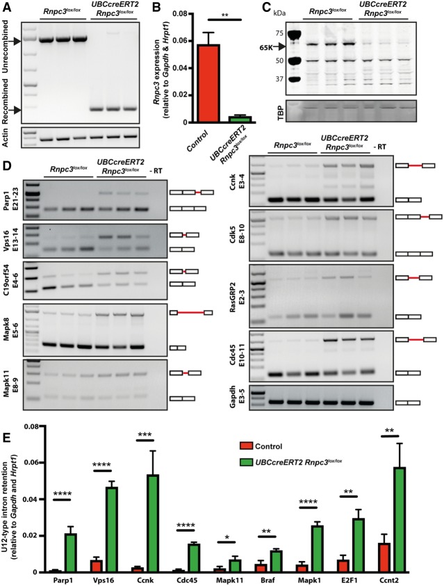
Splicing is an essential step in eukaryotic gene expression. While the majority of introns is excised by the U2-dependent, or major class, spliceosome, the appropriate expression of a very small subset of genes depends on U12-dependent, or minor class, splicing. The U11/U12 65K protein (hereafter 65K), encoded by RNPC3, is one of seven proteins that are unique to the U12-dependent spliceosome, and previous studies including our own have established that it plays a role in plant and vertebrate development. To pinpoint the impact of 65K loss during mammalian development and in adulthood, we generated germline and conditional Rnpc3-deficient mice. Homozygous Rnpc3-/- embryos died prior to blastocyst implantation, whereas Rnpc3+/- mice were born at the expected frequency, achieved sexual maturity, and exhibited a completely normal lifespan. Systemic recombination of conditional Rnpc3 alleles in adult (Rnpc3lox/lox ) mice caused rapid weight loss, leukopenia, and degeneration of the epithelial lining of the entire gastrointestinal tract, the latter due to increased cell death and a reduction in cell proliferation. Accompanying this, we observed a loss of both 65K and the pro-proliferative phospho-ERK1/2 proteins from the stem/progenitor cells at the base of intestinal crypts. RT-PCR analysis of RNA extracted from purified preparations of intestinal epithelial cells with recombined Rnpc3lox alleles revealed increased frequency of U12-type intron retention in all transcripts tested. Our study, using a novel conditional mouse model of Rnpc3 deficiency, establishes that U12-dependent splicing is not only important during development but is indispensable throughout life.

Keywords: RNPC3; U12 intron; development; gastrointestinal epithelium; minor class splicing.


Figures

FIGURE 1.
FIGURE 1.
Heterozygous Rnpc3-deficient mice exhibit a normal lifespan but complete loss of Rnpc3 is indispensable for preimplantation development. (A) The initial targeted allele (Rnpc3neo) used in this study contained an IRES:LacZ and a loxP-flanked promoter-driven neo cassette inserted into intron 3 of the Rnpc3 gene, thereby disrupting its function. (B) Mice harboring conditional alleles in which exons 4 and 5 are flanked by loxP sites were generated by crossing Rnpc3neo mice with an embryonic Flp deleter transgenic line. (C) A germline null allele (Rnpc3) was generated by crossing Rnpc3neo mice with mice carrying a Cre deleter transgene. Diagram modified from Skarnes et al. (2011) with permission from Springer Nature. (D) Longevity of Rnpc3+/− mice is the same as WT littermate controls: n = 58–62, Student's t-test P = 0.1131. (E) Pituitary gland weights of Rnpc3+/− mice (60–70 wk) are comparable to WT littermate controls (n = 11–17, Student's t-test P = 0.4822). (F) Several litters of mice were generated from in-crossing Rnpc3+/− parents and no Rnpc3−/− mice were born or hatched in culture from E3.5 blastocysts. (G) Genotyping of E3.5 blastocysts that had been cultured for 24 h identified all potential genotypes. Rnpc3+/+ and Rnpc3+/− blastocysts appeared normal while Rnpc3−/− exhibited an arrested morula phenotype (right panel). Scale bar in G = 100 µm.
FIGURE 2.
FIGURE 2.
65K is an essential protein in adult mice and is necessary for the integrity of self-renewing tissues. The effect of acute, global 65K deficiency was studied in Rnpc3lox/lox and Rnpc3lox/+ adult mice (8–12 wk) harboring an inducible UBC-CreERT2 allele. These mice were weighed prior to tamoxifen treatment and daily thereafter. At the end of the experiment, tissues and peripheral blood were collected for analysis. (A) UBC-CreERT2;Rnpc3lox/lox (green line, circles) mice had lost weight irreversibly by 6 d post-tamoxifen treatment and reached the ethical end-point requiring euthanasia at 8 d. In contrast, no significant weight loss was experienced by tamoxifen-treated UBC-CreERT2;Rnpc3lox/+ mice (blue line, diamonds) or the three control strains: Rnpc3lox/+ (blue line, inverted triangles), Rnpc3lox/lox (red line, squares), and UBC-CreERT2 (red line, triangles). Data are expressed as mean + SEM (n = 3–9). (*) P < 0.05, two-way ANOVA with Tukey's multiple comparison testing. (B) Analysis of peripheral blood at 8 d post-tamoxifen treatment revealed a significant decrease in the numbers of white blood cells (WBC), lymphocytes, and monocytes in UBC-CreERT2;Rnpc3lox/lox (green) compared to controls (red). (C) Red blood cell (RBC) numbers were also significantly decreased (D) and mice exhibited severe thrombocytopenia. (E) Systemic deletion of 65K induces thymic atrophy. Thymi were collected from control and UBC-CreERT2;Rnpc3lox/lox mice at 8 d post-tamoxifen treatment and weighed. Data are expressed as mean + SEM (n = 7–9). (****) P < 0.0001, (***) P < 0.0002, (**) P < 0.01 Student's t-test. (F) Histological analysis of the thymus displays characteristic cortex (white arrow) and medulla (black arrow) regions in control animals (Rnpc3lox/lox) at 8 d post-tamoxifen treatment. (G) This organization is absent in thymi taken from UBC-CreERT2;Rnpc3lox/lox mice. Scale bar in F–G = 100 µm.
FIGURE 3.
FIGURE 3.
Systemic Rnpc3 deficiency causes severe atrophy of the entire gastrointestinal epithelium. Histological analysis of the epithelial lining of the esophagus, stomach, small intestine, and colon in tamoxifen-treated control (Rnpc3lox/lox) (A–D) and experimental (UBC-CreERT2;Rnpc3lox/lox) mice (E–H) 8 d post-tamoxifen treatment. Whereas the organization and structural integrity of the gastrointestinal epithelium is intact in control organs (A–D), recombination of both Rnpc3lox alleles causes large-scale degeneration of the epithelial layer interspersed with discrete pockets of regenerating crypt-like structures (arrowheads in F, G, and H). Scale bar in A = 50 µm and applies to all images.
FIGURE 4.
FIGURE 4.
65K is expressed in the nuclei of cells in intestinal crypts and is lost following induced Rnpc3 deletion. (A,A′) Immunostaining with 65K antibody highlights positive nuclei at the base of the crypts of the small intestine (A, lower bracket) and colon (B,B′) with weak to absent expression in cells of the small intestinal villi (A, upper bracket) and in the lamina propria. Black arrows denote cells with 65K-positive nuclei in the stem cell compartment of the crypts. (C,D) Four days after tamoxifen (TMX) treatment of UBC-CreERT2;Rnpc3lox/lox mice, 65K protein expression is decreased in the small intestine (C, dashed bracket; magnified in C′) and colon (D,D′). The small intestine epithelial structure is disorganized with shorter villi. (E,F) Six days after TMX treatment, some areas of the small intestinal (E, bracket; magnified in E′) and colonic epithelium (F,F′) in UBC-CreERT2;Rnpc3lox/lox mice have completely lost 65K expression, coinciding with widespread epithelial disruption. Meanwhile, foci of regenerating crypt-like structures arise from cells in which complete recombination has not occurred and 65K protein is still evident (E′, arrows). A′–F′ show regions of interest at higher magnification. Scale bar in A, B, C, D, E, and F = 100 µm. Scale bar in A′, B′, C′, D′, E′, and F′ = 50 µm.
FIGURE 5.
FIGURE 5.
Loss of 65K induces apoptosis of epithelial cells in small intestinal crypts. The impact of acute global 65K deficiency on the integrity of the small intestine was studied in tamoxifen-treated Rnpc3lox/lox adult mice (8–12 wk) harboring an inducible UBC-CreERT2 allele. (A) Imaging of hematoxylin and eosin (H&E)-stained sections of small intestine from UBC-CreERT2;Rnpc3lox/lox mice revealed progressive epithelial damage from 4 d post-tamoxifen treatment, characterized by loss of crypt structure, shortening of villi, and invasion of inflammatory cells. Enlarged, regenerative crypts 6 d and 8 d post-tamoxifen treatment are indicative of a wound healing response. (B) Apo-tag (brown staining) identifies cells undergoing apoptosis. Such cells are rare in the unrecombined epithelium (arrow), but increase markedly in number in UBC-CreERT2;Rnpc3lox/lox mice 2 d and 4 d post-tamoxifen treatment. The apoptotic cells are found mainly in the crypts. (C) At 2, 4, 6, and 8 d post-tamoxifen treatment, mice were injected with BrdU to mark cells in the S-phase of the cell cycle, prior to being euthanized 2 h later. Low magnification images of BrdU-positive cells (brown nuclei) throughout the small intestine highlight the location of proliferative cells in the crypts; such cells are absent from the villi. Scale bar = 500 µm. Upon recombination, BrdU-positive cells are fewer in number and more dispersed, particularly 4 d after tamoxifen treatment. Areas of regenerating proliferating cells are observed from 6- to 8-d post-tamoxifen treatment (arrows). (D) Higher magnification views highlighting proliferative cells in control crypts (brackets) and at sites of regeneration in Rnpc3-depleted intestinal epithelium (arrows). Scale bars in A, B, D = 100 µm (applies to all images in the row).
FIGURE 6.
FIGURE 6.
pERK expression is concordant with 65K expression in the proliferative cells of intestinal crypts and in recovering regions of the epithelium. (A) Immunohistochemistry of pERK1/2 (pERK) reveals staining in the BrdU-positive compartment (cf. Fig. 5C,D) of crypts (boxed region in A is shown at higher magnification in A′ and A″). Meanwhile, pERK staining is absent from the terminally differentiated cells of the villi (bracket A′). The crypt base columnar (CBC) cells (arrowheads A″) strongly express pERK and can be clearly seen wedged between the large, granular, terminally differentiated Paneth cells, which are pERK negative. (B) Six days following tamoxifen treatment to induce recombination of Rnpc3lox alleles, the structural organization of the small intestinal epithelium is lost, save for discontinuous patches of cells that still express 65K due to incomplete recombination of Rnpc3lox alleles (B′ and arrows B″). (C) These same cells also show robust nuclear and cytoplasmic pERK expression (C is a serial section of B). The left box in B is magnified in panels B′ and C′. Regions predominantly lacking 65K (right box in B) also fail to express pERK (see brackets in magnified panels B″ and C″). Arrows denote individual cells in B and C that are both 65K- and pERK-positive (note that the 65K staining is nuclear only, whereas the pERK staining is found in the nucleus and cytoplasm). Scale bars in A, B, and C = 500 µm. Scale bars in A′, B′, C′, B″, and C″ = 50 µm. Scale bar in A″ = 20 µm.
FIGURE 7.
FIGURE 7.
Loss of 65K impairs minor class splicing in colonic epithelial cells. Preparations of gDNA, total RNA, and protein were extracted from purified colon crypt epithelial cells 4 d post-tamoxifen treatment. (A) Allele-specific PCR-based analysis demonstrates almost complete recombination of the Rnpc3 locus in UBC-CreERT2;Rnpc3lox/lox samples, but not in Rnpc3lox/lox samples. (B) RT-qPCR confirms that rnpc3 mRNA levels are significantly reduced in UBC-CreERT2;Rnpc3lox/lox samples (green bars) compared to control (red bars; combination of tamoxifen treated UBC-CreERT2;Rnpc3+/+ and Rnpc3lox/lox samples). (C) Western blot analysis shows a marked reduction in 65K protein in UBC-CreERT2;Rnpc3lox/lox compared to Rnpc3lox/lox controls (relative to TBP loading control). (D) RT-PCR and gel electrophoresis analysis of U12-type intron containing genes in colon crypt epithelial cells of mice shown in A. Schematic representation of spliced and intron-retaining mRNAs is shown on the right, with retained U12-type introns represented by red solid lines between exons (white boxes). (E) RT-qPCR using primers designed to amplify transcripts retaining their U12-type intron in several candidate U12-type intron-containing genes (Parp1, Vps16, Ccnk, Cdc45, Mapk11, Braf, Mapk1, E2F1, and Ccnt2) shows that U12-type intron retention is significantly increased in recombined UBC-CreERT2;Rnpc3lox/lox samples (green bars) compared to controls (red bars; combination of tamoxifen-treated UBC-CreERT2;Rnpc3+/+ and Rnpc3lox/lox samples). Data are expressed as mean + SEM relative to expression of housekeeping genes Gapdh and Hrpt1; n = 3 for each genotype. (*) P < 0.05, (**) P < 0.01, (***) P < 0.001, (****) P < 0.0001 Student's t-test.

References

    1. Alioto TS. 2007. U12DB: a database of orthologous U12-type spliceosomal introns. Nucleic Acids Res 35: D110–D115. - PMC - PubMed
    1. Argente J, Flores R, Gutiérrez-Arumí A, Verma B, Martos-Moreno GÁ, Cuscó I, Oghabian A, Chowen JA, Frilander MJ, Pérez-Jurado LA. 2014. Defective minor spliceosome mRNA processing results in isolated familial growth hormone deficiency. EMBO Mol Med 6: 299–306. - PMC - PubMed
    1. Ashton GH, Morton JP, Myant KK, Phesse TJ, Ridgway RA, Marsh VV, Wilkins JA, Athineos D, Muncan V, Kemp R, et al. 2010. Focal adhesion kinase is required for intestinal regeneration and tumorigenesis downstream of Wnt/c-Myc signaling. Dev Cell 19: 259–269. - PMC - PubMed
    1. Barker N, van Es JH, Kuipers J, Kujala P, van den Born M, Cozijnsen M, Haegebarth A, Korving J, Begthel H, Peters PJ, et al. 2007. Identification of stem cells in small intestine and colon by marker gene Lgr5. Nature 449: 1003–1007. - PubMed
    1. Barker N, van de Wetering M, Clevers H. 2008. The intestinal stem cell. Genes Dev 22: 1856–1864. - PMC - PubMed

Publication types

MeSH terms

Substances