Clinical Trial
. 2018 Dec 15;78(24):6838-6851.
doi: 10.1158/0008-5472.CAN-18-1680. Epub 2018 Sep 25.

Pharmacologic Ascorbate Reduces Radiation-Induced Normal Tissue Toxicity and Enhances Tumor Radiosensitization in Pancreatic Cancer

Affiliations
Clinical Trial

Pharmacologic Ascorbate Reduces Radiation-Induced Normal Tissue Toxicity and Enhances Tumor Radiosensitization in Pancreatic Cancer

Matthew S Alexander et al. Cancer Res. .

Abstract

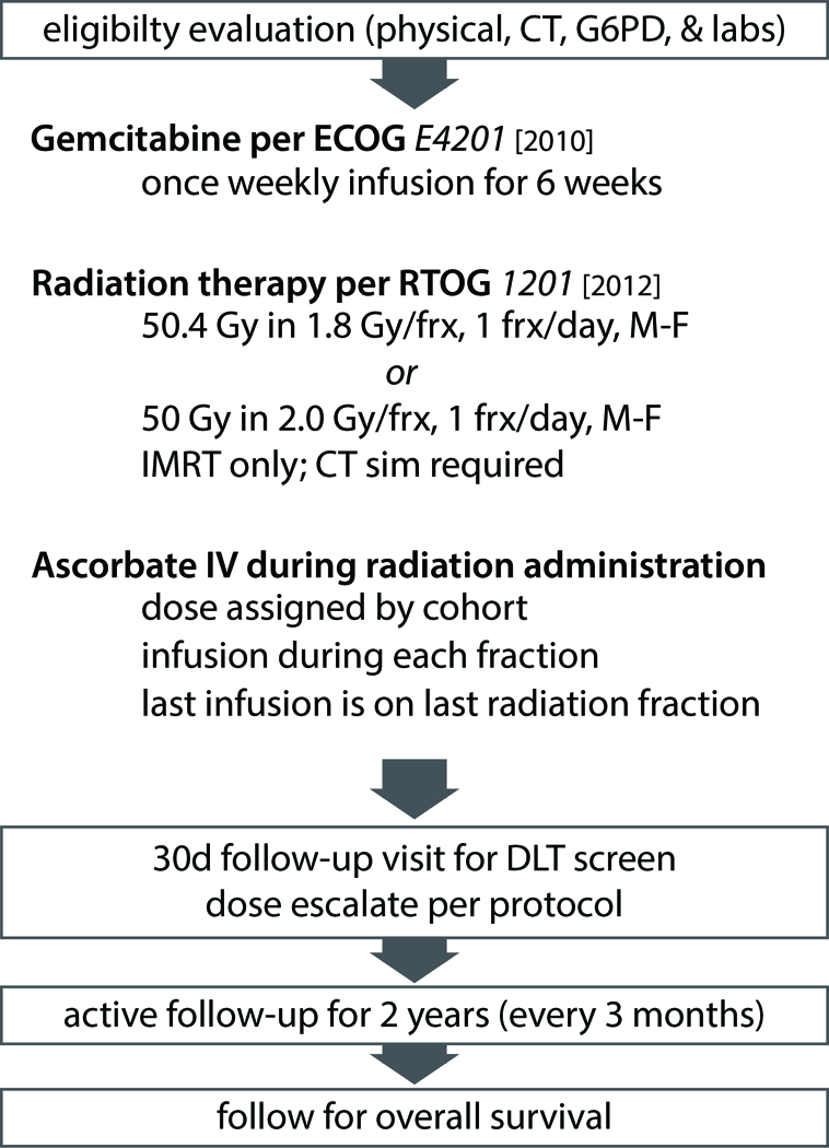
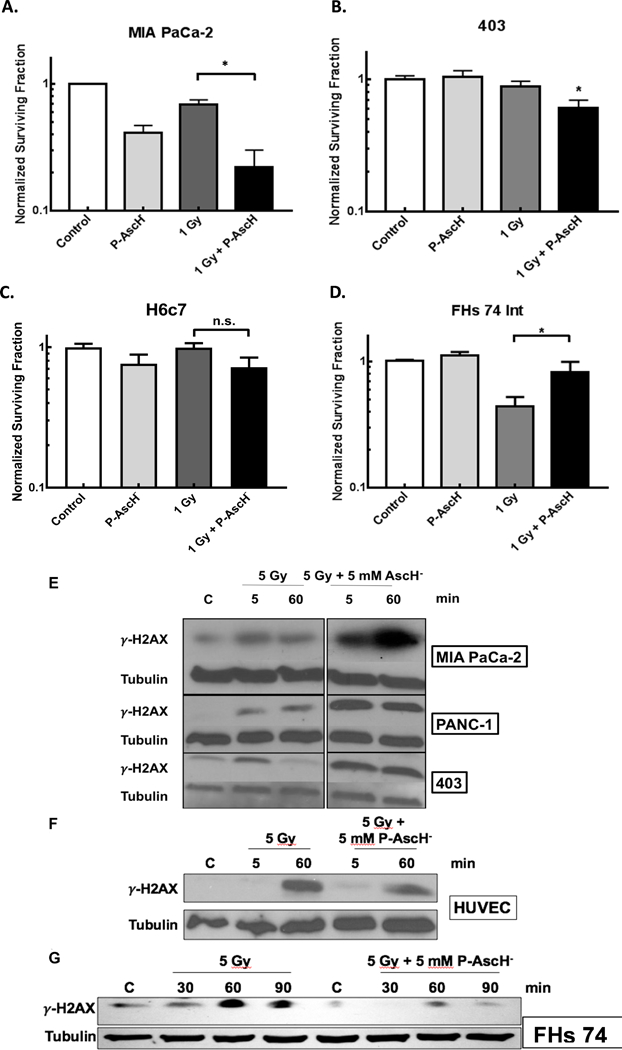
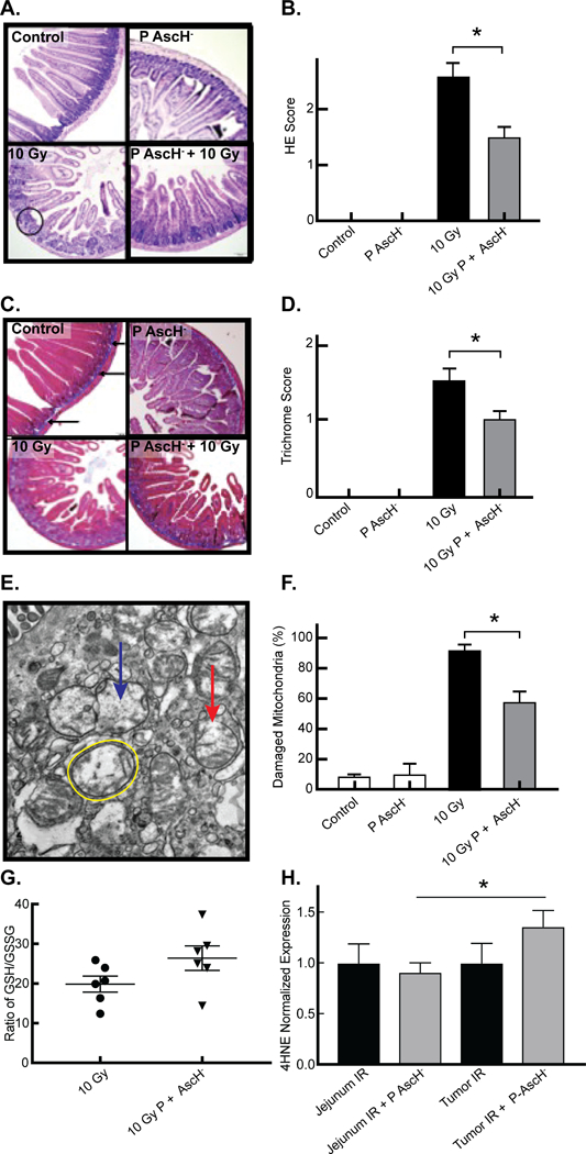
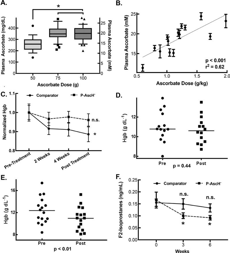
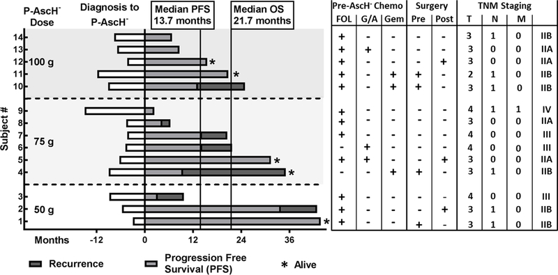
: Chemoradiation therapy is the mainstay for treatment of locally advanced, borderline resectable pancreatic cancer. Pharmacologic ascorbate (P-AscH-, i.e., intravenous infusions of ascorbic acid, vitamin C), but not oral ascorbate, produces high plasma concentrations capable of selective cytotoxicity to tumor cells. In doses achievable in humans, P-AscH- decreases the viability and proliferative capacity of pancreatic cancer via a hydrogen peroxide (H2O2)-mediated mechanism. In this study, we demonstrate that P-AscH- radiosensitizes pancreatic cancer cells but inhibits radiation-induced damage to normal cells. Specifically, radiation-induced decreases in clonogenic survival and double-stranded DNA breaks in tumor cells, but not in normal cells, were enhanced by P-AscH-, while radiation-induced intestinal damage, collagen deposition, and oxidative stress were also reduced with P-AscH- in normal tissue. We also report on our first-in-human phase I trial that infused P-AscH- during the radiotherapy "beam on." Specifically, treatment with P-AscH- increased median overall survival compared with our institutional average (21.7 vs. 12.7 months, P = 0.08) and the E4201 trial (21.7 vs. 11.1 months). Progression-free survival in P-AscH--treated subjects was also greater than our institutional average (13.7 vs. 4.6 months, P < 0.05) and the E4201 trial (6.0 months). Results indicated that P-AscH- in combination with gemcitabine and radiotherapy for locally advanced pancreatic adenocarcinoma is safe and well tolerated with suggestions of efficacy. Because of the potential effect size and minimal toxicity, our findings suggest that investigation of P-AscH- efficacy is warranted in a phase II clinical trial. SIGNIFICANCE: These findings demonstrate that pharmacologic ascorbate enhances pancreatic tumor cell radiation cytotoxicity in addition to offering potential protection from radiation damage in normal surrounding tissue, making it an optimal agent for improving treatment of locally advanced pancreatic adenocarcinoma.


Conflict of interest statement

Conflict of interest statement: The authors have no conflicts of interest to disclose.

Figures

Figure 1.
Figure 1.. Schematic of phase I clinical treatment plan.
Patients were evaluated for G6PD deficiency, as they are pre-disposed to hemolysis with P-AscH-. Gemcitabine and radiation therapy were given per published protocol.[5] After tolerance of a 15-g test dose, patients were escalated to treatment dose (50, 75, or 100 g) and followed for dose-limiting toxicities.
Figure 2.
Figure 2.. P-AscH- exacerbates radiation-induced cell death in cancer cells by increasing radiation-induced double-stranded DNA breaks, but protects normal cells.
A. MIA-PaCa-2 cells demonstrated significantly decreased clonogenic survival when treated with 1 mM (10 pmol cell−1) P-AscH- or 1 Gy radiation alone, with additive effect when radiation and P-AscH- are given in combination. (* p < 0.05) B. Significant decreases in clonogenic survival in the 403 cell line with the combination of 1 mM (10 pmol cell−1) P-AscH- and 1 Gy radiation compared to control (* p < 0.05). C. H6c7 cells demonstrate no significant cytotoxic effect of radiation in combination with P-AscH-. D. FHs 74 Int cells, derived from fetal human small intestinal epithelium, demonstrated significant decreased clonogenic survival when treated with 1 Gy radiation alone, but this effect is significantly reversed when co-treated with P-AscH-, 1 mM (10 pmol cell−1) (* p < 0.05). E. Pancreatic cancer cell lines (MIA PaCa-2, PANC-1, and 403) demonstrate increased γ-H2AX immunoreactivity with 5 Gy Radiation at 5 and 60 min. This effect is increased and prolonged when cells are co-treated with 5 mM P-AscH-. F. HUVEC cells, derived from normal umbilical vein endothelium, demonstrate increased γ-H2AX immunoreactivity 60 min after 5 Gy radiation. This effect is ameliorated when cells are co-treated with 5 mM P-AscH-. G. FHs 74 Int cells demonstrate increased γ-H2AX immunoreactivity 60 and 90 min after radiation. Again, this effect is ameliorated when cells are co-treated with 5 mM P-AscH-.
Figure 3.
Figure 3.. P-AscH- reduces radiation-induced jejunal mucosal damage, intestinal collagen deposition, normal tissue mitochondrial damage, and oxidative stress in vivo.
A. Light microscopy of representative samples of Hematoxylin/Eosin stained mouse jejunal samples demonstrates villous blunting and loss of fusion (circle) and crypt architecture in mice radiated with 10 Gy radiation, and reversal of this effect in mice treated with P-AscH- 2 days prior and 2 days after radiation (4 g kg−1, twice daily). B. Semi-quantitation, performed by a comparative pathologist blinded to the study, confirms increased jejunal damage in radiated mice. Radiation damage is significantly decreased in mice treated with P-AscH- (Means ± SEM, n = 3, * p < 0.05) C. Light microscopy of trichrome-blue stained mouse jejunal samples. Blue staining identifies collagen with normal deposition limited to the submucosa of controls (arrows). There is increased collagen thickness with penetration into the lamina propria in mice radiated with 10 Gy radiation, and inhibition of this effect in mice treated with P-AscH- 2 days prior and 2 days after radiation (4 g kg−1, twice daily). D. Semi-quantitation, performed by a comparative pathologist blinded to the study, confirms increased collagen deposition in radiated mice which is significantly decreased in mice treated with P-AscH- (Means ± SEM, n = 3, * p < 0.05) E. Electron microscopy of representative samples of mouse jejunal epithelial cells samples demonstrates increased mitochondrial damage characterized by mitochondrial swelling (yellow circle), loss of cristae structure (blue arrow), and membrane rupture (red arrow) in mice radiated with 10 Gy radiation, and partial inhibition of this effect in mice treated with P-AscH- 2 days prior and 2 days after radiation (4 g kg−1, twice daily). F. Percentage of damaged mitochondria in control, ascorbate, 10 Gy and 10 Gy + P-AscH- treated mice (Means ± SEM, n = 3, * p < 0.05) G. Ratio of GSH to GSSG in normal mouse jejunum exposed to 10 Gy vs. 10 Gy + P-AscH- (Means ± SEM, n = 5 – 6, *p < 0.05) H. 4HNE expression in mouse jejunum exposed to radiation and radiation + P-AscH- in comparison to 4HNE expression in tumor xenografts exposed to either radiation and radiation + P-AscH- (Means ± SEM, n = 4 – 5, * p < 0.05).
Figure 4.
Figure 4.. P-AscH- produces high plasma ascorbate concentrations, stabilizes hemoglobin concentration during chemoradiotherapy, and decreases plasma F2-Isoprostane levels during chemoradiotherapy.
A. Plasma ascorbate concentrations. Box represents interquartile range with ranges representing 10th and 90th percentiles. Mean plasma ascorbate concentrations for 50-, 75-, and 100-g doses are 265 mg dL-1 (15 mM, n = 17, 95% CI 232 – 299 mg dL-1), 355 mg dL-1 (20 mM, n = 37, 95% CI 333 – 376 mg dL-1), and 356 mg dL-1 (20 mM, n = 32, 95% CI 331 – 380 mg dL-1), respectively (Means ± SEM, n = 17 – 32, * p < 0.05) B. Plasma ascorbate concentration as a function of gram per kilogram dosing. The gram per kilogram dose was calculated as an average weight of the patient over the course of treatment. Linear Regression demonstrates significant non-zero slope and R2 = 0.62. C. A one way repeated measure ANOVA was performed on Hgb (Hgb, g dL−1) values over the course of the chemoradiation therapy cycle for comparator patients and patients receiving P-AscH-. Comparator patients experienced a change in Hgb over this time period (Means ± SEM, n = 13, * p < 0.01) while patients receiving P-AscH- did not (Means ± SEM, n = 14, p = 0.47). D. P-AscH- treated subject hemoglobin pre- and post-chemoradiotherapy were determined by linear regression from all Hgb measurement within each subject to minimized expected variation. Mean change in Hgb −0.34 ± 0.43 g dL−1 (SEM, n = 14, *p < 0.01). E. Hemoglobin levels in subjects not treated with P-AscH- before and after completion of chemoradiotherapy as calculated from linear regression of all subject hemoglobin measurements collected to minimized expected variation. Mean change in hemoglobin −1.47 ± 0.34 g dL−1 (n = 13, p = 0.44). F. Plasma was collected prior to therapy (week 0), after the 3rd week of therapy and at the finish of 6 weeks of chemoradiotherapy. Two-way ANOVA analysis was performed. There was no significant difference between week 0 plasma F2-Isoprostane levels compared to week 3 of treatment (Means ± SEM, n = 4, p = 0.99) or immediately following completion of chemoradiotherapy (Means ± SEM, n = 4, p = 0.88) in control subjects. There was a significant difference between week 0 plasma F2-Isoprostane levels compared to week 3 of treatment (Means ± SEM, n = 14, * p < 0.05) and immediately following completion of chemoradiotherapy (Means ± SEM, n = 14, * p < 0.05) in subjects treated with P-AscH-.
Figure 5.
Figure 5.. Patient Survival
Waterfall plot by dosing cohort demonstrating median progression free survival (13.7 mo) and overall survival (21.7 mo) of all subjects treated with P-AscH- plus combination therapy. Of fourteen subjects enrolled in the trial, five remain alive, three without disease progression. Pre-P-AscH- treatments are outlined indicating which chemotherapy regimens patients received prior to starting the clinical trial. Patients who received pancreas resections are indicated as well. TNM staging data is also available. FOL = FOLFIRINOX, G/A = Gemcitabine + Abraxane, Gem = Gemcitabine alone, Pre = pancreas resection prior to P-AscH- + chemoradiation, Post = pancreas resection following P-AscH- + chemoradiation

References

    1. Siegel RL, Miller KD, Jemal A. Cancer statistics, 2018. CA: A Cancer Journal for Clinicians 2018;68(1):7–30. - PubMed
    1. Ryerson AB, Eheman CR, Altekruse SF, Ward JW, Jemal A, Sherman RL, et al. Annual Report to the Nation on the Status of Cancer, 1975–2012, featuring the increasing incidence of liver cancer. Cancer 2016;122(9):1312–37. - PMC - PubMed
    1. Iacobuzio-Donahue CA, Fu B, Yachida S, Luo M, Abe H, Henderson CM, et al. DPC4 gene status of the primary carcinoma correlates with patterns of failure in patients with pancreatic cancer. Journal of clinical oncology : official journal of the American Society of Clinical Oncology 2009;27(11):1806–13. - PMC - PubMed
    1. Balaban EP, Mangu PB, Khorana AA, Shah MA, Mukherjee S, Crane CH, et al. Locally Advanced, Unresectable Pancreatic Cancer: American Society of Clinical Oncology Clinical Practice Guideline. Journal of Clinical Oncology 2016;34(22):2654–68. - PubMed
    1. Loehrer PJ Sr., Feng Y, Cardenes H, Wagner L, Brell JM, Cella D, et al. Gemcitabine alone versus gemcitabine plus radiotherapy in patients with locally advanced pancreatic cancer: an Eastern Cooperative Oncology Group trial. Journal of clinical oncology : official journal of the American Society of Clinical Oncology 2011;29(31):4105–12. - PMC - PubMed

Publication types

MeSH terms