

Semaphorin 7A Promotes Macrophage-Mediated Lymphatic Remodeling during Postpartum Mammary Gland Involution and in Breast Cancer
- PMID: 30254150
- PMCID: PMC6239927
- DOI: 10.1158/0008-5472.CAN-18-1642
Semaphorin 7A Promotes Macrophage-Mediated Lymphatic Remodeling during Postpartum Mammary Gland Involution and in Breast Cancer
Abstract
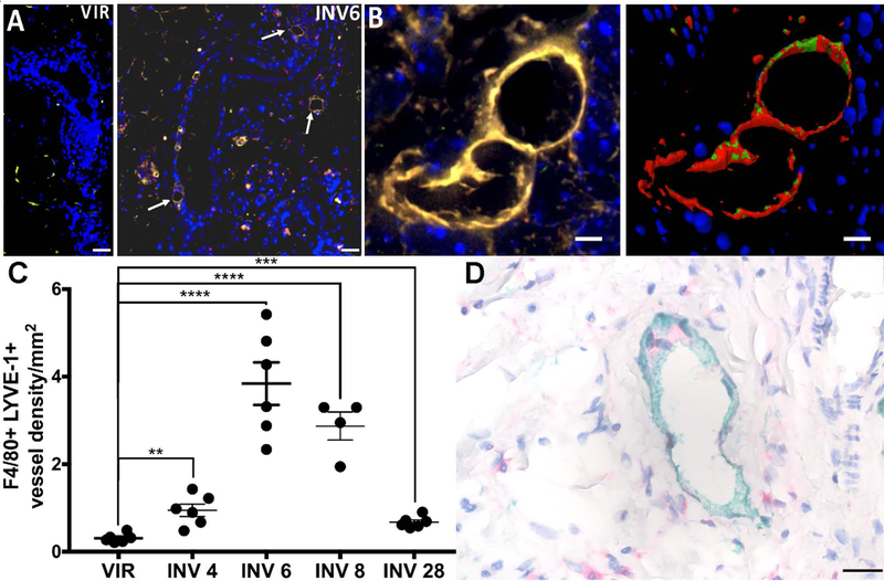
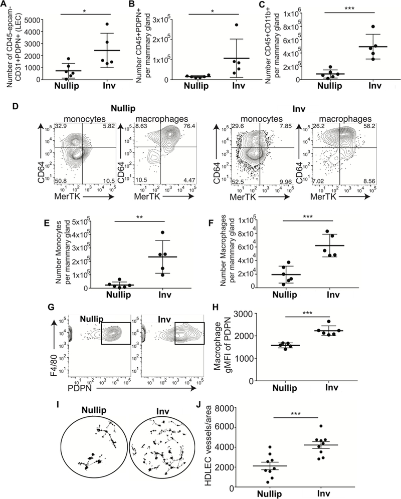
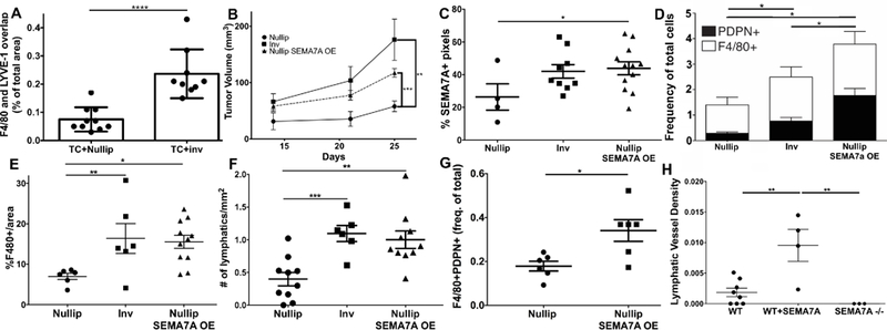
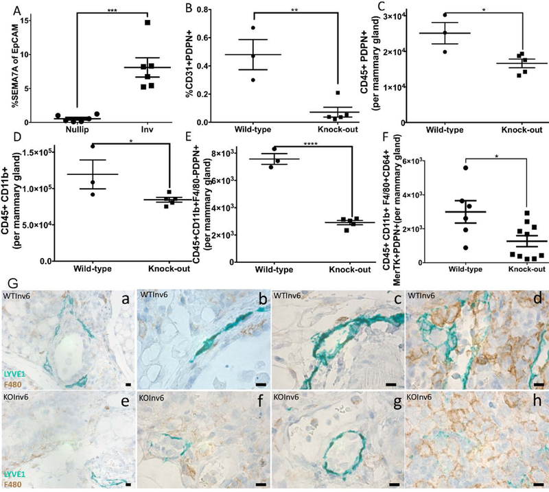
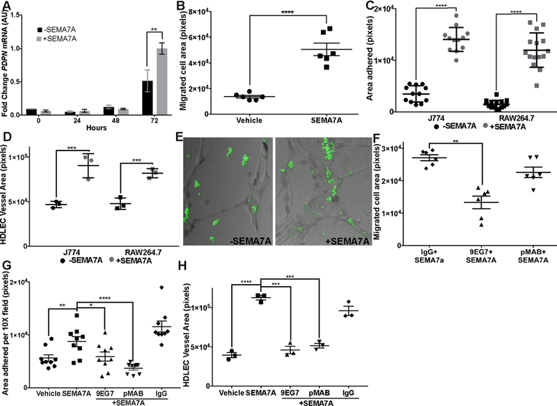
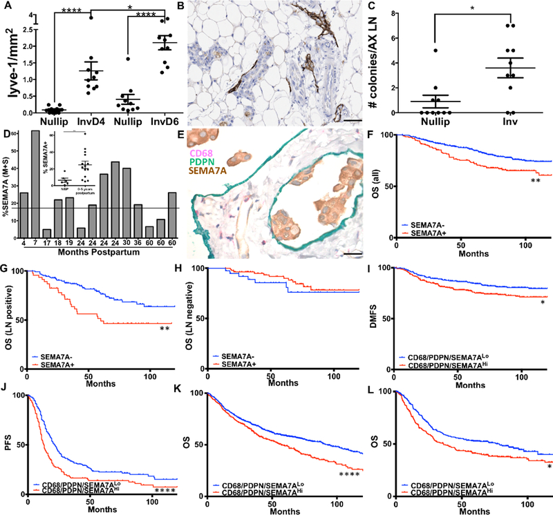
Postpartum mammary gland involution is a tissue remodeling event that occurs in all mammals in the absence of nursing or after weaning to return the gland to the pre-pregnant state. The tissue microenvironment created by involution has proven to be tumor promotional. Here we report that the GPI-linked protein semaphorin 7A (SEMA7A) is expressed on mammary epithelial cells during involution and use preclinical models to demonstrate that tumors induced during involution express high levels of SEMA7A. Overexpression of SEMA7A promoted the presence of myeloid-derived podoplanin (PDPN)-expressing cells in the tumor microenvironment and during involution. SEMA7A drove the expression of PDPN in macrophages, which led to integrin- and PDPN-dependent motility and adherence to lymphatic endothelial cells to promote lymphangiogenesis. In support of this mechanism, mammary tissue from SEMA7A-knockout mice exhibited decreased myeloid-derived PDPN-expressing cells, PDPN-expressing endothelial cells, and lymphatic vessel density. Furthermore, coexpression of SEMA7A, PDPN, and macrophage marker CD68 predicted for decreased distant metastasis-free survival in a cohort of over 600 cases of breast cancer as well as in ovarian, lung, and gastric cancers. Together, our results indicate that SEMA7A may orchestrate macrophage-mediated lymphatic vessel remodeling, which in turn drives metastasis in breast cancer.Signficance: SEMA7A, which is expressed on mammary cells during glandular involution, alters macrophage biology and lymphangiogenesis to drive breast cancer metastasis. Cancer Res; 78(22); 6473-85. ©2018 AACR.
©2018 American Association for Cancer Research.
Conflict of interest statement
Conflicts of Interest:
The authors have no conflicts of interest to disclose.
Figures
Similar articles
-
Postpartum breast cancer progression is driven by semaphorin 7a-mediated invasion and survival.Oncogene. 2020 Mar;39(13):2772-2785. doi: 10.1038/s41388-020-1192-9. Epub 2020 Feb 4. Oncogene. 2020. PMID: 32020054 Free PMC article.
-
Anoikis resistance in mammary epithelial cells is mediated by semaphorin 7a.Cell Death Dis. 2021 Sep 24;12(10):872. doi: 10.1038/s41419-021-04133-5. Cell Death Dis. 2021. PMID: 34561423 Free PMC article.
-
Semaphorin7A and PD-L1 cooperatively drive immunosuppression during mammary involution and breast cancer.Cell Rep. 2025 May 27;44(5):115676. doi: 10.1016/j.celrep.2025.115676. Epub 2025 May 6. Cell Rep. 2025. PMID: 40333186 Free PMC article.
-
Macphatics and PoEMs in Postpartum Mammary Development and Tumor Progression.J Mammary Gland Biol Neoplasia. 2020 Jun;25(2):103-113. doi: 10.1007/s10911-020-09451-6. Epub 2020 Jun 13. J Mammary Gland Biol Neoplasia. 2020. PMID: 32535810 Free PMC article. Review.
-
Mammary gland involution as an immunotherapeutic target for postpartum breast cancer.J Mammary Gland Biol Neoplasia. 2014 Jul;19(2):213-28. doi: 10.1007/s10911-014-9322-z. Epub 2014 Jun 22. J Mammary Gland Biol Neoplasia. 2014. PMID: 24952477 Free PMC article. Review.
Cited by
-
Hormonal Regulation of Semaphorin 7a in ER+ Breast Cancer Drives Therapeutic Resistance.Cancer Res. 2021 Jan 1;81(1):187-198. doi: 10.1158/0008-5472.CAN-20-1601. Epub 2020 Oct 29. Cancer Res. 2021. PMID: 33122307 Free PMC article.
-
Semaphorins as Potential Immune Therapeutic Targets for Cancer.Front Oncol. 2022 Jan 27;12:793805. doi: 10.3389/fonc.2022.793805. eCollection 2022. Front Oncol. 2022. PMID: 35155237 Free PMC article. Review.
-
Postpartum breast cancer progression is driven by semaphorin 7a-mediated invasion and survival.Oncogene. 2020 Mar;39(13):2772-2785. doi: 10.1038/s41388-020-1192-9. Epub 2020 Feb 4. Oncogene. 2020. PMID: 32020054 Free PMC article.
-
Lymphatic transport in anti-tumor immunity and metastasis.J Exp Med. 2025 Mar 3;222(3):e20231954. doi: 10.1084/jem.20231954. Epub 2025 Feb 19. J Exp Med. 2025. PMID: 39969537 Review.
-
Sema6D Regulates Zebrafish Vascular Patterning and Motor Neuronal Axon Growth in Spinal Cord.Front Mol Neurosci. 2022 Apr 7;15:854556. doi: 10.3389/fnmol.2022.854556. eCollection 2022. Front Mol Neurosci. 2022. PMID: 35465091 Free PMC article.
References
-
- Yamada A, Kubo K, Takeshita T, Harashima N, Kawano K, Mine T et al.: Molecular cloning of a glycosylphosphatidylinositol-anchored molecule CDw108. Journal of immunology 1999, 162(7):4094–4100. - PubMed
-
- Pasterkamp RJ, Peschon JJ, Spriggs MK, Kolodkin AL: Semaphorin 7A promotes axon outgrowth through integrins and MAPKs. Nature 2003, 424(6947):398–405. - PubMed
-
- Suzuki K, Okuno T, Yamamoto M, Pasterkamp RJ, Takegahara N, Takamatsu H et al.: Semaphorin 7A initiates T-cell-mediated inflammatory responses through alpha1beta1 integrin. Nature 2007, 446(7136):680–684. - PubMed
-
- Reilkoff RA, Peng H, Murray LA, Peng X, Russell T, Montgomery R et al.: Semaphorin 7a+ regulatory T cells are associated with progressive idiopathic pulmonary fibrosis and are implicated in transforming growth factor-beta1-induced pulmonary fibrosis. American journal of respiratory and critical care medicine 2013, 187(2):180–188. - PMC - PubMed
-
- Scott GA, McClelland LA, Fricke AF: Semaphorin 7a promotes spreading and dendricity in human melanocytes through beta1-integrins. The Journal of investigative dermatology 2008, 128(1):151–161. - PubMed
Publication types
MeSH terms
Substances
Grants and funding
LinkOut - more resources
Full Text Sources
Other Literature Sources
Medical
Molecular Biology Databases
