. 2018 Nov 6;8(11):3637-3659.
doi: 10.1534/g3.118.200641.

HO Endonuclease-Initiated Recombination in Yeast Meiosis Fails To Promote Homologous Centromere Pairing and Is Not Constrained To Utilize the Dmc1 Recombinase

Affiliations

HO Endonuclease-Initiated Recombination in Yeast Meiosis Fails To Promote Homologous Centromere Pairing and Is Not Constrained To Utilize the Dmc1 Recombinase

Lina Yisehak et al. G3 (Bethesda). .

Abstract

Crossover recombination during meiosis is accompanied by a dramatic chromosome reorganization. In Saccharomyces cerevisiae, the onset of meiotic recombination by the Spo11 transesterase leads to stable pairwise associations between previously unassociated homologous centromeres followed by the intimate alignment of homologous axes via synaptonemal complex (SC) assembly. However, the molecular relationship between recombination and global meiotic chromosome reorganization remains poorly understood. In budding yeast, one question is why SC assembly initiates earliest at centromere regions while the DNA double strand breaks (DSBs) that initiate recombination occur genome-wide. We targeted the site-specific HO endonuclease to various positions on S. cerevisiae's longest chromosome in order to ask whether a meiotic DSB's proximity to the centromere influences its capacity to promote homologous centromere pairing and SC assembly. We show that repair of an HO-mediated DSB does not promote homologous centromere pairing nor any extent of SC assembly in spo11 meiotic nuclei, regardless of its proximity to the centromere. DSBs induced en masse by phleomycin exposure likewise do not promote homologous centromere pairing nor robust SC assembly. Interestingly, in contrast to Spo11, HO-initiated interhomolog recombination is not affected by loss of the meiotic kinase, Mek1, and is not constrained to use the meiosis-specific Dmc1 recombinase. These results strengthen the previously proposed idea that (at least some) Spo11 DSBs may be specialized in activating mechanisms that both 1) reinforce homologous chromosome alignment via homologous centromere pairing and SC assembly, and 2) establish Dmc1 as the primary strand exchange enzyme.

Keywords: HO endonuclease; Spo11; chromosome pairing; meiosis; recombination; synapsis.


Figures

Figure 1
Figure 1
Creating strains in which HO endonuclease is the sole source of meiotic DSBs. (A) Illustration indicates the chromosomal positions of a meiosis-specific HO endonuclease gene cassette and various HO cut sites (HO cs). PSPO13 -HO interrupts the LYS2 locus on chromosome II. HO cs sequences together with the natMX4 drug marker were targeted to the indicated chromosome IV coordinates. CENIV (solid circle) corresponds to coordinates 449,711- 449,821bp (Saccharomyces Genome Database). (B) Illustration depicts genotypic and phenotypic MAT locus outcomes of spo11 spo13 meiotic nuclei with or without meiotic expression of HO endonuclease. Meiotic cells undergo a single equational division in spo11 spo13 strains producing a dyad with diploid spores. In the absence of HO-mediated recombination, each dyad spore receives one copy of MATa and one copy of MATα, resulting in two non-mating spores (nm). In the presence of HO endonuclease, interhomolog or intrachromosomal recombination at the MAT locus on any or all of the chromatids (1, 2, 3 or 4) can produce homozygous MATa or MATα spores, which will be phenotypically a or α “maters”.
Figure 2
Figure 2
HO-mediated DSBs at HO cs sequences on chromosome IV in spo11 rad51 meiosis. (A) Southern blot analysis shows DNA cleavage by HO endonuclease at HO cs sequences on chromosome IV in spo11 spo13 rad51 diploid strains (LY491, LY456, LY492, LY481, LY459, LY457 and LY458; see Figure 1 for HO cs positions, Table S4 for strain genotypes). Samples were collected and processed at 0, 12, 18 and 24 hr after placement in sporulation medium. Genomic DNA was digested with restriction enzymes that target sites flanking each of the HO cs loci within a 10 kb region; DNA fragment sizes (kb) are displayed next to blots. Fragments were visualized using a probe that hybridizes to the natMX4 sequence adjacent to each HO cs. In the absence of HO and before entry into meiosis in the presence of HO (0 hr of sporulation), the probe detects a single large fragment that corresponds to an intact fragment of DNA containing the HO cs. In the presence of HO endonuclease, a faster migrating (smaller) fragment is also seen for each of the cut sites beginning at 12hr, except for cs6. The absence of HO-mediated DSBs at cs6 was verified using two different restriction enzymes (See Materials and Methods). (B) Bar graph shows the percent of DNA cut by HO endonuclease within PSPO13-HO spo11 spo13 rad51 meiotic nuclei at the indicated time points. Values were calculated by dividing the intensity of the smaller fragment with the sum of the intensities of the smaller and larger fragment. Bars depict the range given by two experiments.
Figure 3
Figure 3
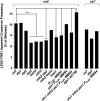
HO-mediated interhomolog recombination during meiosis. (A) Assay for interhomolog recombination at HO cs loci on chromosome IV in spo11 spo13 diploids. LEU2 is inserted at 450 kb (705 bp to the right of CEN4) and THR1 is inserted at 1,416 kb on chromosome IV for all strains, except for cs2 where LEU2 is at 447 kb (2.8 kb to the left of CEN4). A single HO cs is integrated between LEU2 and THR1. spo11 spo13 diploid cells undergo a single equational division during meiosis, resulting in spores carrying a chromatid from each parental homolog. In the absence of HO, each spore receives one LEU2 THR1 chromatid and one chromatid with neither marker (“No Recombination” column). In the presence of HO mediated DSBs, interhomolog reciprocal crossover recombination can result in a spore lacking the THR1 marker on chromosome IV (accompanied by a sister spore with two THR1 markers; upper right dyad). Because half of the crossover events will be invisible by this assay (lower right dyad), the percentage of apparent reciprocal crossovers were calculated by dividing twice the number of observed Thr- spores by the number of total 2-spore viable dyads (Table 2 and Table 4). (B) The upper bar graph plots the percentage of apparent crossing over calculated as described in (A) (n > 100 2-spore viable dyads assayed; Table 2) in various strains that carry no chromosome IV HO cs (LY208), or that carry distinct chromosome IV HO cs locations (left to right: LY208, LY555, LY207, LY324, LY322). The lower bar graph plots the percentage of apparent crossing over in mutant strains carrying HO cs5 (left to right: LY207, LY459, LY290, LY393, LY904, LY939, LY935, LY957 and LY910). Vegetative cultures of control expressing no HO nor HO cs (LY407) as well as LY208, LY555, LY324, LY322 and LY207 were independently evaluated for the uniform presence of the LEU2 and THR1 marker by assessment of growth of >400 single colonies on selective media.
Figure 4
Figure 4
HO-mediated recombination in the absence of Spo11 does not rely heavily on canonical meiotic recombination factors. Bar graph shows the frequency of apparent crossovers in spo11 spo13 strains carrying PSPO13-HO, an HO cs on chromosome IV, and mutant alleles of various recombination factors. Calculations were performed as described in Figure 3 (n > 250; precise values and strain names are reported in Table 4). Left part of graph shows data for strains carrying HO cs5, and the right 3 strains carry HO cs7. Significant deviations, relative to the wild-type value, were determined using Fisher’s Exact test (*P-value ≤ 0.05, **P-value ≤ 0.01, ***P-value ≤ 0.001).
Figure 5
Figure 5
A centromere-proximal or distal HO DSB is not sufficient to pair homologous chromosomes. (A) Cartoons show the locations of lacO DNA sequences 705 bp to the right of centromere IV (green) and tetO DNA sequences at 1,242 kb on chromosome IV in strains used for cytology experiments. GFP-LacI and TetR-mCherry, expressed in trans, bind lacO and tetO respectively. Strains homozygous for a single HO cs (top) or carrying seven active HO cs loci (bottom) were utilized in cytological experiments. (B) Images show surface spread meiotic nuclei from wild-type strains (LY42; left column), and from spo11-Y135F strains expressing meiosis-specific HO endonuclease and carrying HO cs5 (LY887; center and right columns) at 15 hr. of sporulation. The presence of Hop1 (not shown) was used to select meiotic nuclei for pairing analysis. The distance between foci corresponding to LacI-GFP bound to lacO sequences near CEN IV (green), and TetR-mCherry bound to tetO sequences on the arm of chromosome IV were considered paired if foci center to foci center < 0.5 μm apart. Bar, 1μm. (C) Bar graphs display the average frequency of CEN IV pairing or chromosome IV arm pairing at 15 hr of sporulation in control (LY176) and chromosome IV HO cs – carrying strains (left to right: LY176, LY173, LY174, LY175, LY331and LY887). 100 meiotic nuclei per genotype were analyzed in triplicate (n = 300 total per genotype). Bars depict standard error of the mean. (D) Homologous and non-homologous centromere pairing between centromeres indicated on the x axis was assessed in spo11 mutant strains (left to right: LY303, LY176, LY356, LY357 and LY358) at 15 hr of sporulation (n > 100).
Figure 6
Figure 6
Synaptonemal complex does not assemble in response to an HO-mediated meiotic DSB. (A) Representative surface spread meiotic nuclei from SPO11 (top row; YAM424), or spo11 null strains carrying seven active HO cs loci (cs2, cs4, cs5, cs6, cs7, cs8, cs9, cs10; LY371) at 15 hr of sporulation (upper panel, second and third row). Lower panel displays a representative nucleus from the spo11-Y135F strain carrying HO cs5 and overexpressing Rec8 (LY890; lower panel, top row) and two representative nuclei from the spo11 null strain carrying seven active HO cs and overexpressing Rec8 (LY892, lower panel, bottom two rows) at 24 hr of sporulation in ndt80 strains. Zip1 (green) binds diffusely to and also assembles some bright foci on DAPI-stained meiotic chromatin (blue) from these spo11 strains, regardless of HO-induced meiotic DSBs; polycomplex aggregates of Zip1 (white arrowheads) are often observed. The presence of the meiosis-specific Hop1 protein or Rec8-MYC (red) is displayed in the third column. Bar, 1μm. (B) The proportions of nuclei (n = 50) with different Zip1 and Rec8-MYC distribution phenotypes at 24 hr of sporulation are plotted for a control strain missing PSPO13-HO (LY893), the spo11 null strain carrying seven active HO cs loci (LY371), a Rec8-overexpression control spo11-Y135F strain with no chromosome IV HO cs (LY891), the spo11-Y135F strain carrying HO cs5 and overexpressing Rec8 (LY890) and the spo11 null strain carrying seven active HO cs loci and overexpressing Rec8 (LY892). Immunoblot in (C) shows Rec8-MYC levels in meiotic cells at 24 hr of sporulation from a control SPO11 ndt80 strain in which REC8 is untagged and which happens to also be homozygous for PGAL-HOP1 (LY769), a control spo11-Y135F ndt80 strain homozygous for REC8-MYC (LY893), followed by REC8-overexpressing LY890, LY891 and LY892 strains. The same cultures were used to prepare meiotic surface spread nuclei analyzed at the 24 hr time point. Molecular weight indicators are given (kDa) to the left. (D) Graph plots Rec8-MYC protein levels from a strain carrying two endogenous copies of REC8-MYC and strains carrying 2µ-REC8-MYC. Tubulin levels were used to normalize Rec8-MYC levels across samples. The average of 3 replicates is plotted; bars give standard error of the mean.

Similar articles

Cited by

References

    1. Agarwal S., Roeder G. S., 2000. Zip3 provides a link between recombination enzymes and synaptonemal complex proteins. Cell 102: 245–255. 10.1016/S0092-8674(00)00029-5 - DOI - PubMed
    1. Ajimura M., Leem S. H., Ogawa H., 1993. Identification of New Genes Required for Meiotic Recombination in Saccharomyces Cerevisiae. Genetics 133: 51–66. - PMC - PubMed
    1. Alani E., Padmore R., Kleckner N., 1990. Analysis of wild-type and rad50 mutants of yeast suggests an intimate relationship between meiotic chromosome synapsis and recombination. Cell 61: 419–436. 10.1016/0092-8674(90)90524-I - DOI - PubMed
    1. Allers T., Lichten M., 2001a Differential Timing and Control of Noncrossover and Crossover Recombination during Meiosis. Cell 106: 47–57. 10.1016/S0092-8674(01)00416-0 - DOI - PubMed
    1. Allers T., Lichten M., 2001b Intermediates of Yeast Meiotic Recombination Contain Heteroduplex DNA. Mol. Cell 8: 225–231. 10.1016/S1097-2765(01)00280-5 - DOI - PubMed

Publication types

MeSH terms

Substances