Review
. 2018 Sep 25;9(10):1002.
doi: 10.1038/s41419-018-1035-6.

The induction and consequences of Influenza A virus-induced cell death

Affiliations
Review

The induction and consequences of Influenza A virus-induced cell death

Georgia K Atkin-Smith et al. Cell Death Dis. .

Abstract

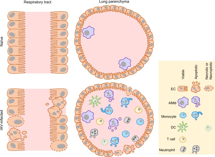
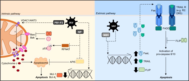
Infection with Influenza A virus (IAV) causes significant cell death within the upper and lower respiratory tract and lung parenchyma. In severe infections, high levels of cell death can exacerbate inflammation and comprise the integrity of the epithelial cell barrier leading to respiratory failure. IAV infection of airway and alveolar epithelial cells promotes immune cell infiltration into the lung and therefore, immune cell types such as macrophages, monocytes and neutrophils are readily exposed to IAV and infection-induced death. Although the induction of cell death through apoptosis and necrosis following IAV infection is a well-known phenomenon, the molecular determinants responsible for inducing cell death is not fully understood. Here, we review the current understanding of IAV-induced cell death and critically evaluate the consequences of cell death in aiding either the restoration of lung homoeostasis or the progression of IAV-induced lung pathologies.


Conflict of interest statement

The authors declare that they have no conflict of interest.

Figures

Fig. 1
Fig. 1. IAV-induced cell death.
During homoeostatic conditions, airway and alveolar epithelial cells (ECs) line the surfaces of the lung and AMΦs can be found within the lung parenchyma. IAV infection induces apoptosis and necrosis of ECs, compromising the integrity of the epithelial cell barrier. Furthermore, immune cells including monocytes, DCs and neutrophils infiltrate into the lung parenchyma and in turn are susceptible to IAV-induced apoptosis and necrotic-like cell death
Fig. 2
Fig. 2. Molecular mechanisms of IAV-induced apoptosis.
IAV can induce host cell apoptosis through either the intrinsic or extrinsic pathway. To induce apoptosis via the intrinsic pathway, IAV infection can promote the downregulation of anti-apoptotic proteins (Mcl-1/Bcl-XL) and p53 stabilisation. In turn, these mechanisms activate bax/bak to facilitate mitochondrial membrane pore formation and allow cytochrome c release. The IAV protein PB1-F2 can interact with VDAC1 and ANT3 to promote mitochondria outer and inner membrane permeabilisation and the release of pro-apoptotic molecules. Additionally, IAV infection can inhibit the anti-apoptotic protein API5 which usually functions to restrict apoptosome formation. IAV infection can also induce apoptosis through the extrinsic pathway by increasing the expression of death receptor ligands including FasL and TRAIL. Furthermore, infection can downregulate the expression of the anti-apoptotic protein FLIP which usually functions to limit Fas-mediated apoptosis. Dotted lines refer to IAV-targeted processes, solid lines refer to host cell processes
Fig. 3
Fig. 3. Functions and consequences of cell debris formation during IAV infection.
a Dead cells and debris generated from IAV-infected cells may be taken up by DCs, where engulfed viral proteins may be cross-presented to IAV-specific CD8+ T cells. b Macrophages can efficiently phagocytose cell debris to remove infectious material. However, if engulfment is impaired apoptotic particles can lyse through secondary necrosis, releasing an abundance of DAMPs that can exacerbate inflammation and lung damage. c Whether apoptotic EVs also contribute to viral pathogenesis is unknown. However, it is possible that such EVs could transfer viral molecules and contribute to antigen presentation or viral propagation

Similar articles

Cited by

References

    1. Galluzzi L, et al. Molecular mechanisms of cell death: recommendations of the Nomenclature Committee on Cell Death 2018. Cell Death Differ. 2018;25:486–541. doi: 10.1038/s41418-017-0012-4. - DOI - PMC - PubMed
    1. Wei MC, et al. Proapoptotic BAX and BAK: a requisite gateway to mitochondrial dysfunction and death. Science. 2001;292:727–730. doi: 10.1126/science.1059108. - DOI - PMC - PubMed
    1. Elmore S. Apoptosis: a review of programmed cell death. Toxicol. Pathol. 2007;35:495–516. doi: 10.1080/01926230701320337. - DOI - PMC - PubMed
    1. Kaufmann T, Strasser A, Jost PJ. Fas death receptor signalling: roles of Bid and XIAP. Cell Death Differ. 2012;19:42–50. doi: 10.1038/cdd.2011.121. - DOI - PMC - PubMed
    1. Atkin-Smith GK, Poon IK. Disassembly of the dying: mechanisms and functions. Trends Cell Biol. 2017;27:151–162. doi: 10.1016/j.tcb.2016.08.011. - DOI - PubMed

Publication types

MeSH terms