

Cervical squamous cell carcinoma-secreted exosomal miR-221-3p promotes lymphangiogenesis and lymphatic metastasis by targeting VASH1
- PMID: 30254211
- PMCID: PMC6363643
- DOI: 10.1038/s41388-018-0511-x
Cervical squamous cell carcinoma-secreted exosomal miR-221-3p promotes lymphangiogenesis and lymphatic metastasis by targeting VASH1
Erratum in
-
Correction to: Cervical squamous cell carcinoma-secreted exosomal miR-221-3p promotes lymphangiogenesis and lymphatic metastasis by targeting VASH1.Oncogene. 2022 Feb;41(8):1231-1233. doi: 10.1038/s41388-021-02165-x. Oncogene. 2022. PMID: 35046534 Free PMC article. No abstract available.
Abstract
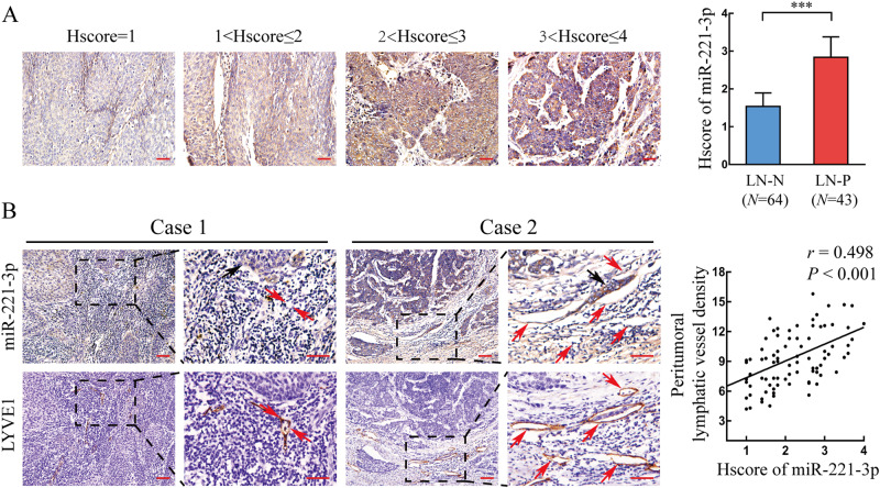
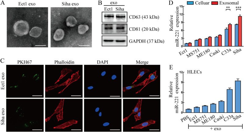
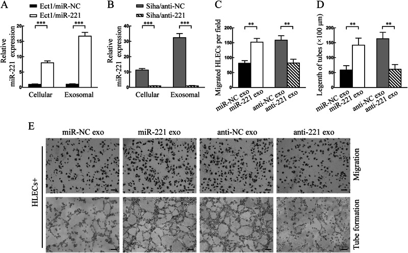
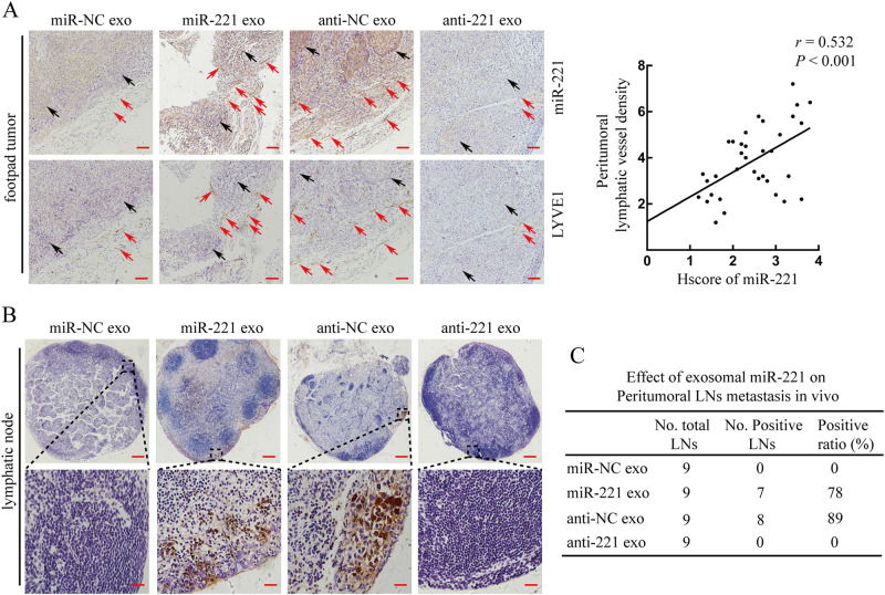
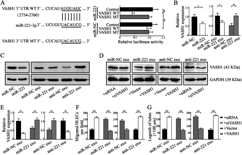
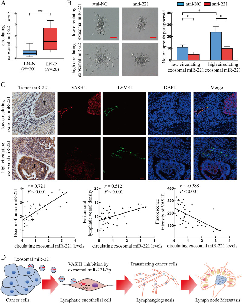
Cancer-secreted exosomal miRNAs are emerging mediators of cancer-stromal cross-talk in the tumor environment. Our previous miRNAs array of cervical squamous cell carcinoma (CSCC) clinical specimens identified upregulation of miR-221-3p. Here, we show that miR-221-3p is closely correlated with peritumoral lymphangiogenesis and lymph node (LN) metastasis in CSCC. More importantly, miR-221-3p is characteristically enriched in and transferred by CSCC-secreted exosomes into human lymphatic endothelial cells (HLECs) to promote HLECs migration and tube formation in vitro, and facilitate lymphangiogenesis and LN metastasis in vivo according to both gain-of-function and loss-of-function experiments. Furthermore, we identify vasohibin-1 (VASH1) as a novel direct target of miR-221-3p through bioinformatic target prediction and luciferase reporter assay. Re-expression and knockdown of VASH1 could respectively rescue and simulate the effects induced by exosomal miR-221-3p. Importantly, the miR-221-3p-VASH1 axis activates the ERK/AKT pathway in HLECs independent of VEGF-C. Finally, circulating exosomal miR-221-3p levels also have biological function in promoting HLECs sprouting in vitro and are closely associated with tumor miR-221-3p expression, lymphatic VASH1 expression, lymphangiogenesis, and LN metastasis in CSCC patients. In conclusion, CSCC-secreted exosomal miR-221-3p transfers into HLECs to promote lymphangiogenesis and lymphatic metastasis via downregulation of VASH1 and may represent a novel diagnostic biomarker and therapeutic target for metastatic CSCC patients in early stages.
Conflict of interest statement
The authors declare that they have no conflict of interest.
Figures
Similar articles
-
Esophageal carcinoma cell-excreted exosomal uc.189 promotes lymphatic metastasis.Aging (Albany NY). 2021 May 23;13(10):13846-13858. doi: 10.18632/aging.202979. Epub 2021 May 23. Aging (Albany NY). 2021. PMID: 34024769 Free PMC article.
-
Exosomal miR-10527-5p Inhibits Migration, Invasion, Lymphangiogenesis and Lymphatic Metastasis by Affecting Wnt/β-Catenin Signaling via Rab10 in Esophageal Squamous Cell Carcinoma.Int J Nanomedicine. 2023 Jan 6;18:95-114. doi: 10.2147/IJN.S391173. eCollection 2023. Int J Nanomedicine. 2023. PMID: 36636641 Free PMC article.
-
Cancer-derived exosomal miR-221-3p promotes angiogenesis by targeting THBS2 in cervical squamous cell carcinoma.Angiogenesis. 2019 Aug;22(3):397-410. doi: 10.1007/s10456-019-09665-1. Epub 2019 Apr 15. Angiogenesis. 2019. Retraction in: Angiogenesis. 2023 Feb;26(1):201. doi: 10.1007/s10456-022-09864-3. PMID: 30993566 Retracted.
-
Control of Gene Expression by Exosome-Derived Non-Coding RNAs in Cancer Angiogenesis and Lymphangiogenesis.Biomolecules. 2021 Feb 9;11(2):249. doi: 10.3390/biom11020249. Biomolecules. 2021. PMID: 33572413 Free PMC article. Review.
-
Double-Face of Vasohibin-1 for the Maintenance of Vascular Homeostasis and Healthy Longevity.J Atheroscler Thromb. 2018 Jun 1;25(6):461-466. doi: 10.5551/jat.43398. Epub 2018 Feb 3. J Atheroscler Thromb. 2018. PMID: 29398681 Free PMC article. Review.
Cited by
-
Exosome-derived miR-142-5p remodels lymphatic vessels and induces IDO to promote immune privilege in the tumour microenvironment.Cell Death Differ. 2021 Feb;28(2):715-729. doi: 10.1038/s41418-020-00618-6. Epub 2020 Sep 14. Cell Death Differ. 2021. PMID: 32929219 Free PMC article.
-
Pancreatic Cancer Cell-Derived Exosomes Promote Lymphangiogenesis by Downregulating ABHD11-AS1 Expression.Cancers (Basel). 2022 Sep 23;14(19):4612. doi: 10.3390/cancers14194612. Cancers (Basel). 2022. PMID: 36230535 Free PMC article.
-
MiR-221-3p targets Hif-1α to inhibit angiogenesis in heart failure.Lab Invest. 2021 Jan;101(1):104-115. doi: 10.1038/s41374-020-0450-3. Epub 2020 Sep 1. Lab Invest. 2021. PMID: 32873879
-
Based on 3D-PDU and clinical characteristics nomogram for prediction of lymph node metastasis and lymph-vascular space invasion of early cervical cancer preoperatively.BMC Womens Health. 2024 Aug 2;24(1):438. doi: 10.1186/s12905-024-03281-y. BMC Womens Health. 2024. PMID: 39090652 Free PMC article.
-
Extracellular vesicles and macrophages in tumor microenvironment: Impact on cervical cancer.Heliyon. 2024 Jul 26;10(15):e35063. doi: 10.1016/j.heliyon.2024.e35063. eCollection 2024 Aug 15. Heliyon. 2024. PMID: 39165926 Free PMC article. Review.
References
-
- Lee YJ, Kim DY, Lee SW, Park JY, Suh DS, Kim JH, et al. A postoperative scoring system for distant recurrence in node-positive cervical cancer patients after radical hysterectomy and pelvic lymph node dissection with para-aortic lymph node sampling or dissection. Gynecol Oncol. 2017;144:536–40. doi: 10.1016/j.ygyno.2017.01.001. - DOI - PubMed
Publication types
MeSH terms
Substances
Grants and funding
LinkOut - more resources
Full Text Sources
Other Literature Sources
Medical
Miscellaneous
