. 2019 Feb;27(2):198-202.
doi: 10.1038/s41431-018-0271-7. Epub 2018 Sep 25.

Fetal fraction evaluation in non-invasive prenatal screening (NIPS)

Affiliations

Fetal fraction evaluation in non-invasive prenatal screening (NIPS)

Matthew S Hestand et al. Eur J Hum Genet. 2019 Feb.

Abstract

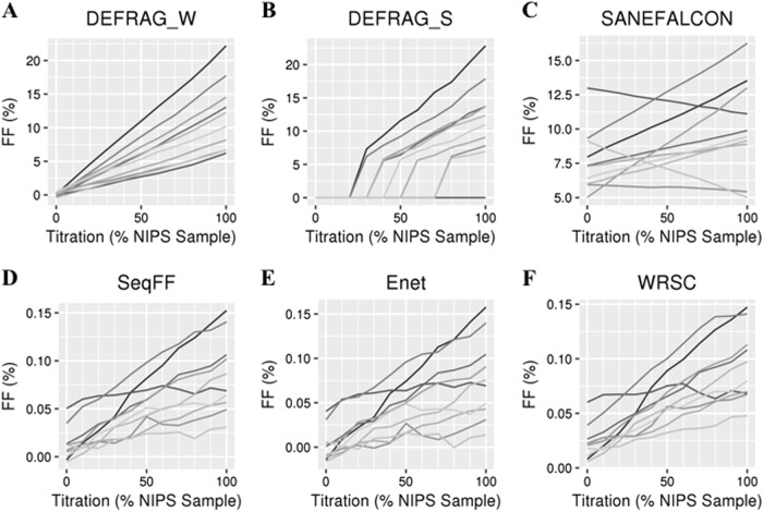
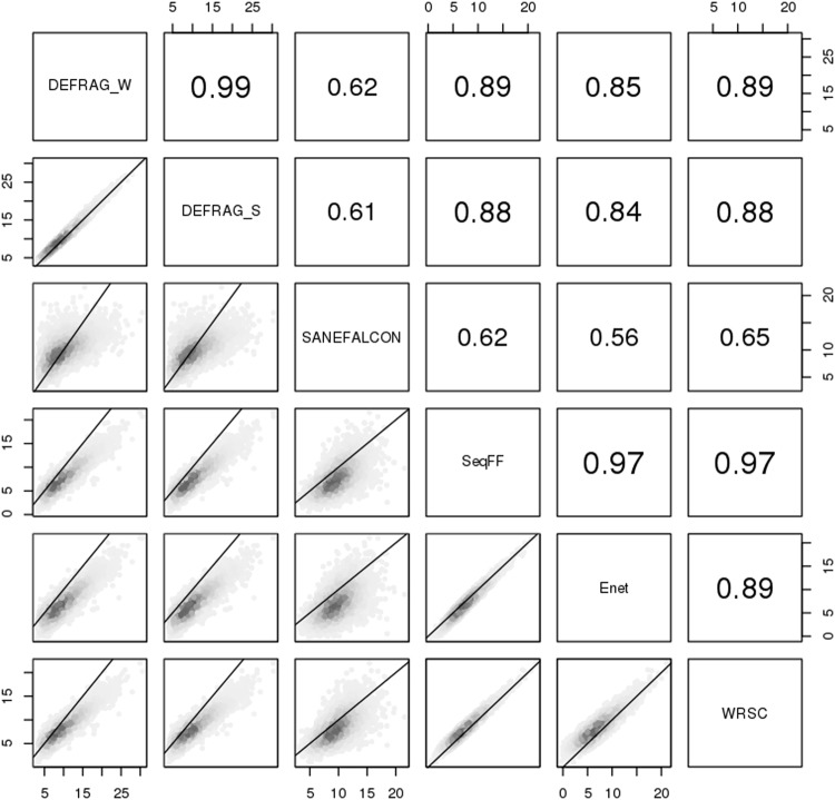
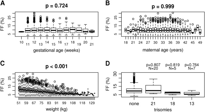
An important factor in quality control of non-invasive prenatal screening (NIPS) or testing (NIPT) is a sufficient percentage of fetal DNA to avoid false-negative results. Here we evaluate 14,379 shallow whole-genome sequenced diagnostic NIPS samples, as well as negative controls, for both technical and biological factors that can influence fetal fraction and its assessment. Technically, bioinformatics analyses can have a profound impact on fetal fraction determination. We found best performance for fetal fraction determination with the Y chromosome based tool DEFRAG for male fetuses and the count based tool SeqFF for female fetuses. Biologically, gestational age of up to 21 weeks and maternal age had no influence on fetal fraction, while an increase in weight and BMI had a negative influence on fetal fraction. While a trend was observed, no statistically significant difference in fetal fraction was found between trisomy and normal samples. Overall, these results confirm the influence of biological factors and give insight into technical factors that can affect fetal fractions in NIPS.


Conflict of interest statement

The authors declare that they have no conflict of interest.

Figures

Fig. 1
Fig. 1
FF determinations by each bioinformatics tool (af) based on ten synthetic titration series (each a different shade of gray) from 100% control (not pregnant, 0% on x-axis) samples to 100% NIPS samples
Fig. 2
Fig. 2
Bioinformatic comparisons for male fetuses FF. Numbers within panels indicate spearman correlations. Axis numbers indicate FF (%). Lines indicate theoretical one-to-one values
Fig. 3
Fig. 3
Male fetuses FFs as determined by DEFRAG_W as a function of gestational time (a), maternal age (b), weight (c), and trisomies (d)

References

    1. Takoudes T, Hamar B. Performance of non-invasive prenatal testing when fetal cell-free DNA is absent. Ultrasound Obstet Gynecol. 2015;45:112. doi: 10.1002/uog.14715. - DOI - PMC - PubMed
    1. Wang Q, Cai Y, Brady P, Vermeesch JR. Real-time PCR evaluation of cell-free DNA subjected to various storage and shipping conditions. Genet Mol Res. 2015;14:12797–804. doi: 10.4238/2015.October.19.23. - DOI - PubMed
    1. Peng XL, Jiang P. Bioinformatics approaches for fetal DNA fraction estimation in noninvasive prenatal testing. Int J Mol Sci. 2017;18:E453. doi: 10.3390/ijms18020453. - DOI - PMC - PubMed
    1. van Beek DM, Straver R, Weiss MM, et al. Comparing methods for fetal fraction determination and quality control of NIPT samples. Prenat Diagn. 2017;37:769–73. doi: 10.1002/pd.5079. - DOI - PMC - PubMed
    1. Song Y, Zhou X, Huang S, et al. Quantitation of fetal DNA fraction in maternal plasma using circulating single molecule amplification and re-sequencing technology (cSMART) Clin Chim Acta. 2016;456:151–6. doi: 10.1016/j.cca.2016.03.005. - DOI - PubMed