. 2018 Sep 25;9(10):1001.
doi: 10.1038/s41419-018-1051-6.

Arginase 1 promotes retinal neurovascular protection from ischemia through suppression of macrophage inflammatory responses

Affiliations

Arginase 1 promotes retinal neurovascular protection from ischemia through suppression of macrophage inflammatory responses

Abdelrahman Y Fouda et al. Cell Death Dis. .

Abstract

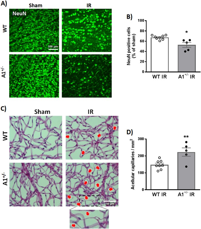
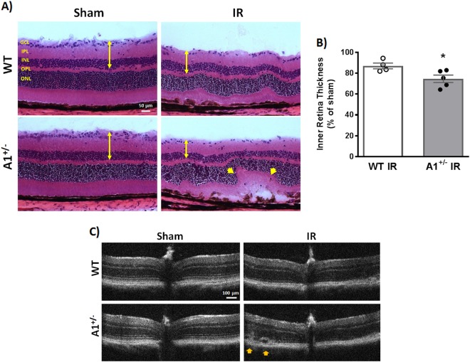
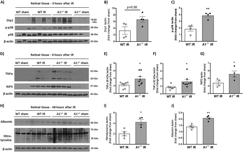
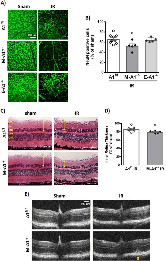
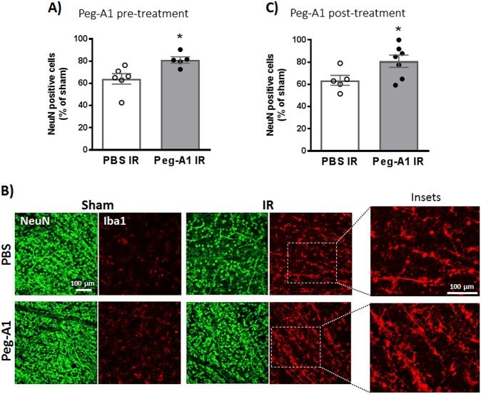
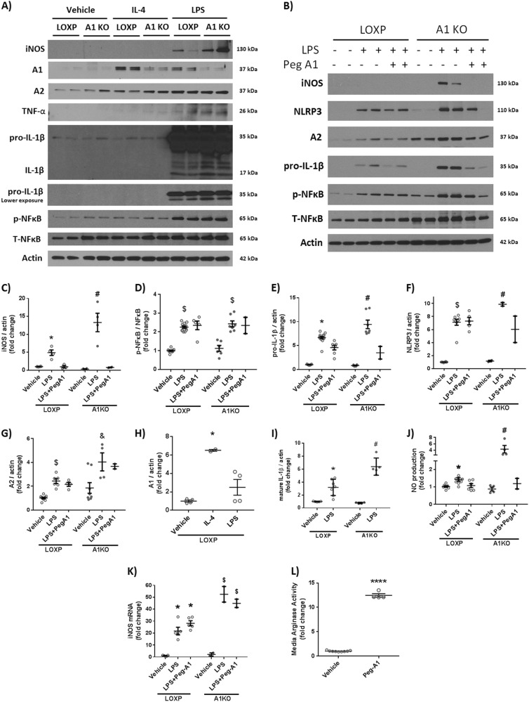
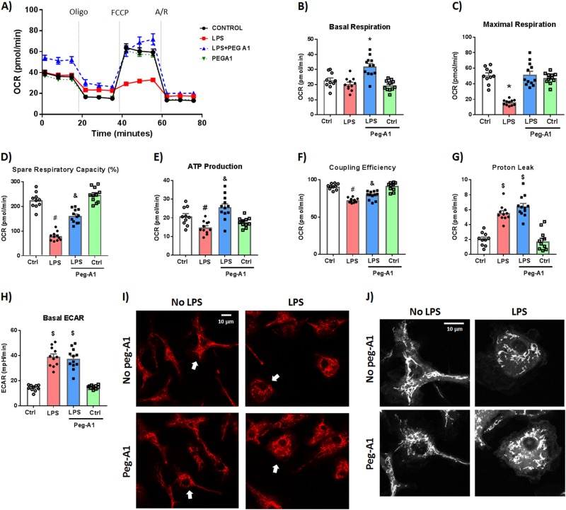
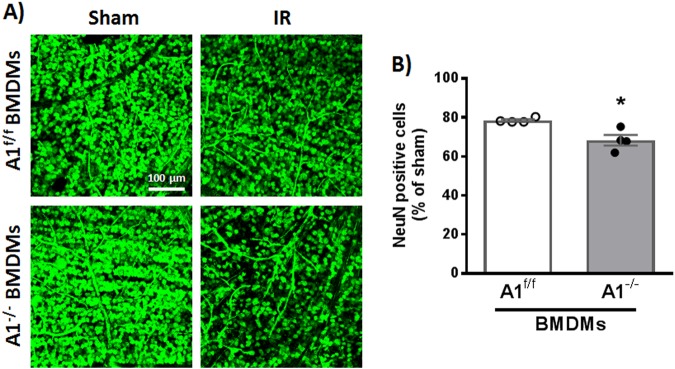
The lack of effective therapies to limit neurovascular injury in ischemic retinopathy is a major clinical problem. This study aimed to examine the role of ureohydrolase enzyme, arginase 1 (A1), in retinal ischemia-reperfusion (IR) injury. A1 competes with nitric oxide synthase (NOS) for their common substrate L-arginine. A1-mediated L-arginine depletion reduces nitric oxide (NO) formation by NOS leading to vascular dysfunction when endothelial NOS is involved but prevents inflammatory injury when inducible NOS is involved. Studies were performed using wild-type (WT) mice, global A1+/- knockout (KO), endothelial-specific A1 KO, and myeloid-specific A1 KO mice subjected to retinal IR injury. Global as well as myeloid-specific A1 KO mice showed worsened IR-induced neuronal loss and retinal thinning. Deletion of A1 in endothelial cells had no effect, while treatment with PEGylated (PEG) A1 improved neuronal survival in WT mice. In addition, A1+/- KO mice showed worsened vascular injury manifested by increased acellular capillaries. Western blotting analysis of retinal tissue showed increased inflammatory and necroptotic markers with A1 deletion. In vitro experiments showed that macrophages lacking A1 exhibit increased inflammatory response upon LPS stimulation. PEG-A1 treatment dampened this inflammatory response and decreased the LPS-induced metabolic reprogramming. Moreover, intravitreal injection of A1 KO macrophages or systemic macrophage depletion with clodronate liposomes increased neuronal loss after IR injury. These results demonstrate that A1 reduces IR injury-induced retinal neurovascular degeneration via dampening macrophage inflammatory responses. Increasing A1 offers a novel strategy for limiting neurovascular injury and promoting macrophage-mediated repair.


Conflict of interest statement

The authors declare that they have no conflict of interest.

Figures

Fig. 1
Fig. 1. A1 deletion worsens neuronal and microvascular degeneration after IR injury.
a WT and A1+/− mice were subjected to retinal IR injury and sacrificed at 7 days. Flat-mount NeuN staining showed neuronal cell loss in WT retinas after IR injury compared to shams, which was further aggravated in A1+/− mice. Scale bar = 100 μm. b Quantification of NeuN-positive cells, n = 5 for WT IR and 8 for A1+/− IR, *p < 0.05. c Vascular digests at 14 days showed increased numbers of acellular capillaries (red arrows) in WT IR injured retinas and this microvascular degeneration was further augmented in A1+/− IR injured retinas. Scale bar = 50 μm. d Quantification of acellular capillaries (empty basement membrane sleeves—enlarged in inset), n = 5 for WT IR and 8 for A1+/− IR, **p < 0.01
Fig. 2
Fig. 2. A1 deletion worsens retinal thinning and distortion after IR injury.
a Hematoxylin and eosin (H&E) staining of retinal frozen sections showed less retinal ganglion cells, distorted morphology, and retinal thinning 7 days after IR injury which was further worsened in A1+/− retinas (yellow arrow heads). Scale bar = 50 μm. GCL ganglion cell layer, IPL inner plexiform layer, INL inner nuclear layer, OPL outer plexiform layer, ONL outer nuclear layer. b Quantification of inner retina thickness (GCL + IPL + INL, denoted by yellow arrows in panel (a)), n = 4 for WT IR and 5 for A1+/− IR, *p < 0.05. c Optical coherence tomography (OCT) in live mice at 7 days corroborated the H&E results with yellow arrow heads pointing at retinal distortion/detachment, n = 3 per group (different cohort of mice than the one used for H&E)
Fig. 3
Fig. 3. A1 deletion increases inflammation, oxidative stress, and necroptosis markers after IR injury.
a Western blotting on retinal tissues collected at 3 h after IR showed higher levels of the stress marker p-p38 in A1+/− mice compared to WT after IR injury. There was also a trend towards higher levels of the mitochondrial fission protein, Drp1. b, c show quantification of Drp1 and p-p38 respectively. d Analysis at 6 h after IR injury showed a similar trend with increased TNF-α (26 kDa, membrane bound and 52 kDa, homotrimeric form), and RIP3 in A1+/− retinas as compared to WT. eg show quantification of TNF-α bands and RIP3 respectively. h A1+/− mice showed increased nitrotyrosine (marker for peroxynitrite-mediated oxidative stress via protein nitration) and albumin extravasation (measure of permeability) at 48 h after IR injury. i, j show quantification of nitrotyrosine and albumin western blotting respectively. *p < 0.05, **p < 0.01
Fig. 4
Fig. 4. Myeloid A1 deletion worsens neuronal loss and retinal thinning after IR injury.
a Retinas of mice with myeloid but not endothelial-specific A1 deletion showed worsened neuron loss compared to floxed control at 7 days after IR injury. Scale bar = 100 μm. b Quantification of NeuN-positive cells, n = 9 for A1f/f IR and 5 for M-A1−/− and E-A1−/− IR, *p < 0.05 vs. A1f/f. c H&E staining at 7 days showed worsened inner retina thinning in M-A1−/− mice compared to control (A1f/f). Scale bar = 50 μm. d Quantification of inner retina thickness, n = 7 for A1f/f IR and 6 for M-A1−/− IR, *p < 0.05. e Optical coherence tomography (OCT) corroborated the H&E results with yellow arrow pointing at retinal detachment
Fig. 5
Fig. 5. A1 treatment protects retinal neurons, and increases microglia/ macrophages after IR injury.
a Mice received intravitreal injection of PEG-A1 (1.7 ng/µl) 3 h before induction of IR injury and were sacrificed at day 7. PEG-A1 treatment preserved retinal neurons (NeuN-positive cells), n = 6 for PBS and 5 for PEG-A1, *p < 0.05. b PEG-A1-treated retinas showed increased microglia/macrophage infiltration as evident by Iba1 staining on retina flat-mounts. c Treatment with PEG-A1 (1.7 ng/µl) 3 h after IR injury achieved similar neuronal preservation to the pretreatment, thus showing post-injury protective effect, n = 5 for PBS and 7 for PEG-A1, *p < 0.05
Fig. 6
Fig. 6. Macrophages lacking A1 show a more pronounced inflammatory response to LPS stimulation in vitro and PEG-A1 treatment mitigates it.
a Western blotting of peritoneal macrophage cell lysates showed increased iNOS expression, TNF-α, and pro-IL-1β upon LPS stimulation which was further augmented in A1 KO macrophages. b PEG-A1 treatment (1 μg/ml) reduced this inflammatory response. ci Quantification of western blot bands. *p < 0.05 vs. loxP vehicle and loxP LPS + PEG-A1, #p < 0.05 vs. loxP LPS, A1KO vehicle and A1KO LPS + PEG-A1, $p < 0.05 vs. respective vehicle. &p < 0.05 vs. loxP LPS. j A1 KO macrophages showed more nitric oxide (NO) release into the media in response to LPS, as measured using NO analyzer and this was ameliorated by PEG-A1, *p < 0.05 vs. vehicle loxP, #p < 0.05 vs. loxP LPS, A1KO vehicle, A1KO LPS + PEG-A1. k RT-PCR on BMDMs showed increased iNOS mRNA expression with LPS that was further increased in A1 KO macrophages. PEG-A1 treatment did not affect iNOS mRNA expression *p < 0.05 vs. loxP vehicle, $p < 0.05 vs. respective loxP group. l Media from wells treated with PEG-A1 show marked elevation of arginase activity (12-fold increase compared to control) at the end of a 24 h incubation, ****p < 0.0001
Fig. 7
Fig. 7. PEG-A1 treatment protects against LPS-induced mitochondrial dysfunction in WT bone marrow-derived macrophages (BMDMs).
WT BMDMs were stimulated with LPS (100 ng/ml) for 24 h ± PEG-A1 (1 μg/ml). Seahorse XFe96 analyzer was used to evaluate mitochondrial function by measuring the oxygen consumption rate (OCR). a Change in OCR with time in response to Mito Stress test inhibitors (oligomycin, FCCP, and rotenone/antimycin A). bg Mitochondrial respiration parameters were decreased with LPS treatment. PEG-A1 significantly rescued this decrease. *p < 0.01 vs. other three groups, $p < 0.01 vs. controls, &p < 0.01 vs. respective control, LPS, #p < 0.01 vs. respective control, and LPS + PEG-A1, n = 12 per group. Representative run from two independent experiments that showed the same results. h Extracellular acidification rate (ECAR), a measure of glycolysis, was increased with LPS stimulation but was not affected by PEG-A1 cotreatment, $p < 0.01 vs. controls, n = 12 per group. i WT macrophages under control condition or PEG-A1 treatment alone exhibited an elongated and interconnected mitochondria (stained with Rhodamine 123). LPS-induced mitochondrial fragmentation and localization around the nucleus consistent with a round activated macrophage morphology. PEG-A1 cotreatment of LPS-stimulated macrophages partially reversed the LPS effect. j Magnification of cells denoted by arrows in panel (i). Images were converted to black and white for clarity. Scale bar = 10 μm
Fig. 8
Fig. 8. Macrophages lacking A1 worsen neurodegeneration after IR injury.
a NeuN staining showing increased neurodegeneration in WT retinas treated with A1 KO BMDMs (2×10⁵ cells, injected intravitreally on day 3 after IR injury), b Quantification of NeuN-positive cells, n = 4 per group, *p < 0.05. Scale bar = 100 μm

References

    1. Osborne NN, et al. Retinal ischemia: mechanisms of damage and potential therapeutic strategies. Prog. Retin. Eye Res. 2004;23:91–147. doi: 10.1016/j.preteyeres.2003.12.001. - DOI - PubMed
    1. D’Onofrio PM, Koeberle PD. What can we learn about stroke from retinal ischemia models? Acta Pharmacol. Sin. 2013;34:91–103. doi: 10.1038/aps.2012.165. - DOI - PMC - PubMed
    1. Hartsock, M. J. et al. A mouse model of retinal ischemia-reperfusion injury through elevation of intraocular pressure. JoVE113, e54065 (2016). - PMC - PubMed
    1. Binet F, et al. Neuronal ER stress impedes myeloid-cell-induced vascular regeneration through IRE1α degradation of netrin-1. Cell Metab. 2013;17:353–371. doi: 10.1016/j.cmet.2013.02.003. - DOI - PubMed
    1. London A, Benhar I, Schwartz M. The retina as a window to the brain-from eye research to CNS disorders. Nat. Rev. Neurol. 2013;9:44–53. doi: 10.1038/nrneurol.2012.227. - DOI - PubMed

Publication types

MeSH terms