. 2018 Sep 25;9(1):3905.
doi: 10.1038/s41467-018-06227-9.

Plasmodium co-infection protects against chikungunya virus-induced pathologies

Affiliations

Plasmodium co-infection protects against chikungunya virus-induced pathologies

Teck-Hui Teo et al. Nat Commun. .

Abstract

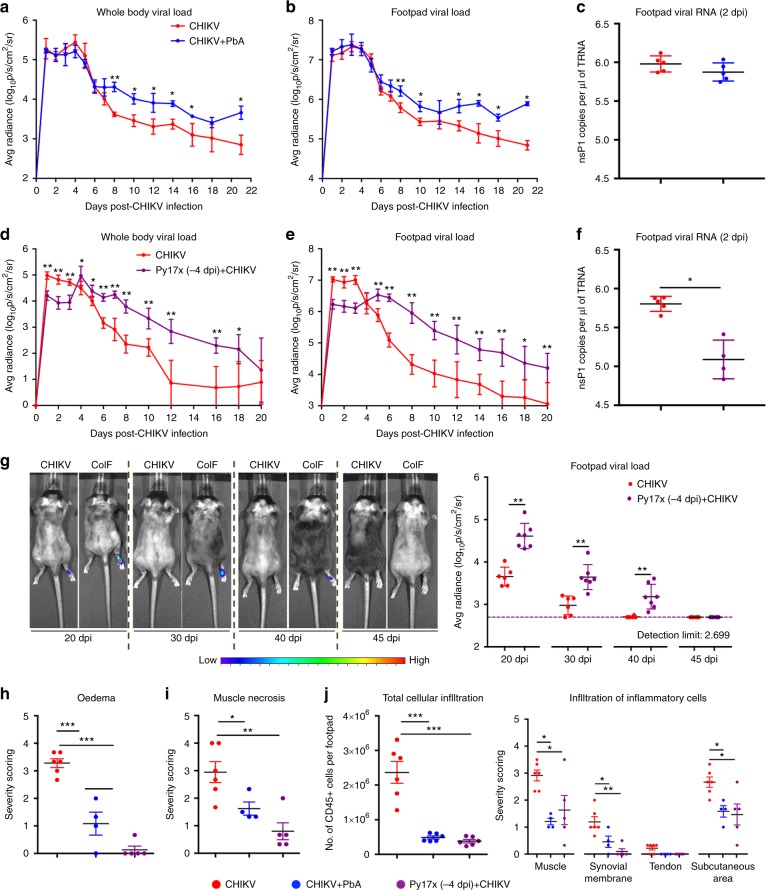
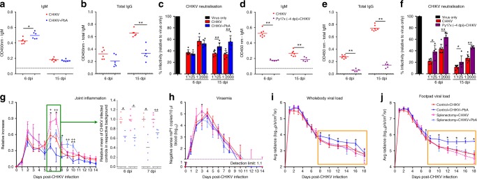
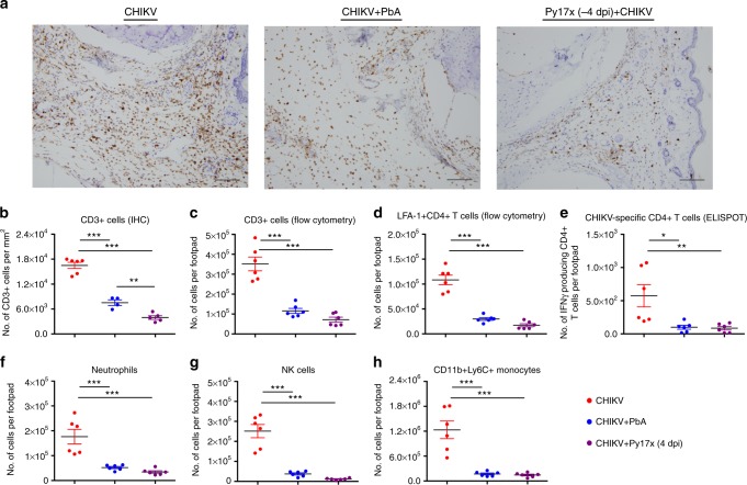
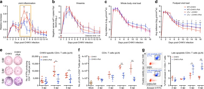
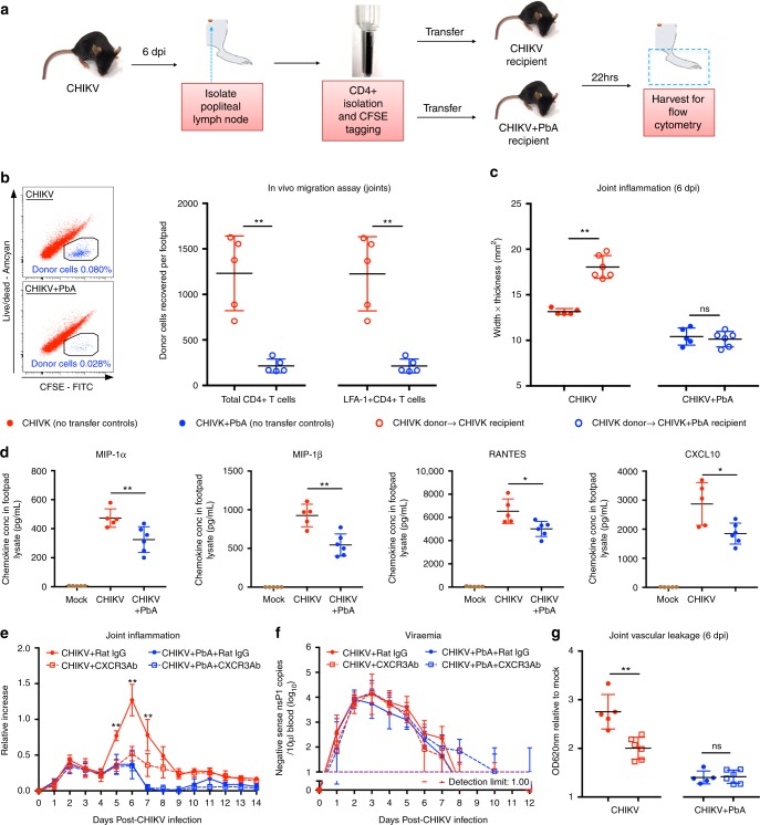
Co-infection with Plasmodium and chikungunya virus (CHIKV) has been reported in humans, but the impact of co-infection on pathogenesis remains unclear. Here, we show that prior exposure to Plasmodium suppresses CHIKV-associated pathologies in mice. Mechanistically, Plasmodium infection induces IFNγ, which reduces viraemia of a subsequent CHIKV infection and suppresses tissue viral load and joint inflammation. Conversely, concomitant infection with both pathogens limits the peak of joint inflammation with no effect on CHIKV viraemia. Reduced peak joint inflammation is regulated by elevated apoptosis of CD4+ T-cells in the lymph nodes and disrupted CXCR3-mediated CD4+ T-cell migration that abolishes their infiltration into the joints. Virus clearance from tissues is delayed in both infection scenarios, and is associated with a disruption of B cell affinity-maturation in the spleen that reduces CHIKV-neutralizing antibody production.


Conflict of interest statement

The authors declare no competing interests.

Figures

Fig. 1
Fig. 1
Co-infection suppresses CHIKV-induced joint swelling and viral replication. a Joint inflammation and viraemia of CHIKV (n = 7) and PbA (−4 dpi) + CHIKV (n = 7) groups. Each blue “†“ symbol represents one mouse that died of experimental cerebral malaria on the respective day. b Joint inflammation and viraemia of CHIKV (n = 5) and Py17x (−4 dpi) + CHIKV (n = 4) groups. For (a) and (b), Plasmodium infection was given 4 days prior to CHIKV infection, as shown in the schematic. c Joint inflammation and viraemia of CHIKV (n = 7) and CHIKV + PbA (n = 7) groups. d Joint inflammation and viraemia of CHIKV (n = 5) and CHIKV + Py17x (n = 5) groups. For (c) and (d), Plasmodium and CHIKV infection occurred concurrently, as shown in the schematic. All data were analyzed by Mann–Whitney two-tailed test (*P< 0.05, **P< 0.01 and ***P< 0.001), and are representative of two independent experiments. Data represent the means ± standard deviation. Parasitaemia were not altered by co-infection and data has been published in Teo et al.. Abbreviations: CHIKV, Chikungunya virus; dpi, days post infection; PbA, Plasmodium berghei-ANKA; Py17x, Plasmodium yoelii 17×
Fig. 2
Fig. 2
Co-infection delays virus resolution in tissues and suppress acute CHIKV-induced joint pathology. Tissue viral load in a the whole body and b the joints of CHIKV (n = 5) and CHIKV + PbA (n = 6) groups. Follow-up was terminated at 22 dpi as co-infected mice died of hyper-parasitaemia between 23 and 26 dpi. c Footpad viral RNA load on 2 dpi in the joints of CHIKV (n = 5) and CHIKV + PbA (n = 5) groups. Tissue viral load in d the whole body and e the joints of CHIKV (n = 7) and Py17x (−4 dpi) + CHIKV (n = 5) groups. f Footpad viral RNA load on 2 dpi in the joints of CHIKV (n = 5) and Py17x (−4 dpi) + CHIKV (n = 4) groups. g Footpad viral load of CHIKV (n = 6) and Py17x (−4 dpi) + CHIKV (n = 7) groups at 20 dpi, 30 dpi, 40 dpi and 45 dpi. Bioluminescence signal from the luciferase-tagged virus in both groups fell below the detection limit at 45 dpi. Representative pseudo-color images of bioluminescence signal at 20 dpi, 30 dpi, 40 dpi and 45 dpi are shown. Histopathological scoring of (h) oedema in the subcutaneous region and (i) muscle necrosis in the joints of CHIKV, CHIKV + PbA and Py17x (−4 dpi) + CHIKV (n ≥ 4 per group) at 6 dpi. j Total CD45 + cellular infiltration (flow cytometry) and infiltration of inflammatory cells in different regions of the joint (histological grading) of CHIKV, CHIKV + PbA and Py17x (−4 dpi) + CHIKV (n ≥ 4 per group) at 6 dpi. Data in ag were analyzed by Mann-Whitney two-tailed test and hj by one-way ANOVA with Tukey post hoc test (*P< 0.05, **P< 0.01 and ***P< 0.001). Data represent the means ± SD. Abbreviations: CHIKV, Chikungunya virus; CoIF, co-infected; dpi, days post infection; PbA, Plasmodium berghei-ANKA; Py17x, Plasmodium yoelii 17×
Fig. 3
Fig. 3
Acute IFNγ abrogates early acute CHIKV-induced joint inflammation, viremia and tissue viral load during sequential infection. Joint inflammation (a), viraemia (b) and viral load in the whole body (e) and footpad (f) of WT + CHIKV (n = 6), WT + Py17x (−4 dpi) + CHIKV (n = 6), IFNγ-/- + CHIKV (n = 6) and IFNγ-/- + Py17x (−4 dpi) + CHIKV (n = 5) groups. c Parasitaemia in WT + Py17x (n = 5,), WT + Py17x (−4 dpi) + CHIKV (n = 5,), IFNγ-/- + Py17x (n = 4) and IFNγ-/- + Py17x (−4 dpi) + CHIKV (n = 5) groups. Py17x or co-infected mice were euthanized at 23 days post Py17x infection as they were suffering from hyperparasitaemia and severe anemia. d Representative pseudo-colored images showing viral load in the tissue from 1–4 dpi. Yellow dotted box highlights acute suppression of the viral load in co-infected (CoIF) WT mice that is absence in CoIF IFNγ-/- mice. Blue box in a, b, e and f denotes acute phenotypes (1–4 dpi) induced in CoIF WT mice that are ameliorated in CoIF IFNγ-/- mice. Mann-Whitney two-tailed test was used to compare CoIF mice against their respective CHIKV-infected or Py17x-infected controls in the same genetic background. Comparisons between WT + CHIKV and WT + Py17x (−4 dpi) + CHIKV are represented by *P< 0.05 and **P< 0.01; comparisons between IFNγ-/- + CHIKV and IFNγ-/- + Py17x (−4 dpi) + CHIKV are represented by +P< 0.05 and ++P< 0.01. Data represent the means ± SD and are representative of two independent experiments. Abbreviations: CHIKV, Chikungunya virus; dpi, days post infection; Py17x, Plasmodium yoelii 17× ; WT, wild type
Fig. 4
Fig. 4
Elevated tissue viral load during is associated with suppression of CHIKV neutralizing antibodies production in the spleen. CHIKV-specific IgM (a) and total IgG (b) titer in the sera of CHIKV (n = 6) and CHIKV + PbA (n = 6) mice at 6 dpi and 15 dpi. c Neutralization of CHIKV infection in HEK293T cells using serum from CHIKV (n = 6) and CHIKV + PbA (n = 6) groups at 6 dpi and 15 dpi. CHIKV-specific IgM (d) and total IgG (e) titer in the sera of CHIKV (n = 7) and Py17x (−4 dpi) + CHIKV (n = 5) on 6 dpi and 15 dpi. f Neutralization of CHIKV infection in HEK293T cells using serum from CHIKV (n = 7) and Py17x (−4 dpi) + CHIKV (n = 5) groups at 6 dpi and 15 dpi. CHIKV-specific IgM and IgG titers were determined at 1:200 and 1:2000 respectively. Black dotted lines represents average OD reading of naïve mice for IgM and IgG. CHIKV neutralization assay was determined at 1:125 and 1:2000 dilution. Data in af were analyzed by Mann–Whitney two-tailed test (*P< 0.05 and **P< 0.01) and are representative of two independent experiments. g Joint inflammation, h, viraemia and viral load in the whole body (i) and footpad (j) of Control + CHIKV (n = 5), Control + CHIKV + PbA (n = 4), Splenectomy + CHIKV (n = 5) and Splenectomy + CHIKV + PbA (n = 5) groups. The green box denotes joint measurement during peak swelling whereby data were transformed to be expressed as relative to the mean of CHIKV infected mice in the respective background. The orange box denotes elevated tissue viral load in the control + CHIKV + PbA mice, which is prevented in the splenectomy + CHIKV + PbA mice. Data from co-infected mice were compared with their respective CHIKV infected controls by Mann-Whitney two-tailed analysis. Control + CHIKV versus Control + CHIKV + PbA comparison (*P< 0.05); Splenectomy + CHIKV versus Splenectomy + CHIKV + PbA (+P< 0.05 and ++P< 0.01). Data represent the means ± SD. Abbreviations: CHIKV, Chikungunya virus; dpi, days post infection; ns, not significant; OD, optical density; PbA, Plasmodium berghei-ANKA; Py17x, Plasmodium yoelii 17×
Fig. 5
Fig. 5
Co-infection limits pathogenic CD4 + T-cell and pro-inflammatory immune-cell infiltration into the joints. a Representative IHC images of CD3 staining in the joints at 6 dpi. Scale bar: 100 μm. Quantification of CD3 + cells (IHC) (b), CD3 + cells (flow cytometry) (c), activated (LFA-1 + ) CD4 + T cells (flow cytometry) (d) and CHIKV-specific CD4 + T cells (ELISPOT) (e) in the joints of CHIKV, CHIKV + PbA and Py17x (−4 dpi) + CHIKV (n ≥ 5 per group) at 6 dpi. Flow cytometric quantification of neutrophils (f), NK cells (g) and CD11b + Ly6C + monocytes (h) in the joints of CHIKV, CHIKV + PbA and Py17x (−4 dpi) + CHIKV (n ≥ 5 per group) at 6 dpi. Data represent the means ± SD and were analyzed by one-way ANOVA with Tukey post hoc test (*P< 0.05, **P< 0.01 and ***P< 0.001). Each data point in the dot plot represents one mouse. Data are representative of two independent experiments. Activated CD4 + T cells are defined as CD45 + /CD3 + CD4 + /LFA-1 + cells. Neutrophils and NK cells are defined as CD45 + /CD11b + /Ly6G + and CD45 + /NK1.1 + cells, respectively. CD11b + /Ly6C + monocytes were gated after gating out all T cells, neutrophils, NK cells and B cells. Abbreviations: CHIKV, Chikungunya virus; dpi, days post infection; IHC, immunohistochemistry; NK, natural killer; ns, not significant; PbA, Plasmodium berghei-ANKA
Fig. 6
Fig. 6
Co-infection induces early apoptosis of CD4 + T cells in the popliteal lymph node (pLN). Joint inflammation (a), viraemia (b) and viral load in the whole body (c) and footpad (d) of WT + CHIKV (n = 5), WT + CHIKV + PbA (n = 5), LTa-/- + CHIKV (n = 4) and LTa-/- + CHIKV + PbA (n = 4) groups. The orange box denotes suppression of joint inflammation (5–8 dpi) in co-infected WT mice compared to WT + CHIKV, which is lost in the co-infected LTa-/- mice. Data from co-infected mice was compared with their respective CHIKV infected controls on the same genetic background using Mann-Whitney two-tailed analysis. Comparisons between WT + CHIKV and WT + CHIKV + PbA are represented by *P< 0.05 and **P< 0.01; comparisons between LTa-/- + CHIKV and LTa-/- + CHIKV + PbA are represented by +P< 0.05. Quantification of CHIKV-specific CD4 + T cells (ELISPOT) (e), total CD4 + T cells (flow cytometry) (f) and late apoptotic CD4 + T cells (g) in the pLN of mock, CHIKV and CHIKV + PbA (n ≥ 5 per group) at 2, 4, 5 and 6 dpi. e Representative ELISPOT images showing spots generated by virus-stimulated CD4 + T cells isolated from 100,000 pLN cells (4 and 5 dpi) and 200,000 pLN cells (6 dpi) in CHIKV-infected and co-infected mice. g Representative dot plots displaying late apoptotic CD4 + T cells gating on 6 dpi. Late apoptotic CD4 + T cells are defined as CD45 + CD3 + CD4 + FVD + AnnexinV + cells. Data shown are pooled from four independent experiments. Data (eg) from the co-infected group were compared against CHIKV-infected controls at the respective time-point using Mann-Whitney two-tailed test (*P< 0.05 and **P< 0.01). Data represent the means ± SD. Abbreviations: CHIKV, Chikungunya virus; dpi, days post infection; PbA, Plasmodium berghei-ANKA
Fig. 7
Fig. 7
Co-infection abrogates CD4 + T-cell migration into the joints. a CD4 + T-cell in vivo migration assay. b In vivo migration assay showing number of recovered CHIKV-infected donor pLN CD4 + T cells (pooled) in the joints of CHIKV-infected or CHIKV + PbA-infected recipients (all groups n = 5). Representative dot plots displaying recovered donor-cell gating in 10,000 CD45 + cells in the footpad. c Joint inflammation of CHIKV (no transfer controls), CHIKV donor transferred to CHIKV recipient, CHIKV + PbA (no transfer controls) and CHIKV donor transferred to CHIKV + PbA recipient (all groups n ≥ 5). d Concentration of MIP-1a, MIP-1b, RANTES and CXCL10 in the joint-footpad cell lysate of mock (n = 5), CHIKV (n = 5) and CHIKV + PbA (n = 6) at 6 dpi. e Joint inflammation and f viraemia of CHIKV + Rat IgG (n = 6), CHIKV + CXCR3Ab (n = 6), CHIKV + PbA + Rat IgG (n = 6) and CHIKV + PbA + CXCR3Ab (n = 8). g Joint vascular leakage for CHIKV + Rat IgG, CHIKV + CXCR3Ab, CHIKV + PbA + Rat IgG and CHIKV + PbA + CXCR3Ab (all groups n ≥ 6). Joint vascular leakage was determined by Evan’s blue assay. All data were analyzed by Mann–Whitney two-tailed test (ns; not significant, *P< 0.05 and **P< 0.01). Each data point in the dot plot corresponds one mouse. Data represent the means ± SD. Abbbreviations: CHIKV, Chikungunya virus; dpi, days post infection; ns, not significant; PbA, Plasmodium berghei-ANKA

References

    1. Suhrbier A, Jaffar-Bandjee MC, Gasque P. Arthritogenic alphaviruses—an overview. Nat. Rev. Rheumatol. 2012;8:420–429. doi: 10.1038/nrrheum.2012.64. - DOI - PubMed
    1. Lwande OW, et al. Global emergence of Alphaviruses that cause arthritis in humans. Infect. Ecol. Epidemiol. 2015;5:29853. doi: 10.3402/iee.v5.29853. - DOI - PMC - PubMed
    1. Powers AM, Brault AC, Tesh RB, Weaver SC. Re-emergence of Chikungunya and O’nyong-nyong viruses: evidence for distinct geographical lineages and distant evolutionary relationships. J. Gen. Virol. 2000;81:471–479. doi: 10.1099/0022-1317-81-2-471. - DOI - PubMed
    1. Storm N, et al. Human cases of Sindbis fever in South Africa, 2006–2010. Epidemiol. Infect. 2014;142:234–238. doi: 10.1017/S0950268813000964. - DOI - PMC - PubMed
    1. Harley D, Sleigh A, Ritchie S. Ross River virus transmission, infection, and disease: a cross-disciplinary review. Clin. Microbiol. Rev. 2001;14:909–932. doi: 10.1128/CMR.14.4.909-932.2001. - DOI - PMC - PubMed

Publication types

MeSH terms

Substances

LinkOut - more resources