

Site-1 protease function is essential for the generation of antibody secreting cells and reprogramming for secretory activity
- PMID: 30254311
- PMCID: PMC6156501
- DOI: 10.1038/s41598-018-32705-7
Site-1 protease function is essential for the generation of antibody secreting cells and reprogramming for secretory activity
Abstract
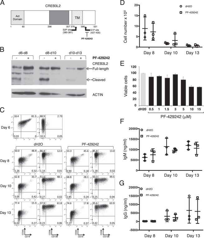
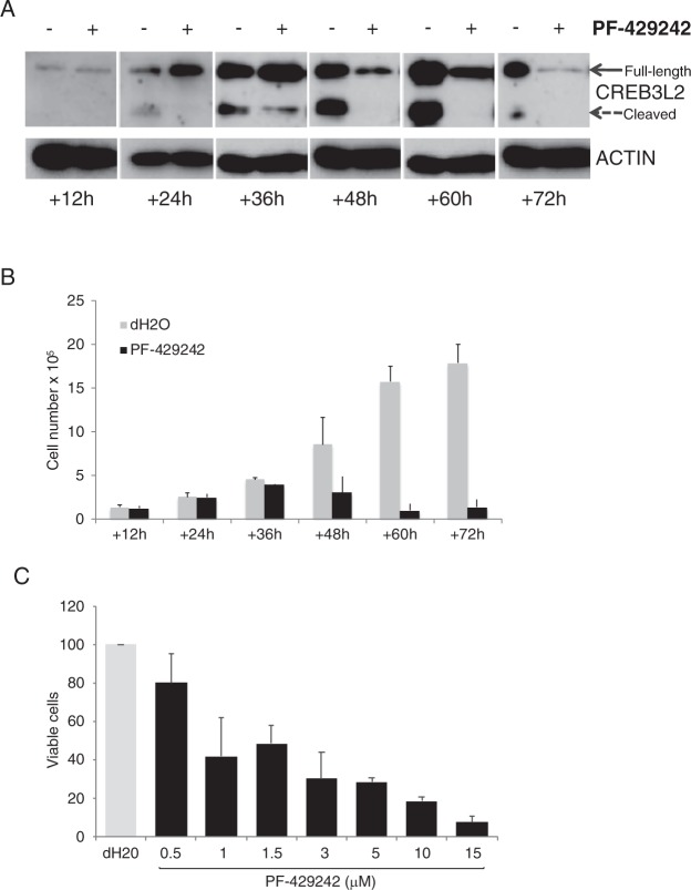
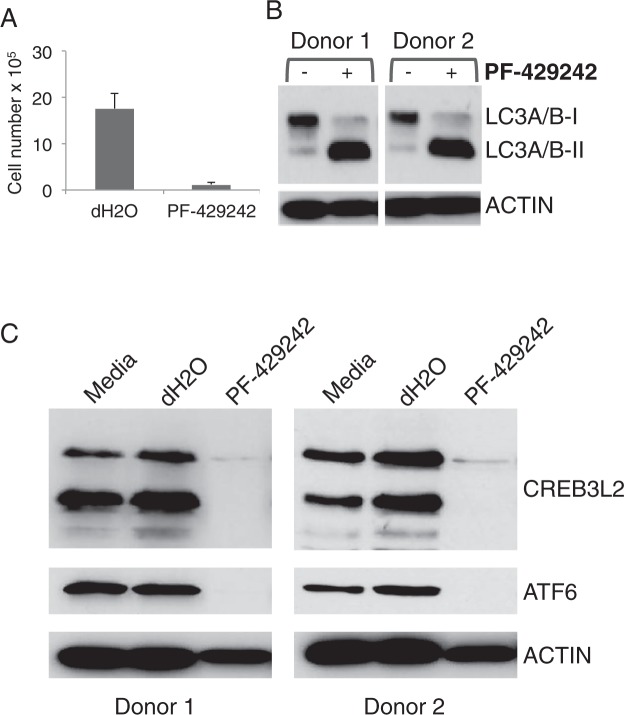
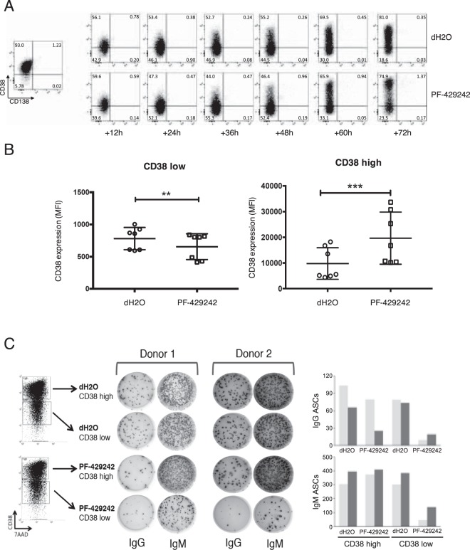
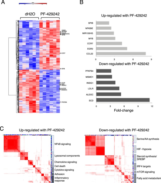
The unfolded protein response (UPR) and activation of XBP1 is necessary for high secretory efficiency and functional differentiation of antibody secreting cells (ASCs). The UPR additionally includes a branch in which membrane-bound transcription factors, exemplified by ATF6, undergo intramembrane-proteolysis by the sequential action of site-1 (MBTPS1/S1P) and site-2 proteases (MBTPS2/S2P) and release of the cytoplasmic domain as an active transcription factor. Such regulation is shared with a family of CREB3-related transcription factors and sterol regulatory element-binding proteins (SREBPs). Of these, we identify that the CREB3 family member CREB3L2 is strongly induced and activated during the transition from B-cell to plasma cell state. Inhibition of site-1 protease leads to a profound reduction in plasmablast number linked to induction of autophagy. Plasmablasts generated in the presence of site-1 protease inhibitor segregated into CD38high and CD38low populations, the latter characterized by a marked reduction in the capacity to secrete IgG. Site-1 protease inhibition is accompanied by a distinctive change in gene expression associated with amino acid, steroid and fatty acid synthesis pathways. These results demonstrate that transcriptional control of metabolic programs necessary for secretory activity can be targeted via site-1 protease inhibition during ASC differentiation.
Conflict of interest statement
The authors declare no competing interests.
Figures

Similar articles
-
Activation of the Arabidopsis membrane-bound transcription factor bZIP28 is mediated by site-2 protease, but not site-1 protease.Plant J. 2017 Aug;91(3):408-415. doi: 10.1111/tpj.13572. Epub 2017 May 29. Plant J. 2017. PMID: 28407373
-
Nelfinavir inhibits regulated intramembrane proteolysis of sterol regulatory element binding protein-1 and activating transcription factor 6 in castration-resistant prostate cancer.FEBS J. 2012 Jul;279(13):2399-411. doi: 10.1111/j.1742-4658.2012.08619.x. Epub 2012 May 21. FEBS J. 2012. PMID: 22540830
-
Site-1 and site-2 proteases: A team of two in regulated proteolysis.Biochim Biophys Acta Mol Cell Res. 2022 Jan;1869(1):119138. doi: 10.1016/j.bbamcr.2021.119138. Epub 2021 Oct 5. Biochim Biophys Acta Mol Cell Res. 2022. PMID: 34619164 Review.
-
Nelfinavir induces liposarcoma apoptosis through inhibition of regulated intramembrane proteolysis of SREBP-1 and ATF6.Clin Cancer Res. 2011 Apr 1;17(7):1796-806. doi: 10.1158/1078-0432.CCR-10-3216. Epub 2011 Feb 25. Clin Cancer Res. 2011. PMID: 21355074
-
Transcription factors activated through RIP (regulated intramembrane proteolysis) and RAT (regulated alternative translocation).J Biol Chem. 2020 Jul 24;295(30):10271-10280. doi: 10.1074/jbc.REV120.012669. Epub 2020 Jun 2. J Biol Chem. 2020. PMID: 32487748 Free PMC article. Review.
Cited by
-
Gene module reconstruction identifies cellular differentiation processes and the regulatory logic of specialized secretion in zebrafish.Dev Cell. 2025 Feb 24;60(4):581-598.e9. doi: 10.1016/j.devcel.2024.10.015. Epub 2024 Nov 25. Dev Cell. 2025. PMID: 39591963 Free PMC article.
-
Nutrient Regulation of Pancreatic Islet β-Cell Secretory Capacity and Insulin Production.Biomolecules. 2022 Feb 20;12(2):335. doi: 10.3390/biom12020335. Biomolecules. 2022. PMID: 35204835 Free PMC article. Review.
-
Gene module reconstruction elucidates cellular differentiation processes and the regulatory logic of specialized secretion.bioRxiv [Preprint]. 2023 Dec 29:2023.12.29.573643. doi: 10.1101/2023.12.29.573643. bioRxiv. 2023. Update in: Dev Cell. 2025 Feb 24;60(4):581-598.e9. doi: 10.1016/j.devcel.2024.10.015. PMID: 38234833 Free PMC article. Updated. Preprint.
-
Single-cell transcriptome analysis and protein profiling reveal broad immune system activation in IgG4-related disease.JCI Insight. 2023 Sep 8;8(17):e167602. doi: 10.1172/jci.insight.167602. JCI Insight. 2023. PMID: 37561593 Free PMC article.
-
Rituximab-resistant splenic memory B cells and newly engaged naive B cells fuel relapses in patients with immune thrombocytopenia.Sci Transl Med. 2021 Apr 14;13(589):eabc3961. doi: 10.1126/scitranslmed.abc3961. Sci Transl Med. 2021. PMID: 33853929 Free PMC article.
References
Publication types
MeSH terms
Substances
Grants and funding
LinkOut - more resources
Full Text Sources
Other Literature Sources
Molecular Biology Databases
Research Materials
Miscellaneous
