. 2018 Sep 25;8(1):14368.
doi: 10.1038/s41598-018-32480-5.

Apoptotic efficacy of multifaceted biosynthesized silver nanoparticles on human adenocarcinoma cells

Affiliations

Apoptotic efficacy of multifaceted biosynthesized silver nanoparticles on human adenocarcinoma cells

Blassan Plackal Adimuriyil George et al. Sci Rep. .

Abstract

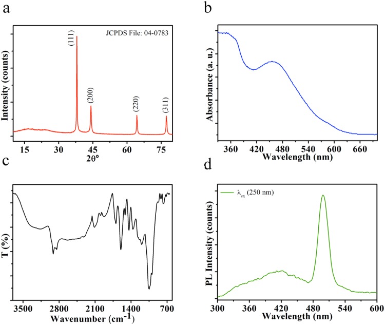
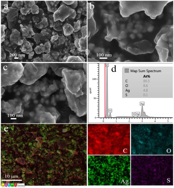
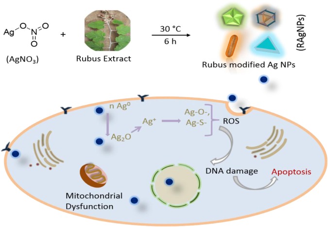
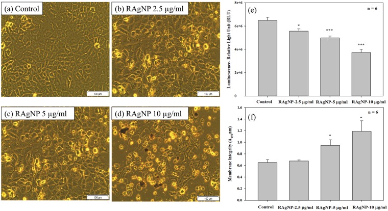
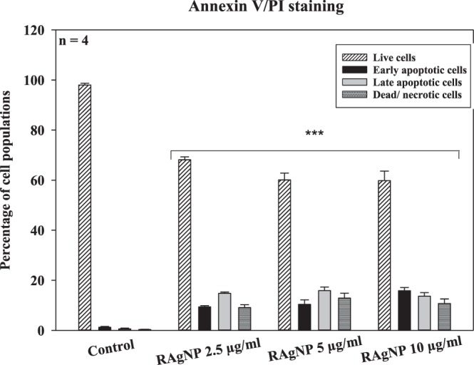
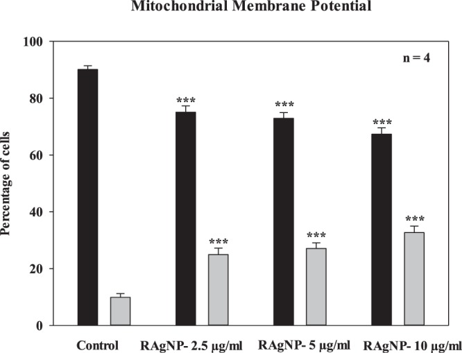
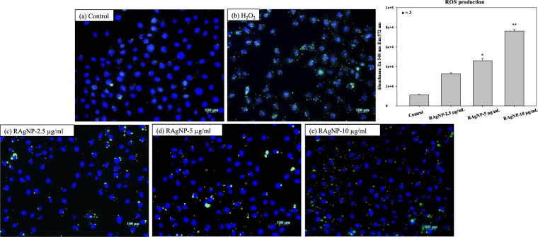
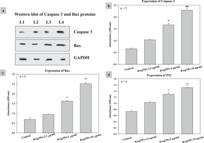
Metallic nanoparticles (NPs) especially silver (Ag) NPs have shown immense potential in medical applications due to their distinctive physio-chemical and biological properties. This article reports the conjugation of Ag NPs with Rubus fairholmianus extract. The modification of Ag NPs was confirmed using various physico-chemical characterization techniques. The cytotoxic effect of Rubus-conjugated Ag NPs (RAgNPs) was studied by LDH assay and proliferation by ATP assay. The apoptotic inducing ability of the NPs were investigated by Annexin V/PI staining, caspase 3/7 analysis, cytochrome c release, intracellular ROS analysis, Hoechst staining and mitochondrial membrane potential analysis using flow cytometry. The expression of apoptotic proteins caspase 3, Bax and P53 were analyzed using ELISA and caspase 3, Bax using western blotting. Cells treated with 10 µg/mL RAgNPs showed an increased number of cell death by microscopic analysis compared to untreated control cells. The RAgNPs induced a statistically significant dose-dependent decrease in proliferation (p < 0.001 for 5 and 10 µg/mL) and increased cytotoxicity in MCF-7 cells. A 1.83 fold increase in cytotoxicity was observed in cells treated with 10 µg/mL (p < 0.05) compared to the untreated cells. Nuclear damage and intracellular ROS production were observed upon treatment with all tested concentrations of RAgNPs and the highest concentrations (5 and 10 µg/mL) showed significant (p < 0.05, p < 0.01) expression of caspase 3, Bax and P53 proteins. The data strongly suggest that RAgNPs induces cell death in MCF-7 cells through the mitochondrial-mediated intrinsic apoptosis pathway. The present investigation supports the potential of RAgNPs in anticancer drug development.


Conflict of interest statement

The authors declare no competing interests.

Figures

Figure 1
Figure 1
The XRD pattern (a), UV-vis spectrum (b), FTIR spectrum (c) and photoluminescence (PL) emission spectrum of RAgNPs (d).
Figure 2
Figure 2
(ac) SEM images of RAgNPs, (d) represents the EDX map spectrum and (e) overlapped EDX map followed by separate map images.
Figure 3
Figure 3
TEM images of RAgNPs: (a) various morphologies of Ag NPs, (b) truncated decahedral particles (Ci) twinned particle, (Cii) nanorods, (Ciii) cuboctahedron, (Civ) truncated triangular nanoplates, (d) HRTEM of icosahedral particle with fringes orientation directions, (e) HRTEM of nanorods and represent also coating surfaces (f) SAED image of RAgNPs. Right side shows the artistic representation of morphologies viz. truncated decahedral particles, nanorods and cuboctahedron (from top to down).
Figure 4
Figure 4
Representative reaction for RAgNPs formation and schematic for nanoparticle induced cancer cell apoptosis.
Figure 5
Figure 5
Morphological changes in MCF-7 cells after RAgNPs treatment. There were no significant visible differences in control (a) and 2.5 µg/mL treated groups (b), the cells did not show any cellular shrinkage and apoptotic bodies after the treatment. However, more dead cells were observed at higher concentrations (5 and 10 µg/mL). The treated cells showed loss of intact membrane, loss of contact with neighbouring cells, condensed, detached from the culture plate showed the features of apoptotic cells. The ATP luminescent cell proliferation assay (e) was used to determine MCF-7 cell proliferation after the treatment with RAgNPs. Control cells showed an increased ATP level, whereas a dose dependent significant (***p < 0.001 and **p < 0.05) decrease in ATP level was observed in experimental groups. The Lactate Dehydrogenase (LDH) cytotoxicity test (f) showed a significant increase in cytotoxicity of cells after the 24 h treatment with RAgNPs compared to control cells. The significant differences between treated and controls groups are shown as *p < 0.05.
Figure 6
Figure 6
Annexin V FITC/PI staining was used to assess the mode of cell death. RAgNPs treated MCF-7 cells showed an increased percentage of apoptotic population after 24 h incubation. The population of early and late apoptotic cells percentage in the control group found to be lower and the live cells percentage was higher in control cells compared with experimental groups. A significant decrease (***p < 0.001) in the live cells and increase in the early, late apoptosis and dead cells percentage were observed in treated MCF-7 cells.
Figure 7
Figure 7
Caspase 3/7 activity (a) was determined as a function of caspase dependent apoptosis in cells after the 24 h treatment. There was a highly significant (p < 0.001) increase in caspase 3/7 activity after 10 µg/mL RAgNPs treatments compared to 2.5 and 5 µg/mLconcentrations and control cells. Cytochrome c release (b) is an important measure of cellular damage. RAgNPs treated cells showed significant (***p < 0.001) release of cytochrome c compared to control cells. The significant differences between treated and controls groups are shown as ***p < 0.001.
Figure 8
Figure 8
Evaluation of mitochondrial membrane potential using the flow cytometric analysis of JC-1 fluorometric stain. Percentage of polarized (black) and depolarized (grey) mitochondrial membrane potential were determined and compared to the percentage of the corresponding mitochondrial membrane potential of untreated cells. Only the PDT-treated cells showed a change in mitochondrial membrane potential (***p < 0.001).
Figure 9
Figure 9
Effect of RAgNPs on ROS production; the green signals showing increased production of ROS upon treatment, in the control (a) slide ROS production is less compared with the RAgNPs treated groups (ce); H2O2 treated group served as the positive control. The quantitative measurement of the ROS (b) showed that there is significant increase in ROS in RAgNPs treated groups in a dose dependent manner.
Figure 10
Figure 10
Effect of RAgNPs on apoptotic protein expression of caspase 3, Bax by western blotting (a) (L1-Control; L2- RAgNPs-2.5 µg/mL; L3- RAgNPs-5 µg/mL; L4- RAgNPs- 10 µg/mL). (bd) shows the effect of RAgNPs on caspase 3, Bax and P53 expression by ELISA. The results showed that the proapoptotic proteins such as P53, caspase 3 and Bax levels were significantly increased in RAgNPs treated groups than control cells.

References

    1. Jahangirian H, Lemraski EG, Webster TJ, Moghaddam RR, Abdollahi Y. A review of drug delivery systems based on nanotechnology and green chemistry: green nanomedicine. Int. J. Nanomed. 2017;12:2957–2978. doi: 10.2147/IJN.S127683. - DOI - PMC - PubMed
    1. Kumar N, George BPA, Abrahamse H, Parashar V, Ngila JC. Sustainable one-step synthesis of hierarchical microspheres of PEGylated MoS2 nanosheets and MoO3 nanorods: Their cytotoxicity towards lung and breast cancer cells. Appl. Surf. Sci. 2017;396:8–18. doi: 10.1016/j.apsusc.2016.11.027. - DOI
    1. Katti KV. Renaissance of nuclear medicine through green nanotechnology: functionalized radioactive gold nanoparticles in cancer therapy- my journey, from chemistry to saving human lives. J. Radioanal. Nucl. Chem. 2016;309:5–14. doi: 10.1007/s10967-016-4888-0. - DOI
    1. Kumar N, et al. A novel approach to low-temperature synthesis of cubic HfO2 nanostructures and their cytotoxicity. Sci. Rep. 2017;7:9351. doi: 10.1038/s41598-017-07753-0. - DOI - PMC - PubMed
    1. Hernandez-Sierra JF, et al. The antimicrobial sensitivity of Streptococcus mutansto nanoparticles of silver, zinc oxide, and gold. Nanomed. Nanotechnol. Biol. Med. 2008;4:237–240. doi: 10.1016/j.nano.2008.04.005. - DOI - PubMed

Publication types