

Platelet Lysate-Derived Neuropeptide y Influences Migration and Angiogenesis of Human Adipose Tissue-Derived Stromal Cells
- PMID: 30254326
- PMCID: PMC6156505
- DOI: 10.1038/s41598-018-32623-8
Platelet Lysate-Derived Neuropeptide y Influences Migration and Angiogenesis of Human Adipose Tissue-Derived Stromal Cells
Abstract
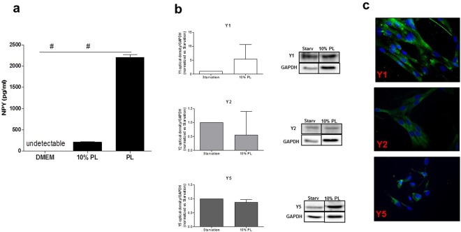
Neuropeptide Y (NPY), a powerful neurotransmitter of the central nervous system, is a key regulator of angiogenesis and biology of adipose depots. Intriguingly, its peripheral vascular and angiogenic powerful activity is strictly associated to platelets, which are source of clinical hemoderivates, such as platelet lysate (PL), routinely employed in several clinical applications as wound healing, and to preserve ex vivo the progenitor properties of the adipose stromal cells pool. So far, the presence of NPY in PL and its biological effects on the adipose stromal cell fraction (ASCs) have never been investigated. Here, we aimed to identify endogenous sources of NPY such as PL-based preparations and to investigate which biological properties PL-derived NPY is able to exert on ASCs. The results show that PL contains a high amount of NPY, which is in part also excreted by ASCs when stimulated with PL. The protein levels of the three main NPY subtype receptors (Y1, Y2, Y5) are unaltered by stimulation of ASCs with PL, but their inhibition through selective pharmacological antagonists, considerably enhances migration, and a parallel reduction of angiogenic features of ASCs including decrease in VEGF mRNA and intracellular calcium levels, both downstream targets of NPY. The expression of VEGF and NPY is enhanced within the sites of neovascularisation of difficult wounds in patients after treatment with leuco-platelet concentrates. Our data highlight the presence of NPY in PL preparations and its peripheral effects on adipose progenitors.
Conflict of interest statement
Prof. G. Frati holds a patent concerning platelet lysate in regenerative medicine (Platelet lysate, uses and method for the preparation thereof. Pub. number WO/2013/042095, International Application number PCT/IB2012/055062).
Figures


Similar articles
-
Neuropeptide Y induces migration, proliferation, and tube formation of endothelial cells bimodally via Y1, Y2, and Y5 receptors.FASEB J. 2006 Sep;20(11):1924-6. doi: 10.1096/fj.05-4770fje. Epub 2006 Aug 4. FASEB J. 2006. PMID: 16891622
-
Neuropeptide Y induces ischemic angiogenesis and restores function of ischemic skeletal muscles.J Clin Invest. 2003 Jun;111(12):1853-62. doi: 10.1172/JCI16929. J Clin Invest. 2003. PMID: 12813021 Free PMC article.
-
IFATS collection: Adipose stromal cells adopt a proangiogenic phenotype under the influence of hypoxia.Stem Cells. 2009 Jan;27(1):266-74. doi: 10.1634/stemcells.2008-0276. Stem Cells. 2009. PMID: 18974212
-
Stress, NPY and vascular remodeling: Implications for stress-related diseases.Peptides. 2007 Feb;28(2):435-40. doi: 10.1016/j.peptides.2006.08.035. Epub 2007 Jan 22. Peptides. 2007. PMID: 17241699 Free PMC article. Review.
-
Atherosclerosis and angiogenesis: what do nerves have to do with it?Pharmacol Rep. 2005;57 Suppl:229-34. Pharmacol Rep. 2005. PMID: 16415503 Review.
Cited by
-
Mechanisms of action of neuropeptide Y on stem cells and its potential applications in orthopaedic disorders.World J Stem Cells. 2020 Sep 26;12(9):986-1000. doi: 10.4252/wjsc.v12.i9.986. World J Stem Cells. 2020. PMID: 33033559 Free PMC article. Review.
-
Oral Plaque from Type 2 Diabetic Patients Reduces the Clonogenic Capacity of Dental Pulp-Derived Mesenchymal Stem Cells.Stem Cells Int. 2019 Jan 14;2019:1516746. doi: 10.1155/2019/1516746. eCollection 2019. Stem Cells Int. 2019. PMID: 30755774 Free PMC article.
-
Palmitoylethanolamide Promotes White-to-Beige Conversion and Metabolic Reprogramming of Adipocytes: Contribution of PPAR-α.Pharmaceutics. 2022 Jan 31;14(2):338. doi: 10.3390/pharmaceutics14020338. Pharmaceutics. 2022. PMID: 35214069 Free PMC article.
-
Updated Role of Neuropeptide Y in Nicotine-Induced Endothelial Dysfunction and Atherosclerosis.Front Cardiovasc Med. 2021 Feb 23;8:630968. doi: 10.3389/fcvm.2021.630968. eCollection 2021. Front Cardiovasc Med. 2021. PMID: 33708805 Free PMC article. Review.
-
Substantial Overview on Mesenchymal Stem Cell Biological and Physical Properties as an Opportunity in Translational Medicine.Int J Mol Sci. 2019 Oct 29;20(21):5386. doi: 10.3390/ijms20215386. Int J Mol Sci. 2019. PMID: 31671788 Free PMC article. Review.
References
-
- Reed E, et al. Paclitaxel, cisplatin, and cyclophosphamide in human ovarian cancer: molecular rationale and early clinical results. Seminars in oncology. 1995;22:90–96. - PubMed
Publication types
MeSH terms
Substances
Grants and funding
LinkOut - more resources
Full Text Sources
Other Literature Sources
Research Materials
Miscellaneous
