. 2019 Jul;26(7):1235-1250.
doi: 10.1038/s41418-018-0202-8. Epub 2018 Sep 25.

TC2N, a novel oncogene, accelerates tumor progression by suppressing p53 signaling pathway in lung cancer

Affiliations

TC2N, a novel oncogene, accelerates tumor progression by suppressing p53 signaling pathway in lung cancer

Xiang-Lin Hao et al. Cell Death Differ. 2019 Jul.

Abstract

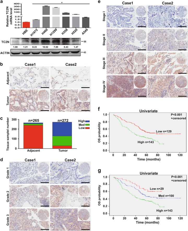
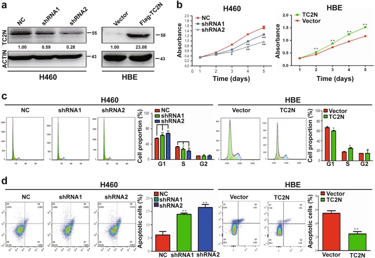
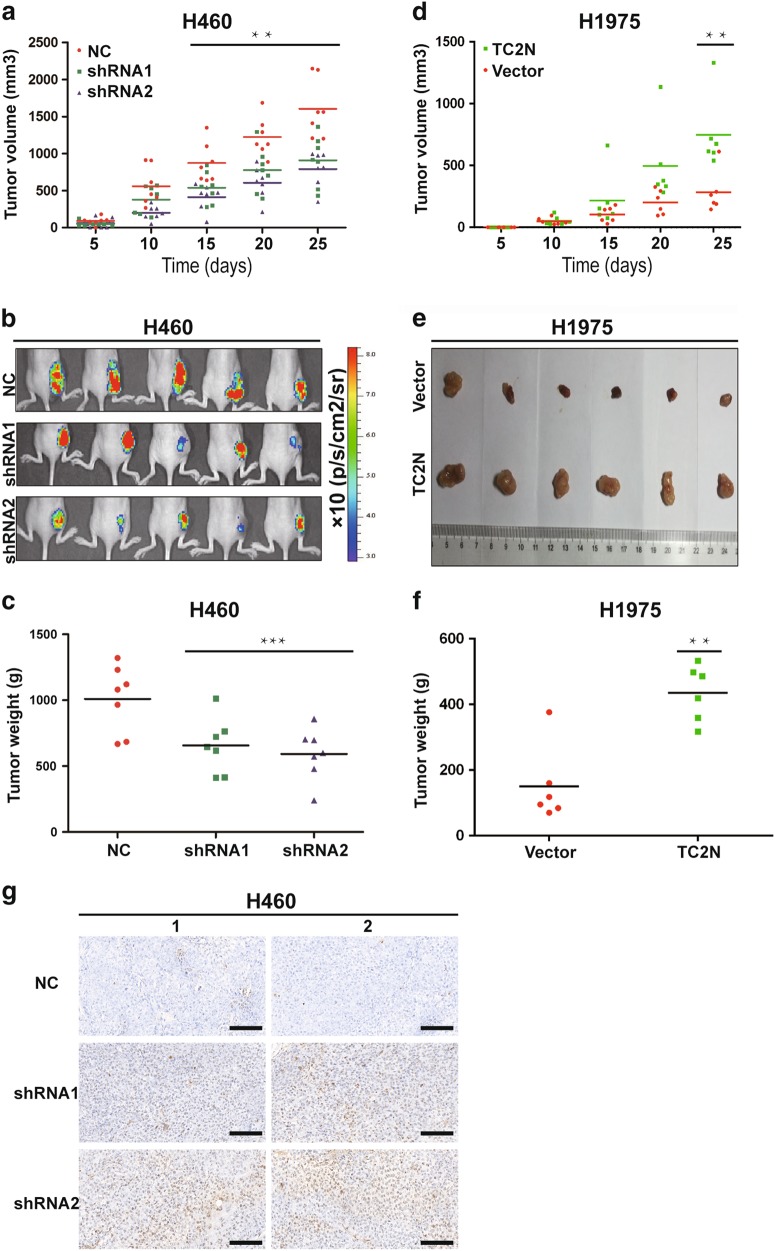
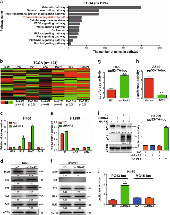
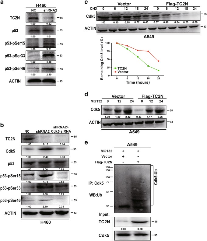
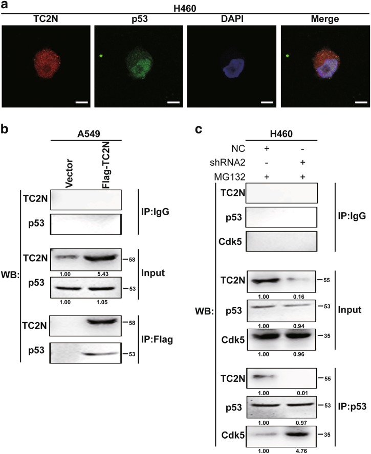
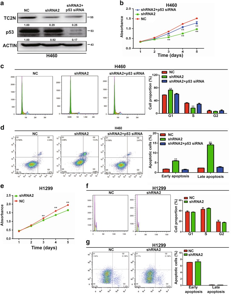
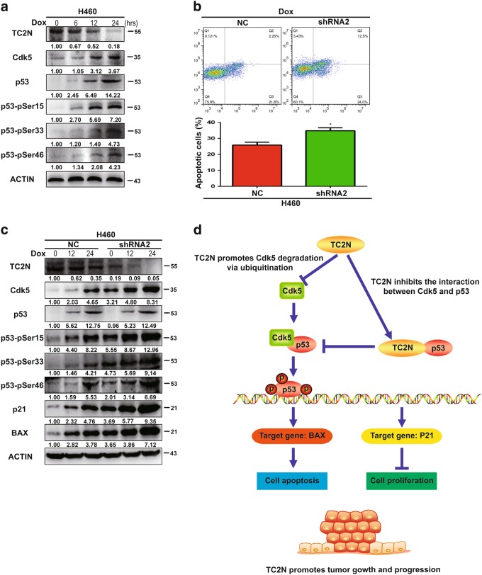
The protein containing the C2 domain has been well documented for its essential roles in endocytosis, cellular metabolism and cancer. Tac2-N (TC2N) is a tandem C2 domain-containing protein, but its function, including its role in tumorigenesis, remains unknown. Here, we first identified TC2N as a novel oncogene in lung cancer. TC2N was preferentially upregulated in lung cancer tissues compared with adjacent normal lung tissues. High TC2N expression was significantly associated with poor outcome of lung cancer patients. Knockdown of TC2N markedly induces cell apoptosis and cell cycle arrest with repressing proliferation in vitro, and suppresses tumorigenicity in vivo, whereas overexpression of TC2N has the opposite effects both in vitro and in vivo. Using a combination of TCGA database and bioinformatics, we demonstrate that TC2N is involved in regulation of the p53 signaling pathway. Mechanistically, TC2N attenuates p53 signaling pathway through inhibiting Cdk5-induced phosphorylation of p53 via inducing Cdk5 degradation or disrupting the interaction between Cdk5 and p53. Moreover, the blockade of p53 attenuates the function of TC2N knockdown in the regulation of cell proliferation and apoptosis. In addition, downregulated TC2N is involved in the apoptosis of lung cancer cells induced by doxorubicin, leading to p53 pathway activation. Overall, these findings uncover a role for the p53 inactivator TC2N in regulating the proliferation and apoptosis of lung cancer cells. Our present study provides novel insights into the mechanism of tumorigenesis in lung cancer.


Conflict of interest statement

The authors declare that they have no conflict of interest.

Figures

Fig. 1
Fig. 1
The expression level of TC2N is correlated with lung cancer progression. a The measurements of TC2N expression in lung cancer cell lines compared with the normal cell line HBE by qRT-PCR and WB. ACTIN is shown as a loading control. The data were analyzed using the formula 2‒Δct. Data represent the mean ± SD of triplicates. b, c The protein expression of TC2N in adjacent and tumor lung samples monitored by immunohistochemistry (IHC) on a tissue array. The anti-TC2N antibody was employed for IHC. IHC scoring was performed blind, prior to association with clinical data. d IHC analysis on a tissue array of lung cancers at different histological grades. e Immunohistochemistry analysis on a tissue array of lung cancers at different clinical stages. Scale bars represent 50 μm. f Kaplan–Meier analysis of TC2N expression in 272 lung cancer patients subdivided into two groups. g Kaplan–Meier analysis of TC2N expression in 272 lung cancer patients subdivided into three groups *P < 0.05
Fig. 2
Fig. 2
Effects of ectopic expression of TC2N on lung cancer cell proliferation and apoptosis in vitro. a Knockdown of TC2N in H460 cells and overexpression of TC2N in HBE cells were identified by WB assay. ACTIN serves as a loading control. b MTS assays were carried out in H460 cells expressing the negative control or shRNA of TC2N and in HBE cells expressing the vector control or TC2N. *P < 0.05; **P < 0.01. c, d Flow cytometry assays were used to examine the effect of TC2N on cell cycle (c) and cell apoptosis (d). Error bars indicate SD (n = 3)
Fig. 3
Fig. 3
TC2N promotes tumorigenesis of lung cancer cells in nude mice. a Examination of tumorigenesis in subcutaneously injected animals using H460 cells stably expressing the negative control or TC2N shRNA. The tumor growth curve of shRNA-expressing cells was compared with negative control cells. b The fluorescent images of xenograft tumors from nude mice subcutaneously injected with H460 stable transfectants were photographed. c Tumor weight from the negative control or TC2N shRNA groups. Each group contained seven mice. d The tumor growth curve was performed in nude mice using H1975 cells stably expressing vector control or TC2N. e Xenograft tumors from (d) were dissected and photographed. f Tumor weight from the vector control and TC2N groups. Each group contained six mice. g Knockdown of TC2N increases the number of apoptotic H460 cells in xenograft tumors. Two representative images were randomly selected from each group (H460-NC, H460-shRNA1 and H460-shRNA2). Scale bars represent 50 μm *P < 0.05; **P < 0.01;***P < 0.001
Fig. 4
Fig. 4
TC2N involves in regulating p53 signaling pathway. a Identification of potential pathways downstream of TC2N with the TCGA lung cancer RNAseq (IlluminaHiSeq; n = 1124) data set, as described in the Materials and methods. b Heatmaps for correlations between TC2N and p53 signaling pathway-related genes in the TCGA lung cancer RNAseq (IlluminaHiSeq; n = 1124) data set. Correlation coefficient R and P values were calculated by Spearman's correlation analysis. c qRT-PCR analysis of P53, P21, BAX and Bcl-2 expression in H460 cells transiently transfected with the negative control or TC2N shRNA. ACTIN serves as an internal control. d The protein levels of TC2N, p53, P21, BAX and Bcl-2 were monitored by WB after knockdown of TC2N in H460 cells. e qRT-PCR analysis of P21, BAX and Bcl-2 expression in H1299 cells transiently transfected with the negative control or TC2N shRNA. f The protein levels of TC2N, p53, P21, BAX and Bcl-2 were monitored by WB after knockdown of TC2N in H1299 cells. ACTIN serves as an internal control. g The effects of TC2N knockdown on the p53 response reporter construct pp53-TA-Luc in H460 cells. h The effects of TC2N overexpression on the p53 response reporter construct pp53-TA-Luc in A549 cells. i H1299 (p53 null) cells were co-transfected with p53 response reporter construct pp53-TA-Luc, negative control or TC2N shRNA and HA-p53 expression vectors. Luciferase activity was measured at 24 h after the transfection. j H460 cells were co-transfected with PG13-luc or MG15-luc reporters and TC2N expression vectors as indicated, and luciferase activity was measured 24 h after transfection. Results are means ± SEM of three independent experiments **P < 0.01
Fig. 5
Fig. 5
TC2N blocks Cdk5-mediated p53 phosphorylation by promoting the degradation of Cdk5. a H460 cells were transfected with negative control or TC2N shRNA for 48 h and then cells were lysed and were subjected to WB using anti-phospho-p53 antibodies. b H460 cells were co-transfected with negative control or TC2N shRNA and Cdk5 siRNA. At 48 h after transfection, the cells were lysed and were subjected to WB using indicated antibodies. c Lysates from A549 vector control or TC2N cells treated with 20 µM cycloheximide (CHX) for the indicated times were subjected to WB (Up). Relative Cdk5 levels were quantified by ImageJ software (Down). d Lysates from A549 vector control or TC2N cells were treated with 20 µM MG132 for the indicated times were subjected to WB. e A549 vector control or TC2N cells were transfected with 7.5 µg of ubiquitin-expressing plasmids. At 24 h after the transfection, cells were treated 20 µM MG132 for 24 h. Cell lysates were IP with anti-Cdk5 antibody and analyzed by WB with anti-ubiquitin antibody, and normal IgG was used as a negative control. Whole-cell lysates were used as a positive control (Input)
Fig. 6
Fig. 6
TC2N inhibits the interaction between Cdk5 and p53. a Immunofluorescence and microscope analysis of TC2N (red) and endogenous p53 (green) localization in nucleus of H460 cells. DAPI (blue) serves as a nuclear counterstain. Right panels show merged images. Scale bars represent 10 μm. b Lysates from A549 vector control or TC2N cells were IP with anti-Flag antibody, and normal IgG was used as a negative control. Whole-cell lysates were used as a positive control (Input). c H460 negative control or TC2N shRNA cells were treated with 20 µM MG132 for 24 h. Cell lysates were IP with anti-p53 antibody and analyzed by WB using indicated antibody
Fig. 7
Fig. 7
p53 is a functionally important target protein of TC2N in lung cancer cells. a TC2N and p53 knockdown were confirmed by WB in H460 cells. b MTS assays were performed to analyze cell proliferation of H460 cells co-transfected with P53 siRNA and TC2N shRNA. **P < 0.01. c, d Assessment of the effects of the expression of TC2N and P53, respectively, on cell cycle (c) and cell apoptosis (d) in H460 cells. e MTS assays were performed to analyze cell proliferation of H1299 cells transfected with negative control or TC2N shRNA. f, g Assessment of the effects of the overexpression of TC2N on cell cycle (f) and cell apoptosis (g) in H1299 cells
Fig. 8
Fig. 8
Inhibition of TC2N increases cell apoptosis with doxorubicin treatment. a H460 cells were incubated with 1 µM Dox for 0 to 24 h, as indicated. Cells were harvested, and then indicated protein levels were determined by WB. b H460 cells stably expressing the negative control or TC2N shRNA were treated with Dox (1 µM) for 24 h and then analyzed by flow cytometry for apoptosis. Results are the mean ± SEM of three independent experiments. *P < 0.05. c The protein expression determined by WB were depicted for the cells treated as the same in (b). d Schematic diagram for the potential mechanisms of TC2N-mediated promotion of tumorigenesis in lung cancer

Similar articles

Cited by

References

    1. Torre LA1, Bray F, Siegel RL, Ferlay J, Lortet-Tieulent J, Jemal A. Global cancer statistics, 2012. CA Cancer J Clin. 2015;65:87–108. doi: 10.3322/caac.21262. - DOI - PubMed
    1. Yuan S, Yu Z, Liu Q, Zhang M, Xiang Y, Wu N, et al. GPC5, a novel epigenetically silenced tumor suppressor, inhibits tumor growth by suppressing Wnt/beta-catenin signaling in lung adenocarcinoma. Oncogene. 2016;35:6120–31. doi: 10.1038/onc.2016.149. - DOI - PubMed
    1. Neri S, Yoshida J, Ishii G, Matsumura Y, Aokage K, Hishida T, et al. Prognostic impact of microscopic vessel invasion and visceral pleural invasion in non-small cell lung cancer: a retrospective analysis of 2657 patients. Ann Surg. 2014;260:383–8. doi: 10.1097/SLA.0000000000000617. - DOI - PubMed
    1. Zhang J, Gold KA, Lin HY, Swisher SG, Xing Y, Lee JJ, et al. Relationship between tumor size and survival in non-small-cell lung cancer (NSCLC): an analysis of the surveillance, epidemiology, and end results (SEER) registry. J Thorac Oncol. 2015;10:682–90. doi: 10.1097/JTO.0000000000000456. - DOI - PMC - PubMed
    1. Wanders R, Steevens J, Botterweck A, Dingemans AM, Reymen B, Av Baardwijk, et al. Treatment with curative intent of stage III non-small cell lung cancer patients of 75 years: a prospective population-based study. Eur J Cancer. 2011;47:2691–2197. doi: 10.1016/j.ejca.2011.06.023. - DOI - PubMed

Publication types

MeSH terms