. 2018 Sep 25;9(1):3916.
doi: 10.1038/s41467-018-06449-x.

Caspase-1 inhibition alleviates cognitive impairment and neuropathology in an Alzheimer's disease mouse model

Affiliations

Caspase-1 inhibition alleviates cognitive impairment and neuropathology in an Alzheimer's disease mouse model

Joseph Flores et al. Nat Commun. .

Abstract

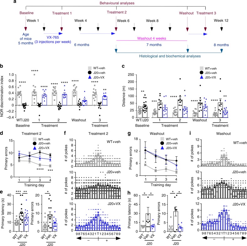
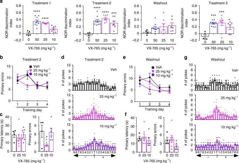
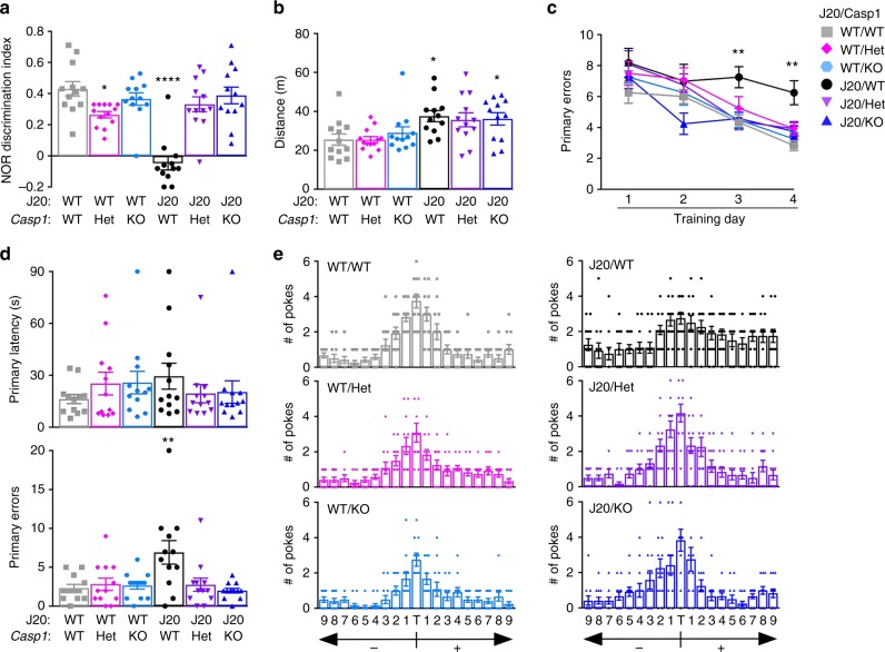
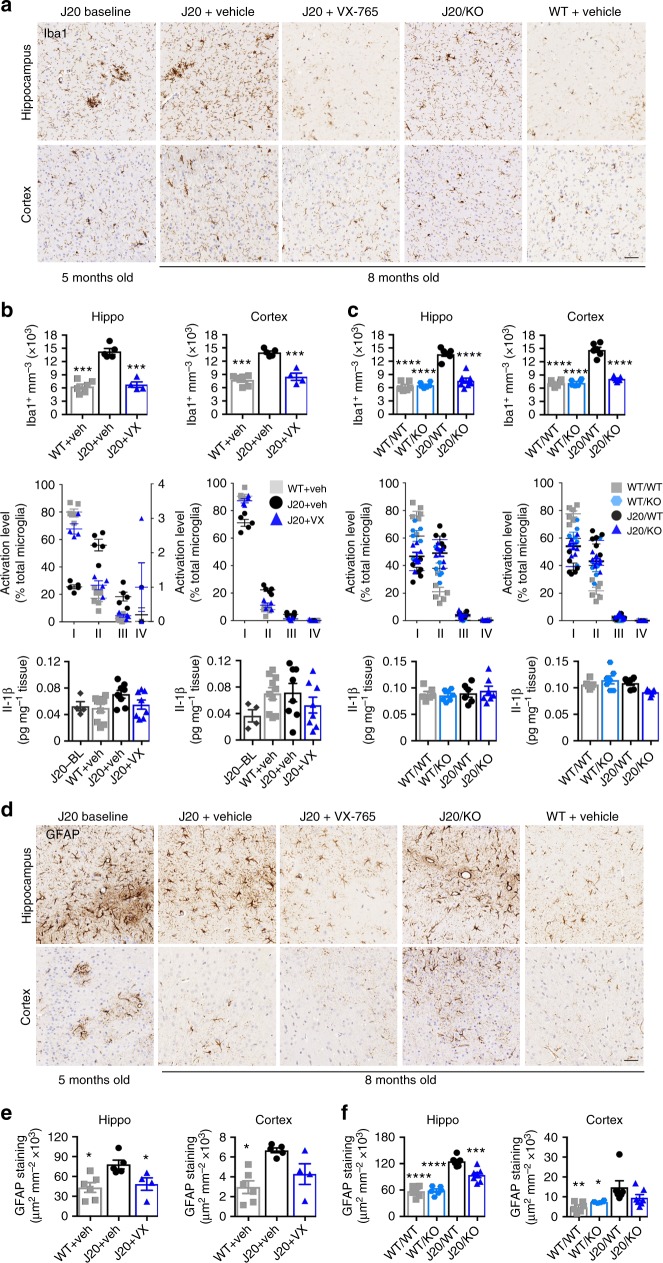
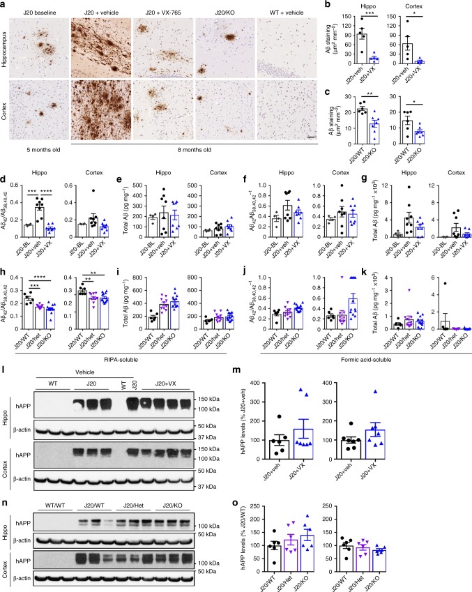
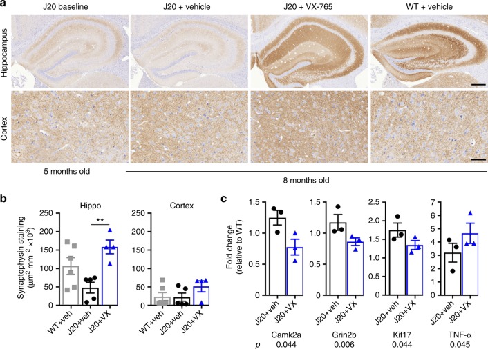
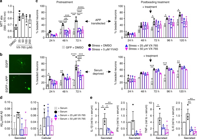
Alzheimer's disease (AD) is an intractable progressive neurodegenerative disease characterized by cognitive decline and dementia. An inflammatory neurodegenerative pathway, involving Caspase-1 activation, is associated with human age-dependent cognitive impairment and several classical AD brain pathologies. Here, we show that the nontoxic and blood-brain barrier permeable small molecule Caspase-1 inhibitor VX-765 dose-dependently reverses episodic and spatial memory impairment, and hyperactivity in the J20 mouse model of AD. Cessation of VX-765 results in the reappearance of memory deficits in the mice after 1 month and recommencement of treatment re-establishes normal cognition. VX-765 prevents progressive amyloid beta peptide deposition, reverses brain inflammation, and normalizes synaptophysin protein levels in mouse hippocampus. Consistent with these findings, Caspase-1 null J20 mice are protected from episodic and spatial memory deficits, neuroinflammation and Aβ accumulation. These results provide in vivo proof of concept for Caspase-1 inhibition against AD cognitive deficits and pathologies.


Conflict of interest statement

The authors declare no competing interests.

Figures

Fig. 1
Fig. 1
VX-765 treatment restores J20 mice cognitive function. a Experimental treatment paradigm. b NOR Discrimination index from vehicle-treated WT (grey squares), vehicle-treated J20 (black circles) and VX-765-treated J20 (blue triangles). Each mouse tested is represented by one symbol. Data represent mean and s.e.m. (Treatment, F(2,20) = 85.8, p < 0.0001; Time, F(4,80) = 4.188, p = 0.0039; Treatment × Time, F(8,80) = 3.599, p = 0013). c Distance travelled during open field task (Treatment, F(2,19) = 11.47, p = 0.0005; Treatment × Time, F(8,76) = 5.69, p < 0.0001). b, c Two-way repeated-measures ANOVA and Dunnett’s post-hoc versus J20 + vehicle, *p < 0.05, **p < 0.01, ***p < 0.001, ****p < 0.0001. di Barnes maze: learning acquisition # of primary errors during d T2 (Treatment, F(2,212) = 19.93, p < 0.0001) and g WO (Treatment, F(2,56) = 10.86, p = 0.001; Training day, F(3,56) = 3.392, p < 0.0241). d, g Two-way repeated-measures ANOVA and Dunnett’s post-hoc versus J20 + vehicle *p < 0.05, ***p < 0.001, ****p < 0.0001. Probe primary latency and errors during e T2 and h WO. Target preference: # of pokes of each hole labelled +1 to +9 to the right or −1 to −9 to the left of the target (T) during the probe after f T2 and i WO (T2 primary latency, F(2,52) = 5.879, p = 0.0050; T2 primary errors, F(2,52) = 9.998, p = 0.0002; WO primary latency, F(2,22) = 4.076, p = 0.0312; WO primary errors, F(2,22) = 10.84, p = 0.0005). e, h ANOVA, Tukey’s post-hoc, *p < 0.05, **p < 0.01, ***p < 0.001
Fig. 2
Fig. 2
VX-765 dose-dependently restores cognitive function in J20 mice. a NOR discrimination index at T1 (F(3,24) = 20.42, p < 0.0001), T2 (F(3,23) = 12.83, p < 0.0001), WO, and T3 (F(3,23) = 8.828, p = 0.0004). Vehicle-treated (black circles), and 50 mg kg−1 (blue triangles), 25 mg kg−1 (pink triangles), or 10 mg kg−1 (purple hexagon) VX-765-treated J20 mice. Each mouse tested is represented by one symbol. Data represent mean and s.e.m. b−g Barnes maze during b−d T2 and e−g WO. Learning acquisition at b T2 and e WO; probe and target preference at c, d T2 and f, g WO. Learning acquisition # of primary errors during b T2 (Treatment, F(2,56) = 3.377, p = 0.0412; Training day, F(3,56) = 7.102, p = 0.0004), and e WO (Treatment, F(2,56) = 5.434, p = 0.007; Training day, F(3,56) = 11.52, p < 0.0001), probe primary errors at c T2 (F(2,14) = 5.69, p = 0.0155). a, c, f ANOVA and b, e Two-way repeated-measures ANOVA, Dunnett’s post-hoc versus J20 + vehicle, *p < 0.05, **p < 0.01, ***p < 0.001, ****p < 0.0001
Fig. 3
Fig. 3
Casp1 KO restores cognitive function in J20 mice. a−e Genotypes: J20−/−/Casp1−/− WT/WT (grey squares), J20−/−/Casp1+/− WT/Het (pink diamonds), J20−/−/Casp1−/− WT/KO (blue symbol), J20−/+/Casp1+/+ J20/WT (black circles), J20−/+/Casp1−/+ J20/Het (purple triangles), J20−/+/Casp1−/− J20/KO (blue triangles). Each mouse tested is represented by one symbol. Data represent mean and s.e.m. a NOR discrimination index (F(5,67) = 16.22, p < 0.0001) and b distance travelled during open field task (F(5,67) = 3.717, p = 0.005). c−e Barnes maze: c # of primary errors during learning acquisition (Genotype, F(5,263) = 6.469, p < 0.0001; Training day, F(3,263) = 29.44, p < 0.0001), d probe primary latency and errors (Primary errors, F(5,66) = 4.8, p = 0008) and e target preference. a, b, d ANOVA and c two-way ANOVA, Dunnett’s post-hoc versus WT/WT. *p < 0.05, **p < 0.01, ****p < 0.0001
Fig. 4
Fig. 4
VX-765 reverses neuroinflammation in J20 mice. a Iba1-immunopositive microglia in hippocampal SLM and cortical S1 regions. b, c, e Vehicle-treated J20−/−/Casp1−/− WT (grey squares), vehicle-treated J20−/−/Casp1−/− WT/KO (blue hexagons), J20−/+ at 5 months baseline (J20-BL; dark grey hexagons), vehicle-treated J20−/+ (black circles), and VX-765-treated J20−/+ (purple triangles). b Stereological quantification of Iba1-positive microglia from hippocampal pyramidal cell layer to the SLM (F(2,11) = 58, p < 0.0001) and cortex (F(2,11) = 39.95, p < 0.0001) (top panels), average % distribution of morphological microglial subtypes I, II, III and IV (middle panels), and Il-1-β levels (bottom panels) in 5-month-old baseline J20 and in 8-month-old WT + vehicle, J20 + vehicle, and J20 + VX-765 WT mice. ANOVA, Dunnett’s post-hoc versus J20 + vehicle, ***p < 0.001. c Stereological quantification of Iba1-positive microglia in the hippocampal CA1 (F(3,21) = 50.53, p < 0.0001) and cortex (F(3,21) = 96.21, p < 0.0001) (top panels), average % distribution of morphological microglial subtypes I, II, III and IV (middle panels), and Il-1-β levels (bottom panels) in WT/WT, WT/Casp1−/− (WT/KO), J20/WT, and J20/Casp1−/− (J20/KO) mice. ANOVA, Dunnett’s post-hoc versus J20/WT, ****p < 0.0001. d Micrographs of GFAP immunopositive astrocytes. e GFAP immunostaining density in vehicle-treated WT and J20 and in VX-765-treated J20 brain hippocampi (F(2,12) = 5.234, p = 0.0232) and cortex (F(2,12) = 8.582, p = 0.0049). f GFAP immunostaining density in J20/KO and littermate control brain hippocampi (F(3,21) = 41.15, p < 0.0001) and cortex (F(3,21) = 4.746, p = 0.0111). ANOVA, Dunnett’s post-hoc versus J20 + vehicle (e) or J20/WT (f), *p < 0.05, ***p < 0.001, ****p < 0.0001. Scale bar in a, d = 50 µm
Fig. 5
Fig. 5
VX-765 prevents progressive Aβ accumulation in J20 mice. a Aβ micrographs of hippocampal SLM and S1 cortex (scale bar = 50 µm). b Quantitative analysis comparing Aβ immunostaining density between J20 + vehicle (black circles) and J20 + VX-765 (blue triangles) mice in the CA1 hippocampal pyramidal cell layer to the SLM (p = 0.0029) and cortex (p = 0.0314, unpaired t test). c Quantitative analysis comparing Aβ immunostaining density between J20−/+/Casp1+/+ J20/WT (black circles) and J20−/+/Casp1−/− J20/KO (blue triangles) mice in the CA1 hippocampal cell layer (p = 0.0040) and cortex (0.0276, unpaired t test). dg RIPA- (d, e) and formic acid- (f, g) soluble Aβ levels in the hippocampus and cortex of 5-month baseline J20−/+ (black diamonds), J20 + vehicle (black circles), and J20 + VX-765 (blue triangles) mice. d, f42/Aβ38 + Aβ40 + Aβ42 levels (RIPA soluble, hippo: F(2,17) = 27.85, p < 0.0001) and e, g total Aβ ANOVA, Dunnett’s post-hoc versus J20 + vehicle, ***p < 0.001, ****p < 0.0001. hk J20−/+/Casp1+/+ J20/WT (black circles), J20−/+/Casp1−/+ J20/Het (purple triangles), and J20−/+/Casp1−/− J20/KO (blue triangles) RIPA- (h, i) and formic acid- (j, k) soluble Aβ levels in the hippocampus and cortex of J20/WT, J20/Casp1+/− (J20/Het), and J20/Casp1−/− J20/KO mice. h, j42/Aβ38 + Aβ40 + Aβ42 levels (RIPA-soluble, hippo: F(2,26) = 16.03, p < 0.0001; RIPA-soluble, cortex: F(2,26) = 6.602, p < 0.0048) and i, k total Aβ ANOVA, Dunnett’s post-hoc versus J20/WT, **p < 0.01, ***p < 0.001, ****p < 0.0001. l−o Human APP protein levels (6E10 immunostaining) and quantification in the hippocampus and cortex of l, m J20 + vehicle (black circles) and J20 + VX-765 mice (blue triangles), and no J20/WT (black circles), J20/Het (purple triangles), and J20/KO (blue triangles) mice
Fig. 6
Fig. 6
VX-765 reverses loss of synaptophysin in J20 mice. a Synaptophysin immunopositive micrographs. Scale bar = 200 µm for hippocampus and 50 µm for cortex. b Synaptophysin immunopositive density in WT + vehicle (grey squares, n = 4), J20 + vehicle (black circles, n = 4), and J20 + VX-765 (blue triangles, n = 4) hippocampus (F(2,9) = 7.974, p = 0.0102, ANOVA, Tukey’s post-hoc, **p < 0.01) and cortex. c Significantly altered (Kruskall−Wallis) synaptic protein mRNA levels in vehicle or VX-765-treated J20 mice hippocampi (n = 3 per group)
Fig. 7
Fig. 7
VX-765 protects human neurons against stress-mediated neuritic beading. a MTT assay. b EGFP fluorescent micrographs of EGFP- or APP+ EGFP-transfected neurons. c % beading in 25 or 50 µM VX-765 or 5 µM Z-YVAD-fmk Casp1 peptide inhibitor pretreated (1 h) or post-treated (48 h) APP-transfected or serum-deprived neurons. DMSO vehicle treatment of EGFP-transfected neurons (grey squares), DMSO- (black circles), 5 µM YVAD- (purple hexagons), 25 µM VX-765- (blue triangles) or 50 µM VX-765- (purple triangles)-treated stressed (APP transfection in upper panels or serum deprived in bottom panel) neurons. Two-way repeated-measures ANOVA, Dunnett’s post hoc versus APPWT or serum-deprived, *p < 0.05, **p < 0.01, ***p < 0.001, ****p < 0.0001. Pretreatment: APPWT (Treatment F(4,10) = 10.17, p = 0.0015; Time F(2,20) = 93.32, p < 0.0001; Treatment × Time F(8,20) = 6.736, p = 0.0003), serum-deprived (Treatment F(4,10) = 10.2, p = 0.0015; Time F(2,20) = 37.65, p < 0.0001). Postbeading treatment: APPWT (Time F(4,40) = 102.7, p < 0.0001), serum-deprived (Treatment F(4,23) = 14.12, p < 0.0001; Time F(4,23) = 24.35, p < 0.0001). d, e Serum-treated normal (grey square), serum-deprived (black circles), serum-deprived and treated with 25 µM VX-765 (blue triangles), and serum-deprived and treated with 50 µM VX-765 (pink triangles) neurons. d Secreted or cellular Aβ42/ Aβ38 + Aβ40 + Aβ42. e Secreted Il-1β (F(3,8) = 6.636, p = 0.0146), IFN-γ, TNF-α (F(3,8) = 3.931, p = 0.054), and IL-6 (F(3,8) = 10.24, p = 0.0041) relative to untreated (+Serum) HPN. ANOVA, Dunnett’s post-hoc versus +Serum, *p<0.05. a, c, d, e. Each individual neuron preparation tested is represented by one symbol. Data represent mean and s.e.m.

References

    1. Heppner FL, Ransohoff RM, Becher B. Immune attack: the role of inflammation in Alzheimer disease. Nat. Rev. Neurosci. 2015;16:358–372. doi: 10.1038/nrn3880. - DOI - PubMed
    1. Shi Qiaoqiao, Chowdhury Saba, Ma Rong, Le Kevin X., Hong Soyon, Caldarone Barbara J., Stevens Beth, Lemere Cynthia A. Complement C3 deficiency protects against neurodegeneration in aged plaque-rich APP/PS1 mice. Science Translational Medicine. 2017;9(392):eaaf6295. doi: 10.1126/scitranslmed.aaf6295. - DOI - PMC - PubMed
    1. Olmos-Alonso A, et al. Pharmacological targeting of CSF1R inhibits microglial proliferation and prevents the progression of Alzheimer’s-like pathology. Brain. 2016;139:891–907. doi: 10.1093/brain/awv379. - DOI - PMC - PubMed
    1. Spangenberg EE, et al. Eliminating microglia in Alzheimer’s mice prevents neuronal loss without modulating amyloid-beta pathology. Brain. 2016;139:1265–1281. doi: 10.1093/brain/aww016. - DOI - PMC - PubMed
    1. Heneka MT, et al. NLRP3 is activated in Alzheimer’s disease and contributes to pathology in APP/PS1 mice. Nature. 2013;493:674–678. doi: 10.1038/nature11729. - DOI - PMC - PubMed

Publication types

MeSH terms