Review
. 2018 Sep 21;24(35):3980-3999.
doi: 10.3748/wjg.v24.i35.3980.

Pediatric hepatocellular carcinoma

Affiliations
Review

Pediatric hepatocellular carcinoma

Rajeev Khanna et al. World J Gastroenterol. .

Abstract

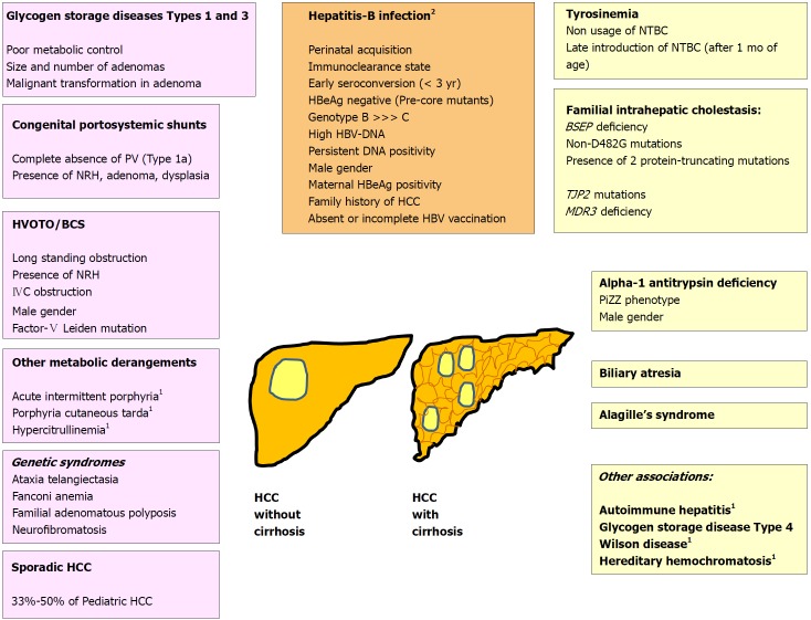
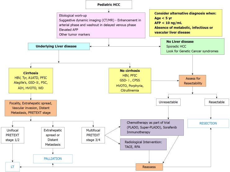
Pediatric hepatocellular carcinoma (HCC) is the second common malignant liver tumor in children after hepatoblastoma. It differs from the adult HCC in the etiological predisposition, biological behavior and lower frequency of cirrhosis. Perinatally acquired hepatitis-B virus, hepatorenal tyrosinemia, progressive familial intrahepatic cholestasis, glycogen storage disease, Alagille's syndrome and congenital portosystemic shunts are important predisposing factors. Majority of children (87%) are older than 5 years of age. Following mass immunization against hepatitis-B, there has been a drastic fall in the incidence of new cases of pediatric HCC in the Asia-Pacific region. Management is targeted on complete surgical removal either by resection or liver transplantation. There is a trend towards improving survival of children transplanted for HCC beyond Milan criteria. Chemotherapeutic regimens do not offer good results but may be helpful for down-staging of advanced HCC. Surveillance of children with chronic liver diseases with ultrasound and alpha-fetoprotein may be helpful in timely detection, intervention and overall improvement in outcome of HCC.

Keywords: Children; Hepatocellular carcinoma; Liver transplantation; Outcome; Risk-factors.


Conflict of interest statement

Conflict-of-interest statement: There are no potential conflicts of interest.

Figures

Figure 1
Figure 1
Risk factors for pediatric hepatocellular carcinoma. 1Conditions cause HCC in adults, and very rarely in children; 2HBV related HCC may occur in presence or absence of cirrhosis. BCS: Budd-Chiari syndrome; HCC: Hepatocellular carcinoma; HVOTO: Hepatic venous outflow tract obstruction; IVC: Inferior vena cava; NRH: Nodular regenerative hyperplasia; BSEP: Bile salt export pump; HCC: Hepatocellular carcinoma; MDR3: Multidrug resistance protein-3; NTBC: [2-(2-nitro-4-trifluoromethylbenzoyl)-1,3-cyclohexanedione (Nitisinone); PiZZ: Homozygous PiZ phenotype of alpha-1 antitrypsin; PV: Portal vein, TJP: Tight junction protein.
Figure 2
Figure 2
Algorithmic management of pediatric hepatocellular carcinoma. A1ATD: Alpha-1 antitrypsin deficiency; AFP: Alpha-fetoprotein; AIH: Autoimmune hepatitis; CPSS: Congenital portosystemic shunt; GSD: Glycogen storage disorder; HBV: Hepatitis-B virus; HVOTO: Hepatic venous outflow tract obstruction; LT: Liver transplantation; PFIC: Progressive familial intrahepatic cholestasis; PLADO: Cisplatin + Doxorubicin; PRETEXT: Pretreatment tumor extent evaluation; PSC: Primary sclerosing cholangitis; RFA: Radiofrequency ablation; Super-PLADO: Cisplatin, Carboplatin and Doxorubicin; TACE: Transarterial chemo-embolization; Tyr: Tyrosinemia; WD: Wilson disease.
Figure 3
Figure 3
Radiological images in children with hepatocellular carcinoma. A and B: 14 and half years old boy with chronic hepatitis-B (Immunoclearance phase), HBeAg+, DNA 5 log, AFP = 3 ng/mL (normal), with a segment 3 lesion (2.5 cm × 2.4 cm), BCLC stage A. Contrast-enhanced CT (CECT) image showing enhancement of the lesion (arrow) in the arterial phase (A) and washout in delayed venous phase (B) in a background of non-cirrhotic liver; C: 10 mo old boy with infantile cholestasis, bile salt export pump (BSEP) deficiency on immunostaining, incidentally found to have lesion in segment II and IVa abutting the capsule, BCLC stage B. The lesion was T2 hyperintense with enhancement in the arterial phase (arrows) and washout in the delayed phase; D and E: 8-1/2 yr old boy with decompensated chronic liver disease, chronic hepatitis-B (Immunoclearance phase), HBeAg+, DNA 7 log, AFP = 250000 ng/mL, with multifocal HCC, BCLC stage D, Child-Pugh C. Contrast-enhanced CT (CECT) image showing heterogenous lesion (arrows, D and E) involving whole of the right lobe with enhancement in the arterial phase in few lesions (arrows, E) in a background of cirrhotic liver with nodular margins (open black arrows, E), ascites (star, D) and collaterals (black solid arrows, E) and splenomegaly (star, E); F: 14 yr old girl with hepatic venous outflow tract obstruction, ascites, portal hypertension, incidentally found to have a lesion in segment 4 (1 cm × 1 cm), BCLC stage D, Child-Pugh C. Contrast-enhanced CT (CECT) image showing enhancement of lesion in the arterial phase (arrow).
Figure 4
Figure 4
Liver histology images in children with hepatocellular carcinoma. A and B: 15 yr old boy with HBeAg negative chronic hepatitis-B, DNA 4 log, precore mutant, AFP = 150000 ng/mL, with a large HCC in right lobe, with portal vein thrombosis, BCLC stage C, Child Pugh A, underwent right hepatectomy. Low power view showing tumor nodules separated by fibrous bands (arrow), adjacent non-malignant parenchyma (star) (100 ×, HE stain) (A). High power view showing malignant nuclear details with prominent nucleoli and eoisnophilic cytoplasm (400 ×, HE stain) (B); C and D: 14 and half years old boy with chronic hepatitis-B (Immunoclearance phase), HBeAg+, DNA 5 log, AFP = 3 ng/mL (normal), with a segment 3 lesion (2.5 cm × 2.4 cm), BCLC stage A, underwent non-anatomic resection. Low power view of tumor showing broadened trabeculae (arrows) (100 ×, HE stain) (C). Trabeculae with malignant cytological features - nuclear atypia and anisocytosis (200 ×, HE stain) (D).

References

    1. Hadzic N, Finegold MJ. Liver neoplasia in children. Clin Liver Dis. 2011;15:443–462, vii-vix. - PubMed
    1. Kelly D, Sharif K, Brown RM, Morland B. Hepatocellular carcinoma in children. Clin Liver Dis. 2015;19:433–447. - PubMed
    1. Otte JB. Progress in the surgical treatment of malignant liver tumors in children. Cancer Treat Rev. 2010;36:360–371. - PubMed
    1. Ng VL, Fecteau A, Shepherd R, Magee J, Bucuvalas J, Alonso E, McDiarmid S, Cohen G, Anand R; Studies of Pediatric Liver Transplantation Research Group. Outcomes of 5-year survivors of pediatric liver transplantation: report on 461 children from a north american multicenter registry. Pediatrics. 2008;122:e1128–e1135. - PubMed
    1. Darbari A, Sabin KM, Shapiro CN, Schwarz KB. Epidemiology of primary hepatic malignancies in US children. Hepatology. 2003;38:560–566. - PubMed

MeSH terms