. 2018 Sep 11:13:5335-5345.
doi: 10.2147/IJN.S170680. eCollection 2018.

Effect of young exosomes injected in aged mice

Affiliations

Effect of young exosomes injected in aged mice

Bo-Ram Lee et al. Int J Nanomedicine. .

Abstract

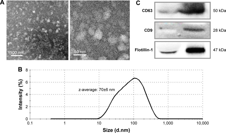
Introduction: Exosomes, nanosized extracellular vesicles, are known to circulate through the blood stream to transfer molecular signals from tissue to tissue.

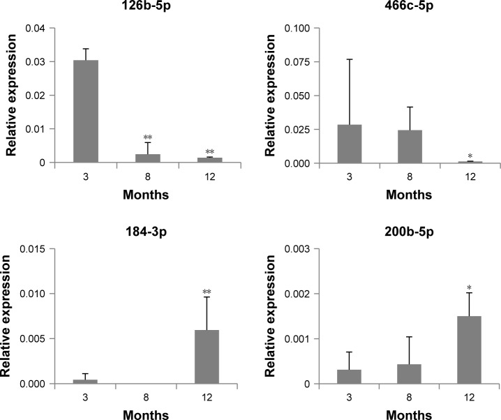
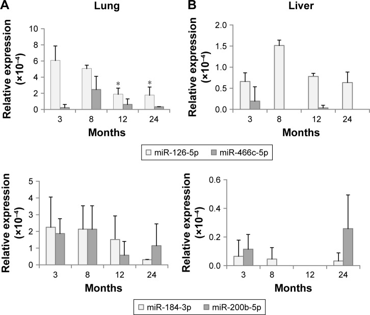
Methods: To determine whether exosomes affect aging in animals, we primarily identified the changes in exosomal miRNA contents during the aging process. In exosomes from 12-month-old mice, mmu-miR-126-5p and mmu-miR-466c-5p levels were decreased and mmu-miR-184-3p and mmu-miR-200b-5p levels were increased significantly compared with those of 3-month-old mice. Their levels in exosomes were partially correlated with those in tissues: levels of only mmu-miR-126-5p and mmu-miR-466c-5p in lungs and/or liver were decreased, but those of mmu-miR-184-3p and mmu-miR-200b-5p in tissues did not coincide with those of exosomes.

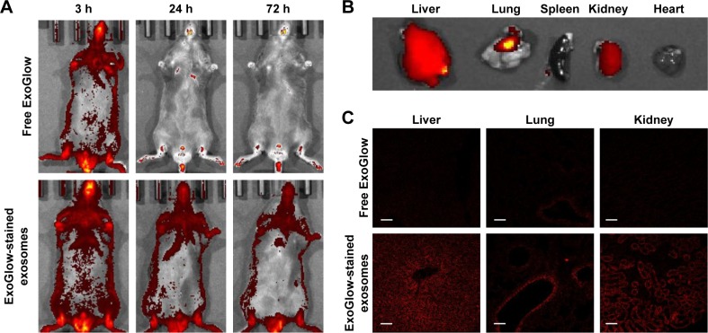
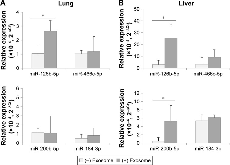
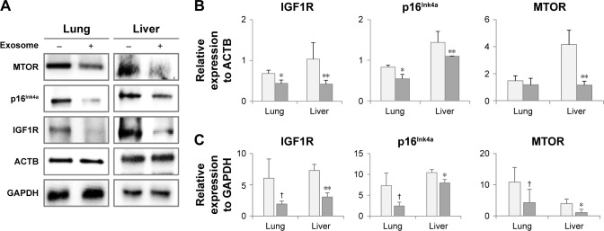
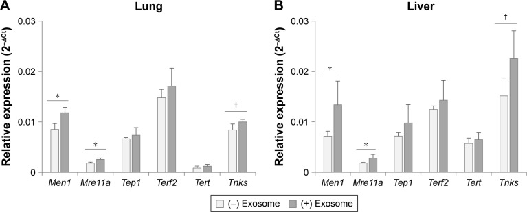
Results and discussion: In the aged tissues injected with young exosomes isolated from serum, mmu-miR-126b-5p levels were reversed in the lungs and liver. Expression changes in aging-associated molecules in young exosome-injected mice were obvious: p16Ink4A, MTOR, and IGF1R were significantly downregulated in the lungs and/or liver of old mice. In addition, telomerase-related genes such as Men1, Mre11a, Tep1, Terf2, Tert, and Tnks were significantly upregulated in the liver of old mice after injection of young exosomes.

Conclusion: These results indicate that exosomes from young mice could reverse the expression pattern of aging-associated molecules in aged mice. Eventually, exosomes may be used as a novel approach for the treatment and diagnosis of aging animals.

Keywords: biomarker; exosome; injection; molecular therapy; reverse aging; telomerase.


Conflict of interest statement

Disclosure The authors report no conflicts of interest in this work.

Figures

Figure 1
Figure 1
Characterization of the isolated exosomes by transmission electron microscopy (A), dynamic light scattering (B), and Western blotting for exosome-specific biomarkers (C).
Figure 2
Figure 2
Expression of miRNAs determined as differentially expressed miRNAs in exosomes, using droplet digital PCR. Notes: The relative expression of miRNAs at each age was determined in comparison with that of 5S rRNA. The statistical significance of expression changes at 8 and 12 months of 126b-5p, 184-3p, and 200b-5p were determined by comparison with those of 3 months, but the significance for miR-466c-5p was compared to that of 8 months. *P<0.05; **P<0.01.
Figure 3
Figure 3
Expression of differentially expressed miRNAs in tissues, determined using droplet digital PCR. Notes: (A) Expression in the lungs; (B) Expression in the liver. The statistical significance was determined by comparison with the expression at 3 months. *P<0.05.
Figure 4
Figure 4
Distribution of exosomes after injection of exogenous exosomes labeled with an exosome-specific dye (ExoGlow™-RNA, System Biosciences, Inc.). Notes: (A) Whole-body imaging for 3 days after exosome administration; (B) ex vivo imaging of exosome distribution in mouse tissues; (C) distribution of exosomes in the section of liver, lungs, and kidney observed using a confocal microscope.
Figure 5
Figure 5
Expression of differentially expressed miRNAs in lungs (A) and liver (B) after the injection of exosomes from 3-month-old to 18-month-old mice. Note: *P<0.05.
Figure 6
Figure 6
Expression of aging-associated biomarkers following the injection of exosomes from 3-month-old to 18-month-old mice. Notes: Following the analysis of protein expression by Western blotting (A), relative quantity of protein expression was determined by comparison with ACTB expression (B) and GAPDH expression (C). P<0.1; *P<0.05; **P<0.01.
Figure 7
Figure 7
Expression of telomerase-related genes in lungs (A) and liver (B) after the injection of exosomes from 3-month-old to 18-month-old mice. Notes: P<0.1; *P<0.05.

Similar articles

Cited by

References

    1. Baker DJ, Wijshake T, Tchkonia T, et al. Clearance of p16Ink4a-positive senescent cells delays ageing-associated disorders. Nature. 2011;479(7372):232–236. - PMC - PubMed
    1. Gladyshev TV, Gladyshev VN. A disease or not a disease? Aging as a pathology. Trends Mol Med. 2016;22(12):995–996. - PMC - PubMed
    1. Lal A, Kim HH, Abdelmohsen K, et al. p16(INK4a) translation suppressed by miR-24. PLoS One. 2008;3(3):e1864. - PMC - PubMed
    1. Noguchi S, Mori T, Otsuka Y, et al. Anti-oncogenic microRNA-203 induces senescence by targeting E2F3 protein in human melanoma cells. J Biol Chem. 2012;287(15):11769–11777. - PMC - PubMed
    1. Overhoff MG, Garbe JC, Koh J, Stampfer MR, Beach DH, Bishop CL. Cellular senescence mediated by p16INK4A-coupled miRNA pathways. Nucleic Acids Res. 2014;42(3):1606–1618. - PMC - PubMed