. 2018 Sep 11:12:309.
doi: 10.3389/fncel.2018.00309. eCollection 2018.

Differential Regulation of Adhesion and Phagocytosis of Resting and Activated Microglia by Dopamine

Affiliations

Differential Regulation of Adhesion and Phagocytosis of Resting and Activated Microglia by Dopamine

Yang Fan et al. Front Cell Neurosci. .

Abstract

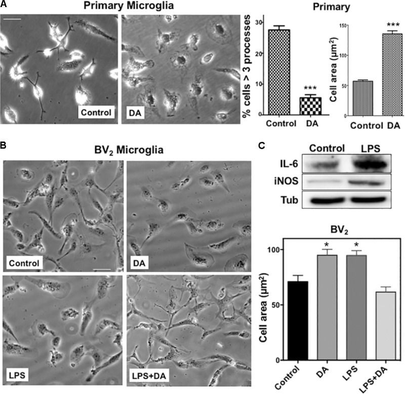
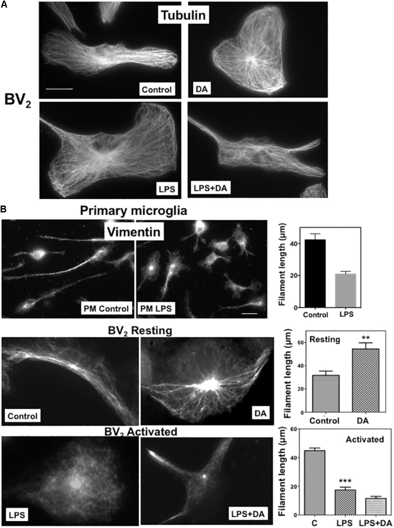
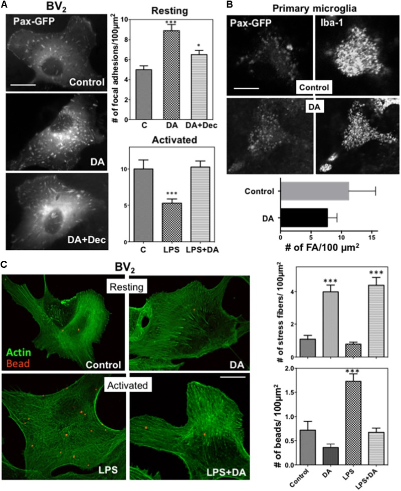
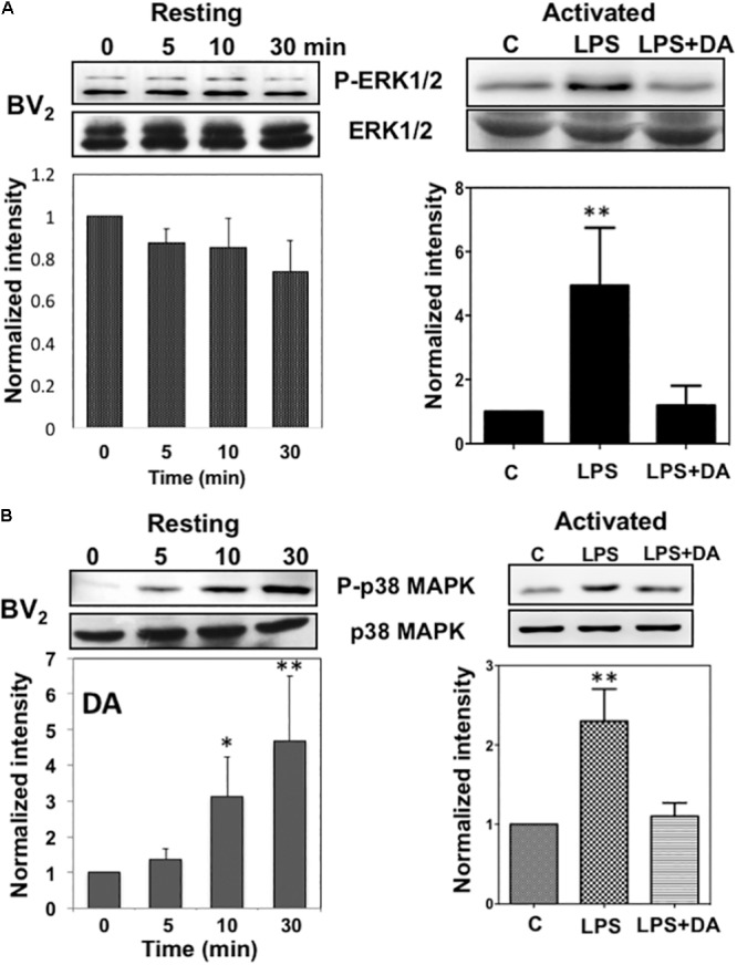
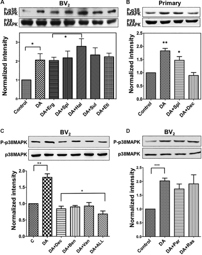
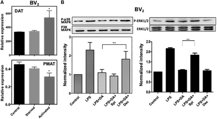
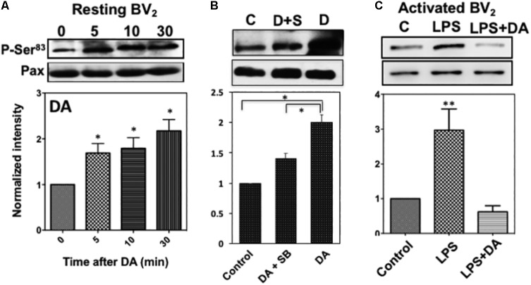
Microglia, the immune competent cells of the central nervous system (CNS), normally exist in a resting state characterized by a ramified morphology with many processes, and become activated to amoeboid morphology in response to brain injury, infection, and a variety of neuroinflammatory stimuli. Many studies focused on how neurotransmitters affect microglia activation in pathophysiological circumstances. In this study, we tried to gain mechanistic insights on how dopamine (DA) released from neurons modulates cellular functions of resting and activated microglia. DA induced the reduction of the number of cellular processes, the increase of cell adhesion/spreading, and the increase of vimentin filaments in resting primary and BV2 microglia. In contrast to resting cells, DA downregulated the cell spreading and phagocytosis of microglia activated by LPS. DA also significantly downregulated ERK1/2 phosphorylation in activated microglia, but not in resting microglia. Downregulation of ERK1/2 by DA in activated microglia required receptor signaling. In contrast, we found a significant increase of p38MAPK activity by DA treatment in resting, but not in activated microglia. These latter effects required the uptake of DA through the high-affinity transporter but did not require receptor signaling. Activation of p38MAPK resulted in the increase of focal adhesion number via phosphorylation of paxillin at Ser83. These results indicate that DA might have a differential, depending upon the activation stage of microglia, impact on cellular functions such as adhesion and phagocytosis.

Keywords: dopamine; microglia; p38MAPK; paxillin; phagocytosis.


Figures

FIGURE 1
FIGURE 1
(A) Changes in the morphology of primary microglia upon DA treatment. Primary microglia cells showed small cell body and multiple processes. Upon DA (2 μM) treatment for 30 min, they acquired flattened morphology and area of cells were increased significantly, accompanied by a decreased number of cells having multiple processes (unpaired t-test: ∗∗∗P < 0.005). Values are means ± standard error of 15–20 cells. Right panels show the measurements of cellular process number and cell area. (B) BV2 microglia were stimulated with LPS and DA, and their morphology (left panels) and cell area (right panel) are shown (ANOVA: P < 0.05 compared to vehicle control ##P < 0.05 compared to LPS). Scale bar = 20 μm. (C) Increased expression of IL-6 and iNOS upon LPS stimulation in BV2 cells.
FIGURE 2
FIGURE 2
Changes in microglia cytoskeleton upon DA stimulation. (A) Microtubule organization shown by immunofluorescence staining with a β-tubulin antibody. Resting and activated BV2 microglia with or without DA (2 μM) treatment showed little changes in microtubule organization. Scale bar = 10 μm. (B) Changes of vimentin filaments in cells treated with DA for 30 min. The number and length of vimentin filaments in primary (PM) and BV2 microglia were visualized by immunofluorescence staining. Values are means ± standard error of 10–15 cells. DA (2 μM) induces the increase of number and length of vimentin filaments in resting BV2 cells. Loss of vimentin filaments and cell spreading upon the activation of microglia by LPS (100 ng/ml) were observed in both primary and BV2 microglia. DA could not rescue the loss of vimentin filaments in activated microglia. (ANOVA: ∗∗∗P < 0.005, ∗∗P < 0.01compare to control). Scale bar = 10 μm.
FIGURE 3
FIGURE 3
(A) Changes in focal adhesions in response to DA. BV2 cells were transfected with paxillin-GFP, and focal adhesions were imaged using fluorescence microscopy. The number of focal adhesions in resting cells was significantly increased in response to DA, which can be blocked by decynium (ANOVA: ∗∗∗P < 0.005 compared to control; P < 0.05 compared to DA). Values are means ± standard error of 10–15 cells. Scale bar = 10 μm. (B) Primary astroglia mixture cultures were transfected with paxillin-GFP. Microglia cells were identified with Iba-1 staining and focal adhesions, visualized with paxillin-GFP, were imaged using confocal microscopy. Scale bar = 5 μm. (C) Phagocytic activity of resting and activated microglia was measured by examining the phagocytosis of beads labeled with Alexa 594 (1 μm diameter)) by cells for 30 min, followed by phalloidin staining. Treatment of DA results in a significant increase in the number of stress fibers in both resting and activated BV2 cells. A significant decrease of phagocytic activity was observed in activated microglia treated with DA. Values are means ± standard error of 10–15 cells. (ANOVA: ∗∗∗P < 0.005 compare to control ###P < 0.005 compare to LPS). Scale bar = 10 μm.
FIGURE 4
FIGURE 4
Immunoblot analysis of the activation of ERK1/2 and p38MAPK in resting and activated microglia. (A) Activation of ERK1/2 upon DA stimulation in resting and activated Bv2 microglia. Total cell lysates were collected at various time points (5, 10, and 30 min) after DA (2 μM) stimulation. The lysates were separated by SDS-PAGE, transferred onto PVDF membranes, and immunoblotted with the anti-phospho-ERK1/2 antibody. Immunoblots with anti-ERK1/2 antibody were used for loading control. ERK1/2 were measured as fold increase of the control. Values are means ± standard error of three or four independent experiments. (B) Activation of p38MAPK in resting, but not in activated microglia in response to 2 μM of DA. Both the resting and activated BV2 microglia were treated with 2 μM of DA for 30 min. Total cell lysates were separated by SDS-PAGE and immunoblotted with an anti-phospho-p38MAPK antibody. Values are means ± standard error of four independent experiments. (ANOVA: P < 0.05, ∗∗P < 0.01 compared to control).
FIGURE 5
FIGURE 5
(A) Activation of p38MAPK by DA in resting microglia cannot be blocked with DA receptor antagonists. Resting BV2 microglia cells were pre-treated with various DA receptor antagonists (2 μM each, specificity of each antagonist was mentioned in section “Materials and Methods”) for 10 min and then treated with 2 μM DA for 30 min. Activation of p38MAPK was measured as fold increase to the control using immunoblotting with the anti-phospho-p38MAPK antibody. Values are means ± standard error of five independent experiments (ANOVA: P < 0.05). (B) Activation of p38MAPK by DA in primary microglia. Cultured primary microglia cells from mouse brain were pre-treated with spiperone and decynium and treated with 2 μM DA for 30 min. Activation of p38MAPK was assessed by immunoblotting (ANOVA: P < 0.05, ∗∗P < 0.01 compared to control). (C) p38MAPK activation by DA can be inhibited by selective DAT blockers (benztropine and vanoxerine; 2 μM each) and PMAT blocker (Decynium; 2 μM). Combination of DAT and PMAT blockers further inhibited the activation of p38MAPK (ANOVA: P < 0.05, ∗∗P < 0.01). (D) Inhibitors of monoamine oxidase did not show any significant effect on p38MAPK activation by DA. Neither pargyline nor rasagiline (2 μM) showed a significant effect on p38MAPK activation by DA in resting microglia (ANOVA: ∗∗∗P < 0.005).
FIGURE 6
FIGURE 6
(A) Expression of DAT and PMAT in resting and activated microglia. Quantitative RT-PCR detected mRNA for DAT and PMAT in resting and activated BV2 microglia. The expression level of DAT increased significantly in activated microglia while PMAT expression decreased. (ANOVA: P < 0.05 compared to control). (B) Activation of p38MAPK resulted from the activation of microglia by LPS can be blocked by decynium while activation of ERK1/2 can be blocked by spiperone (ANOVA: ∗∗P < 0.01, ∗∗∗P < 0.005).
FIGURE 7
FIGURE 7
(A) Phosphorylation of paxillin at Ser83 via the activation of p38MAPK in resting microglia in response to DA. Paxillin phosphorylation was detected by immunoblots with the anti-phospho-paxillin Ser83 antibody. Immunoblots with anti-paxillin antibody were used as a loading control. Representative images of western blots for the phosphorylation of paxillin at Ser83 are shown. Values are means ± standard error of four independent experiments (ANOVA: P < 0.05). (B) Inhibition of Ser83 phosphorylation by the p38MAPK inhibitor. Resting Bv2 microglia cells were pre-treated with 0.5 μM SB203580 (p38MAPK inhibitor) for 10 min and then 2 μM DA for 30 min. Values are means ± standard error of three independent experiments (ANOVA: P < 0.05). (C) DA downregulates paxillin phosphorylation at Ser83 in activated microglia (ANOVA: ∗∗P < 0.01).

Similar articles

Cited by

References

    1. Avshalumov M. V., MacGregor D. G., Sehgal L. M., Rice M. E. (2004). The glial antioxidant network and neuronal ascorbate: protective yet permissive for H(2)O(2) signaling. Neuron Glia Biol. 1 365–376. 10.1017/S1740925X05000311 - DOI - PMC - PubMed
    1. Barcia C., Ros C. M., Ros-Bernal F., Gómez A., Annese V., Carrillo-de Sauvage M. A., et al. (2013). Persistent phagocytic characteristics of microglia in the substantia nigra of long-term Parkinsonian macaques. J. Neuroimmunol. 261 60–66. 10.1016/j.jneuroim.2013.05.001 - DOI - PubMed
    1. Bergquist J., Ohlsson B., Tarkowski A. (2000). Nuclear factor-kappa B is involved in the catecholaminergic suppression of immunocompetent cells. Ann. N. Y. Acad. Sci. 917 281–289. 10.1111/j.1749-6632.2000.tb05394.x - DOI - PubMed
    1. Biber K., Neumann H., Inoue K., Boddeke H. W. (2007). Neuronal ’On’ and ’Off’ signals control microglia. Trends Neurosci. 30 596–602. 10.1016/j.tins.2007.08.007 - DOI - PubMed
    1. Bottalico B., Larsson I., Brodszki J., Hernandez-Andrade E., Casslén B., Marsál K., et al. (2004). Norepinephrine transporter (NET), serotonin transporter (SERT), vesicular monoamine transporter (VMAT2) and organic cation transporters (OCT1, 2 and EMT) in human placenta from pre-eclamptic and normotensive pregnancies. Placenta 25 518–529. 10.1016/j.placenta.2003.10.017 - DOI - PubMed

LinkOut - more resources