

Usefulness of EEG Techniques in Distinguishing Frontotemporal Dementia from Alzheimer's Disease and Other Dementias
- PMID: 30254710
- PMCID: PMC6140274
- DOI: 10.1155/2018/6581490
Usefulness of EEG Techniques in Distinguishing Frontotemporal Dementia from Alzheimer's Disease and Other Dementias
Abstract
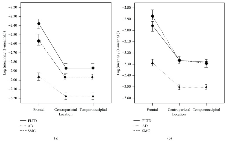
The clinical distinction of frontotemporal dementia (FTD) and Alzheimer's disease (AD) may be difficult. In this narrative review we summarize and discuss the most relevant electroencephalography (EEG) studies which have been applied to demented patients with the aim of distinguishing the various types of cognitive impairment. EEG studies revealed that patients at an early stage of FTD or AD displayed different patterns in the cortical localization of oscillatory activity across different frequency bands and in functional connectivity. Both classical EEG spectral analysis and EEG topography analysis are able to differentiate the different dementias at group level. The combination of standardized low-resolution brain electromagnetic tomography (sLORETA) and power parameters seems to improve the sensitivity, but spectral and connectivity biomarkers able to differentiate single patients have not yet been identified. The promising EEG findings should be replicated in larger studies, but could represent an additional useful, noninvasive, and reproducible diagnostic tool for clinical practice.
Figures


Similar articles
-
Differences in quantitative EEG between frontotemporal dementia and Alzheimer's disease as revealed by LORETA.Clin Neurophysiol. 2011 Sep;122(9):1718-25. doi: 10.1016/j.clinph.2011.02.011. Epub 2011 Mar 10. Clin Neurophysiol. 2011. PMID: 21396882
-
Quantitative EEG in the Differential Diagnosis of Dementia Subtypes.J Geriatr Psychiatry Neurol. 2024 Sep;37(5):368-378. doi: 10.1177/08919887241227410. Epub 2024 Jan 13. J Geriatr Psychiatry Neurol. 2024. PMID: 38217438
-
Quantitative EEG and LORETA: valuable tools in discerning FTD from AD?Neurobiol Aging. 2012 Oct;33(10):2343-56. doi: 10.1016/j.neurobiolaging.2011.12.011. Epub 2012 Jan 13. Neurobiol Aging. 2012. PMID: 22244088
-
Alzheimer's Disease or Behavioral Variant Frontotemporal Dementia? Review of Key Points Toward an Accurate Clinical and Neuropsychological Diagnosis.J Alzheimers Dis. 2020;73(3):833-848. doi: 10.3233/JAD-190924. J Alzheimers Dis. 2020. PMID: 31884475 Free PMC article. Review.
-
Functional changes in brain oscillations in dementia: a review.Rev Neurosci. 2022 Jun 21;34(1):25-47. doi: 10.1515/revneuro-2022-0010. Print 2023 Jan 27. Rev Neurosci. 2022. PMID: 35724724 Review.
Cited by
-
Early dementia diagnosis, MCI-to-dementia risk prediction, and the role of machine learning methods for feature extraction from integrated biomarkers, in particular for EEG signal analysis.Alzheimers Dement. 2022 Dec;18(12):2699-2706. doi: 10.1002/alz.12645. Epub 2022 Apr 7. Alzheimers Dement. 2022. PMID: 35388959 Free PMC article.
-
Characteristics of Resting State EEG Power in 80+-Year-Olds of Different Cognitive Status.Front Aging Neurosci. 2021 Aug 11;13:675689. doi: 10.3389/fnagi.2021.675689. eCollection 2021. Front Aging Neurosci. 2021. PMID: 34456708 Free PMC article.
-
Neural biomarker diagnosis and prediction to mild cognitive impairment and Alzheimer's disease using EEG technology.Alzheimers Res Ther. 2023 Feb 10;15(1):32. doi: 10.1186/s13195-023-01181-1. Alzheimers Res Ther. 2023. PMID: 36765411 Free PMC article.
-
Accurate deep-learning model to differentiate dementia severity and diagnosis using a portable electroencephalography device.Sci Rep. 2025 Jul 20;15(1):26304. doi: 10.1038/s41598-025-12526-1. Sci Rep. 2025. PMID: 40685486 Free PMC article.
-
Predicting future risk of developing cognitive impairment using ambulatory sleep EEG: Integrating univariate analysis and multivariate information theory approach.J Alzheimers Dis. 2025 Mar 4:13872877251319742. doi: 10.1177/13872877251319742. Online ahead of print. J Alzheimers Dis. 2025. PMID: 40035493
References
Publication types
MeSH terms
LinkOut - more resources
Full Text Sources
Other Literature Sources
Medical
