. 2018 Sep 21:3:25.
doi: 10.1038/s41392-018-0027-4. eCollection 2018.

Loss of mdig expression enhances DNA and histone methylation and metastasis of aggressive breast cancer

Affiliations

Loss of mdig expression enhances DNA and histone methylation and metastasis of aggressive breast cancer

Chitra Thakur et al. Signal Transduct Target Ther. .

Abstract

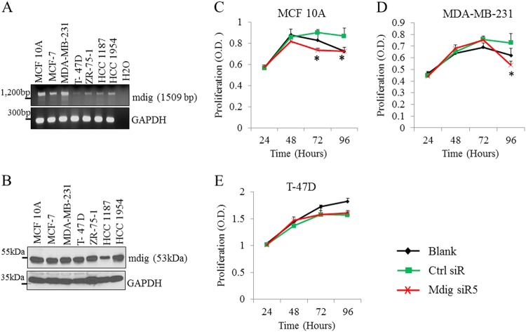
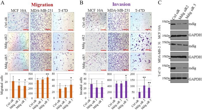
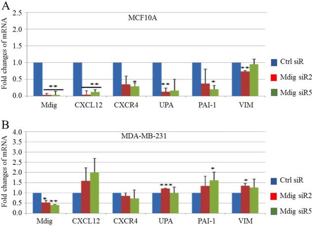
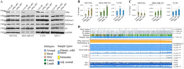
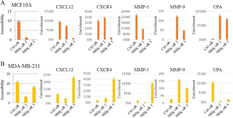
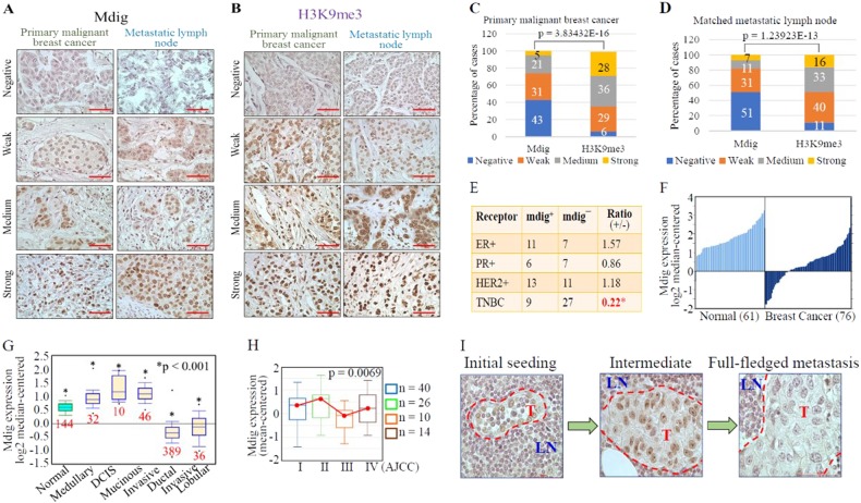
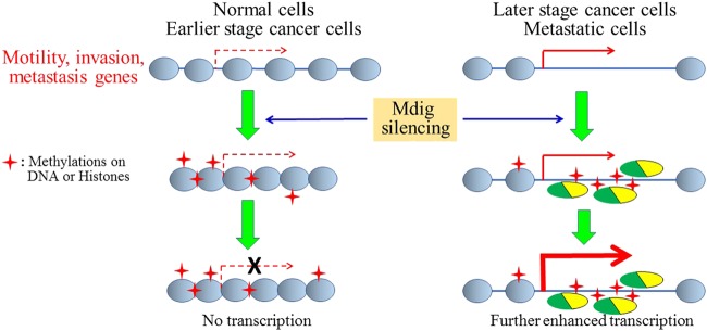
We previously reported that expression of an environmentally induced gene, mineral dust-induced gene (mdig), predicts overall survival in breast cancer patients. In the present report, we further demonstrate the differential roles of mdig between earlier- and later-stage breast cancers. In noncancerous breast, mdig is a proliferation factor for cell growth and cell motility. In breast cancer, however, higher levels of mdig negatively regulate the migration and invasion of cancer cells. Assessment of global DNA methylation, chromatin accessibility and H3K9me3 heterochromatin signature suggests that silencing mdig enhances DNA and histone methylation. Through immunostaining and data mining, we found that mdig is significantly upregulated in noninvasive and/or earlier-stage breast cancers. In contrast, in triple-negative and other invasive breast cancers, diminished mdig expression was noted, indicating that the loss of mdig expression could be an important feature of aggressive breast cancers. Taken together, our data suggest that mdig is a new biomarker that likely promotes tumor growth in the early stages of breast cancer while acting as a tumor suppressor to inhibit invasion and metastasis in later-stage tumors.


Conflict of interest statement

The authors declare no competing interests.

Figures

Fig. 1
Fig. 1
Mdig regulates cell proliferation. RT-PCR (a) and western blotting (b) show expression of mdig gene and protein, respectively, in MCF10A noncancerous human breast cells and breast cancer cell lines MCF-7, MDA-MB-231, T-47D, ZR-75-1, HCC 1187 and HCC 1954. c, d and E, MTT assays of indicated cells for 24, 48, 72, and 96 h post transfection with control siRNA (Ctrl siR) or a siRNA targeting mdig, mdig siR5 (*p < 0.05, n = 3)
Fig. 2
Fig. 2
Mdig affects cell motility and invasion of noncancerous breast and breast cancer cells. Transwell migration (a) and Matrigel invasion (b) assay of indicated cell lines following transfection with Ctrl siRNA, mdig siR2 or mdig siR5 24 h post transfection. Scale bar = 200 μM. Quantification of number of migrated cells and invaded cells depicted by a bar graph below each of the figure panels. Ten randomly selected fields were counted for migrated and invasive cells (n = 10). * p < 0.05; **p < 0.01. c. Western blotting shows a decrease in mdig protein for indicated cell lines 24 h after knockdown of mdig by siRNAs
Fig. 3
Fig. 3
Mdig regulates expression of genes implicated in cell motility and invasiveness of breast cancer. Real time PCR for determination of genes involved in cell motility and invasion in MCF10A noncancerous breast cells (upper panel) and MDA-MB-231 breast cancer cells (lower panel) after transfection with indicated siRNAs. Fold changes were calculated relative to Ctrl siR and the housekeeping gene GAPDH. Data represents ± S.D., n = 3
Fig. 4
Fig. 4
Mdig silencing enhances histone and DNA methylation. a Protein levels of mdig, H3K9me, H3K9me2, H3K9me3, H3, and GAPDH were determined by western blotting in the indicated cell lines after transfection with indicated siRNAs. b, c Total DNA methylation in indicated cell lines transfected with siRNAs were determined by measuring 5-methylcytosine (5-mC, B) and 5-hydroxymethycytosine (5-hmC, C) as indicated in the Materials and Methods. d Visualization of TCGA data containing 871 cases of invasive breast cancer for expression and DNA methylation of mdig genes using MEXPRESS (http://mexpress.be/). Samples are ordered by mdig expression values
Fig. 5
Fig. 5
Mdig regulates chromatin accessibility. a, b Chromatin accessibility assay coupled with real time PCR for the indicated genes were performed in MCF10A (a) and MDA-MB-231 (b) with or without mdig silencing. Fold enrichment was calculated using the formula FE = 2ˆ(NseCT-noNseCT) × 100%. Error bars = percentage error of value 5
Fig. 6
Fig. 6
Loss of mdig expression in invasive and metastatic breast cancers. a Mdig expression pattern in primary malignant breast carcinoma and matched metastatic carcinomas in lymph nodes. b H3K9me3 staining pattern in primary malignant breast carcinoma and matched metastatic carcinoma in lymph nodes. Data represent 154 cases, ×40 magnification, scale bar = 50 µm. c, d Quantification of scored images for mdig and H3K9me3 in breast cancer (c) and matched metastatic lymph nodes (D), respectively. e, f Loss of mdig expression in TNBC and invasive breast cancer (data source: http://www.oncomine.org). g Relative levels of mdig expression in breast cancers with different histological subtypes (CURTIS Breast, n = 2136, Oncomine). h Relative levels of mdig expression in breast cancers with different AJCC stages (UCSC Xena, http://xena.ucsc.edu). i Loss of mdig expression in breast cancer cells that metastasized to lymph nodes. Representative images from the metastatic lymph nodes showing the characteristic phenotype of cells positive for mdig. Left panel, very few tumor cells that are positive for mdig were detected in lymph nodes; middle panel, increased number of tumor cells in the lymph nodes, some of which showed weak staining for mdig; right panel, full-fledged tumor within the lymph node showing loss of mdig in tumor cells. LN = lymph node area, T = Tumor area; ×40 magnification. Scale bar = 50 µm
Fig. 7
Fig. 7
Schematic diagram showing opposing effects of mdig silencing on expression of genes for cell motility and invasion between earlier and later stage cancer cells. In normal cells or earlier stage cancer cells, the chromatin of these genes is partially open. Mdig silencing increases both H3K9me3 and DNA methylation, causing further condensation of the chromatin and inhibition of these genes. In later stage and metastatic cancer cells, the chromatin configuration of these genes is widely open. Mdig silencing enhances DNA and histone methylation in the intron or gene body of these genes, and a few of the nucleosome histone proteins, which may result in recruitment of transcriptional regulatory proteins for enhanced transcription of genes involved in cell motility, invasion and metastasis

References

    1. Zhang Y, et al. The Human mineral dust-induced gene, mdig, is a cell growth regulating gene associated with lung cancer. Oncogene. 2005;24:4873–4882. doi: 10.1038/sj.onc.1208668. - DOI - PubMed
    1. Tsuneoka M, Koda Y, Soejima M, Teye K, Kimura H. A novel myc target gene, mina53, that is involved in cell proliferation. J. Biol. Chem. 2002;277:35450–35459. doi: 10.1074/jbc.M204458200. - DOI - PubMed
    1. Thakur C, Chen F. Current understanding of mdig/MINA in human cancers. Genes Cancer. 2015;6:288–302. - PMC - PubMed
    1. Lu Y, et al. Lung cancer-associated JmjC domain protein mdig suppresses formation of tri-methyl lysine 9 of histone H3. Cell Cycle. 2009;8:2101–2109. doi: 10.4161/cc.8.13.8927. - DOI - PubMed
    1. Mori T, et al. Ablation of Mina53 in mice reduces allergic response in the airways. Cell Struct. Funct. 2013;38:155–167. doi: 10.1247/csf.13006. - DOI - PubMed

Publication types

LinkOut - more resources