

A three-step evaluation for safer curettage treatment of cesarean scar pregnancy
- PMID: 30254898
- PMCID: PMC6135174
- DOI: 10.1016/j.gmit.2017.02.003
A three-step evaluation for safer curettage treatment of cesarean scar pregnancy
Abstract
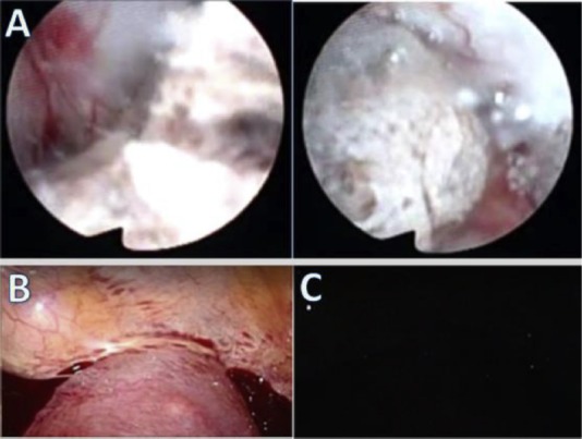
Dilation and curettage is one of the treatment options for cesarean scar pregnancy, however, it sometimes requires a salvage therapy. Few reports discuss the methods of evaluating cesarean scar pregnancy before therapeutic procedures. We aimed to present a case study in which a three-step approach using a combination of preoperational sonohysterography, hysteroscopy, and laparoscopy was performed to evaluate cesarean scar pregnancy. A 33-year-old, G2P2, Japanese female with a history of two elective cesarean sections was diagnosed with viable cesarean scar pregnancy. We used the three-step approach right after undergoing bilateral uterine artery embolization and confirmed that there was a low possibility of fatal complications and we performed dilation and curettage. These steps could be done safely even if the cesarean scar pregnancy was viable. To perform safer curettage on cesarean scar pregnancy patients, these three steps seem to be useful.
Keywords: cesarean scar pregnancy; cesarean section; endoscopy; pregnancy loss.
Conflict of interest statement
Conflicts of interest: The authors have no conflicts of interest relevant to this article.
Figures

Similar articles
-
[Comparison of hysteroscopy and curettage in incomplete pregnancy with cesarean scar pregnancy].Zhonghua Yi Xue Za Zhi. 2018 Jan 16;98(3):217-221. doi: 10.3760/cma.j.issn.0376-2491.2018.03.012. Zhonghua Yi Xue Za Zhi. 2018. PMID: 29374918 Chinese.
-
Cesarean Scar Pregnancy, Incidence, and Recurrence: Five-Year Experience at a Single Tertiary Care Referral Center.Obstet Gynecol. 2018 Nov;132(5):1285-1295. doi: 10.1097/AOG.0000000000002940. Obstet Gynecol. 2018. PMID: 30303911
-
Cesarean Scar Ectopic Pregnancy: Laparoscopic Resection and Total Scar Dehiscence Repair.J Minim Invasive Gynecol. 2018 Feb;25(2):297-298. doi: 10.1016/j.jmig.2017.01.022. Epub 2017 Feb 4. J Minim Invasive Gynecol. 2018. PMID: 28179198
-
Hysteroscopy and suction evacuation of cesarean scar pregnancies: a case report and review.J Obstet Gynaecol Res. 2014 Mar;40(3):853-7. doi: 10.1111/jog.12260. Epub 2013 Dec 10. J Obstet Gynaecol Res. 2014. PMID: 24320609 Review.
-
Treatment of viable cesarean scar ectopic pregnancy with suction curettage.Int J Gynaecol Obstet. 2005 May;89(2):163-6. doi: 10.1016/j.ijgo.2004.12.038. Int J Gynaecol Obstet. 2005. PMID: 15847889 Review.
References
-
- Arslan M, Pata O, Dilek TU, Aktas A, Aban M, Dilek S. Treatment of viable cesarean scar ectopic pregnancy with suction curettage. Int J Gynaecol Obstet. 2005;89(2):163–166. - PubMed
-
- Rotas MA, Haberman S, Levgur M. Cesarean scar ectopic pregnancies: etiology, diagnosis, and management. Obstet Gynecol. 2006;107(6):1373–1381. - PubMed
-
- Sugawara J, Senoo M, Chisaka H, Yaegashi N, Okamura K. Successful conservative treatment of a cesarean scar pregnancy with uterine artery embolization. Tohoku J Exp Med. 2005;206(3):261–265. - PubMed
-
- Vial Y, Petignat P, Hohlfeld P. Pregnancy in a cesarean scar. Ultrasound Obstet Gynecol. 2000;16(6):592–593. - PubMed
-
- Fylstra DL. Hysteroscopy and suction evacuation of cesarean scar pregnancies: a case report and review. J Obstet Gynaecol Res. 2014;40(3):853–857. - PubMed
Publication types
LinkOut - more resources
Full Text Sources
Other Literature Sources