

Case Reports
doi: 10.1016/j.ijvsm.2018.02.005.
eCollection 2018 Jun.
Liver enzyme elevation caused by a compression of infiltrative lipoma in a dog
Affiliations
- PMID: 30255090
- PMCID: PMC6147384
- DOI: 10.1016/j.ijvsm.2018.02.005
Item in Clipboard
Case Reports
Liver enzyme elevation caused by a compression of infiltrative lipoma in a dog
Int J Vet Sci Med.
.
No abstract available
Figures

Transverse post-contrast abdominal CT image at the level of the kidneys (A), Reformatted, left parasagittal post-contrast plane image (B), and a reformatted, dorsal post-contrast plane image (C), acquired with a soft tissue algorithm (window width, 330 Household Units [HU]; window level, 30 HU; slice thickness, 2 mm). (A): there is a large, fat-attenuating mass (M) that distorts and displaces the left kidney, spleen and other abdominal structures (white arrows). The mass separates the relationship of the internal and external abdominal oblique and transverse abdominis muscle layers (black arrows). Infiltration of the transverse abdominis muscle is visualized in the center of the muscular discontinuity (asterisk). (B): the mass causes cranioventral deviation of left lob of liver and spleen (white arrows). (C): the large fat-attenuating mass spreads across the left abdominal wall, displacing abdominal organs rightward. G = Stomach. LK = left kidney. RK = right kidney L = Liver. M = abdominal wall mass. S = Spleen. C = colon. D = Dorsal. L = Left. R = Right. V = Ventral.
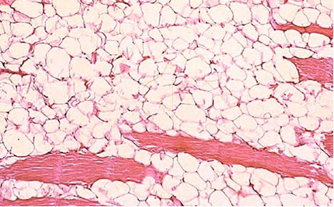
Histopathology image reveals well-differentiated adipocytes infiltrating the adjacent muscle fibers as well as causing myodegeneration, consistent with the infiltrative lipoma. Hematoxylin and Eosin stain.
References
-
- Hendrick M.J. Tumors of adipose tissue. In: Hendrick M.J., Mahaffey E.A., Moore F.M., Vos J.H., Walder E.J., editors. Histological classification of mesenchymal tumors of skin and soft tissues of domestic animals. Armed Forces Institute of Pathology in cooperation with the American Registry of Pathology and the World Health Organization Collaborating Center for Worldwide Reference on Comparative Oncology; Washington, D.C.: 1998. pp. 11–60.
-
- Gleiser C.A., Jardine J.H., Raulston G.L., Gray K.N. Infiltrative lipoma in the dog. Vet Pathol. 1979;16:623–624. - PubMed
-
- Morgan L.W., Toal R., Siemering G., Gavin P. Imaging diagnosis – infiltrative lipoma causing spinal cord compression in a dog. Vet Radiol Ultrasound. 2007;48:35–37. - PubMed
-
- Agut A., Anson A., Navarro A., Murciano J., Soler M., Belda E. Imaging diagnosis-infiltrative lipoma causing spinal cord and lumbar nerve root compression in a dog. Vet Radiol Ultrasound. 2013;54:381–383. - PubMed
-
- Jennifer L., O’Driscoll, John J.M. What is your neurologic diagnosis? J Am Vet Med Assoc. 2006;229:933–935. - PubMed
Publication types
LinkOut - more resources
Full Text Sources
Other Literature Sources
