

MRI Findings of Early Myositis Ossificans without Calcification or Ossification
- PMID: 30255094
- PMCID: PMC6140134
- DOI: 10.1155/2018/4186324
MRI Findings of Early Myositis Ossificans without Calcification or Ossification
Abstract
Purpose: To characterize and evaluate the MR imaging features of early myositis ossificans (MO) without calcification or ossification.
Methods: The MRI manifestations of seven patients with pathologically proven early MO were retrospectively analyzed with regard to tumor location, size, margins, signal intensity, and enhancement appearance in MR images. Additionally, the surrounding soft-tissue edema and adjacent bone change were assessed.
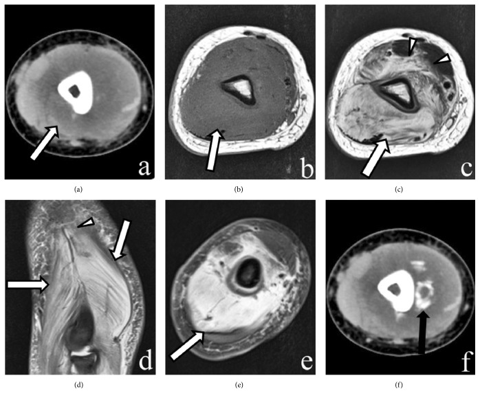
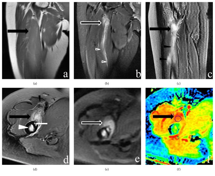
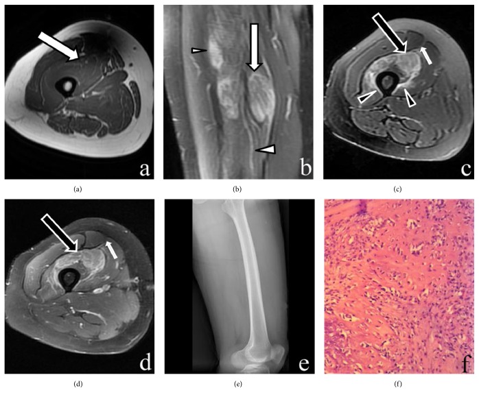
Results: All cases (n=7) had intramuscular tumor-like masses without calcifications. The lesions appeared as isointense in T1-weighted images (T1-WI) and inhomogeneous hyperintense in T2-weighted MR images (T2-WI). On T2-WI and postcontrast T1-WI, the heterogeneously high signal intensity in the expanded muscle interspersed with a few hypointense linear structures consistent with intact muscle fibers showed "striate pattern" in the plane parallel with muscle fibers. The relatively hypointense areas with geometrical pattern consistent with the bundles of intact muscle fibers are found within the lesion with diffuse high signal intensity, displaying the "checkerboard-like pattern" in the plane vertical to muscle fibers. A "striate pattern" (n = 7) and "checkerboard-like pattern" (n = 3) in the lesion appeared in T2-WI. In contrast-enhanced MRI images, all cases showed diffuse "striate pattern" enhancement. Among them, one case demonstrated "checkerboard-like pattern" enhancement. All cases had diffuse and prominent muscle edema that preserved the muscle fascicles. For two lesions located in the deep muscle group, the adjacent bone showed bone marrow edema.
Conclusion: MR imaging has unique advantages for diagnosis of early MO without calcification or ossification: the "striate pattern" and "checkerboard-like pattern" appearance shown in T2-WI and contrast-enhanced MRI images can be helpful for differential diagnosis. MRI can delineate the extent of the tumor and provides accurate anatomical information, which is important in diagnosis, treatment, and follow-up.
Figures
Similar articles
-
MR imaging of myositis ossificans: variable patterns at different stages.J Magn Reson Imaging. 1995 May-Jun;5(3):287-92. doi: 10.1002/jmri.1880050312. J Magn Reson Imaging. 1995. PMID: 7633105
-
Magnetic resonance imaging of myositis ossificans: analysis of seven cases.Skeletal Radiol. 1992;21(8):503-7. doi: 10.1007/BF00195231. Skeletal Radiol. 1992. PMID: 1465642
-
Myositis ossificans: MR appearance with radiologic-pathologic correlation.AJR Am J Roentgenol. 1991 Dec;157(6):1243-8. doi: 10.2214/ajr.157.6.1950874. AJR Am J Roentgenol. 1991. PMID: 1950874
-
Magnetic resonance imaging of thymic epithelial tumors.Crit Rev Diagn Imaging. 1996 Aug;37(3):191-259. Crit Rev Diagn Imaging. 1996. PMID: 8872410 Review.
-
Magnetic resonance imaging of intramuscular myxoma with histological comparison and a review of the literature.Skeletal Radiol. 2005 Jan;34(1):19-28. doi: 10.1007/s00256-004-0848-9. Epub 2004 Oct 22. Skeletal Radiol. 2005. PMID: 15538560 Review.
Cited by
-
Malignant peripheral nerve sheath tumor in the pelvis: a case report.Surg Case Rep. 2023 Sep 6;9(1):157. doi: 10.1186/s40792-023-01733-5. Surg Case Rep. 2023. PMID: 37672135 Free PMC article.
-
A nontraumatic myositis ossificans case of the forearm: Case report and literature review.Exp Ther Med. 2021 May;21(5):531. doi: 10.3892/etm.2021.9963. Epub 2021 Mar 23. Exp Ther Med. 2021. PMID: 33815604 Free PMC article.
-
Case Report: Unusual Presentation of Myositis Ossificans of the Elbow in a Child Who Underwent Excessive Postoperative Rehabilitation Exercise.Front Pediatr. 2021 Nov 12;9:757147. doi: 10.3389/fped.2021.757147. eCollection 2021. Front Pediatr. 2021. PMID: 34869112 Free PMC article.
-
[A case of progressive ossifying myositis caused by ACVR1 gene mutation].Zhongguo Dang Dai Er Ke Za Zhi. 2024 Sept 15;26(9):961-966. doi: 10.7499/j.issn.1008-8830.2401060. Zhongguo Dang Dai Er Ke Za Zhi. 2024. PMID: 39267512 Free PMC article. Chinese.
-
Aggressive atraumatic myositis ossificans in a toddler.Radiol Case Rep. 2022 Sep 28;17(12):4550-4555. doi: 10.1016/j.radcr.2022.09.032. eCollection 2022 Dec. Radiol Case Rep. 2022. PMID: 36193266 Free PMC article.
References
MeSH terms
LinkOut - more resources
Full Text Sources
Other Literature Sources
Medical
