Review
. 2019 Apr;29(4):1762-1777.
doi: 10.1007/s00330-018-5668-8. Epub 2018 Sep 25.

Breast density implications and supplemental screening

Affiliations
Review

Breast density implications and supplemental screening

Athina Vourtsis et al. Eur Radiol. 2019 Apr.

Abstract

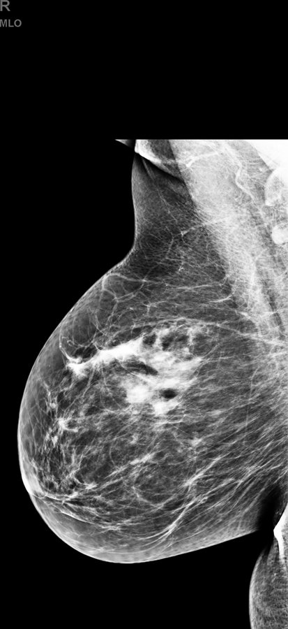
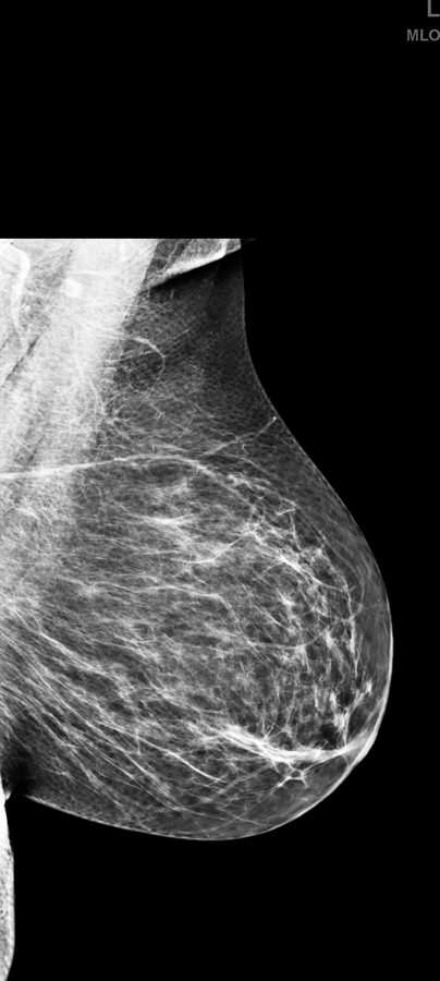
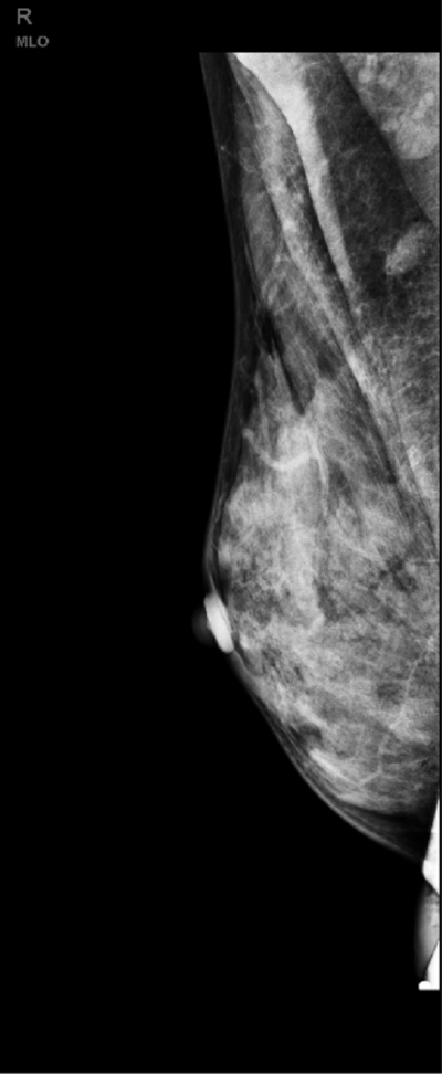
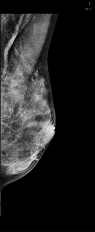
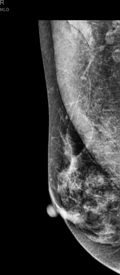
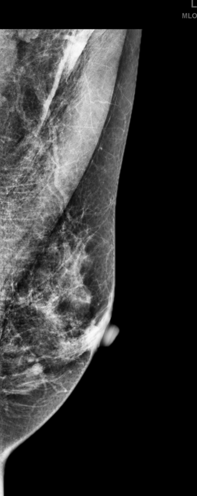
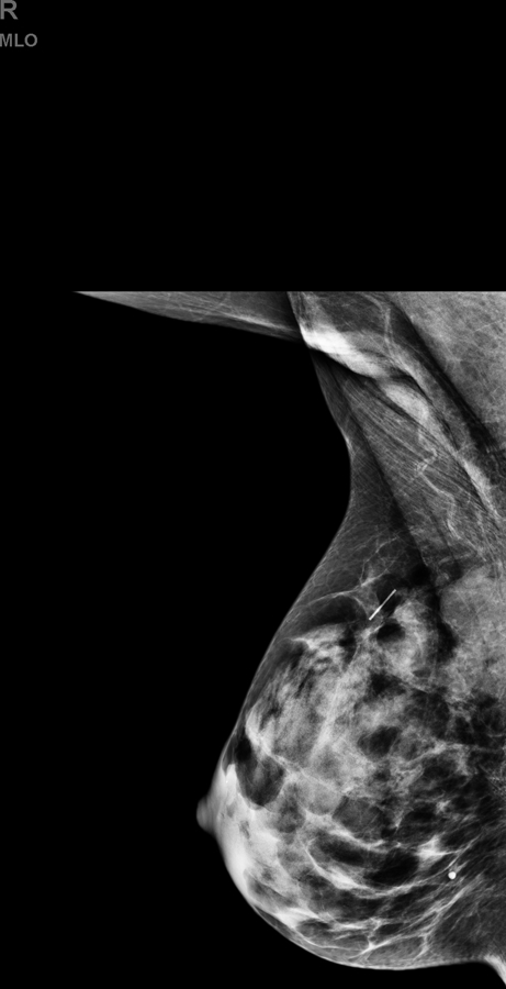
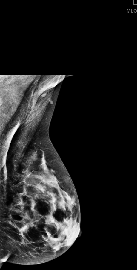
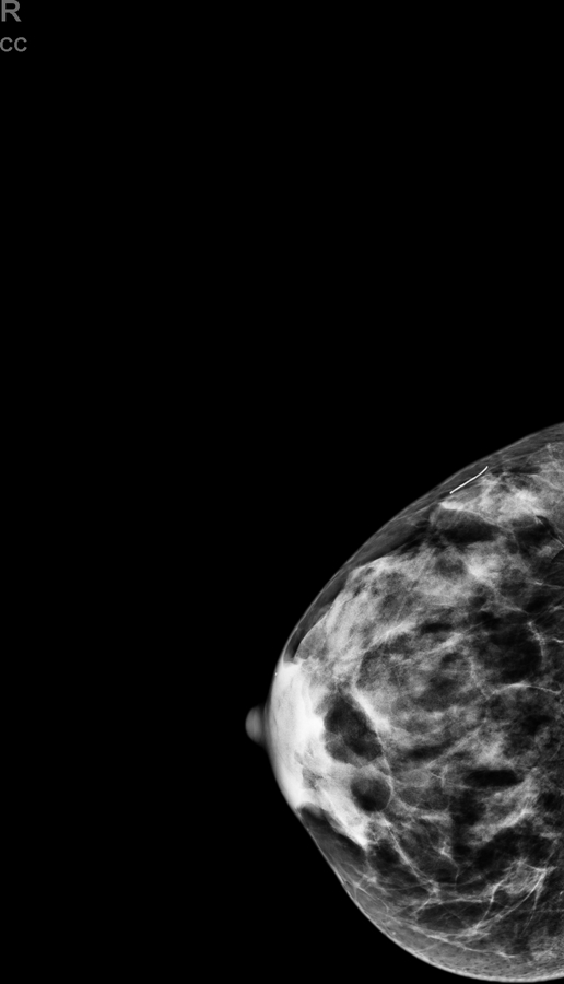
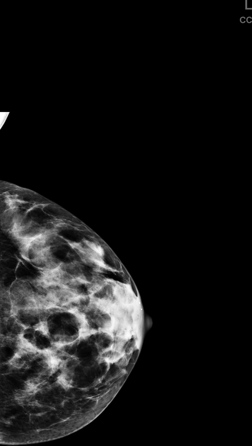
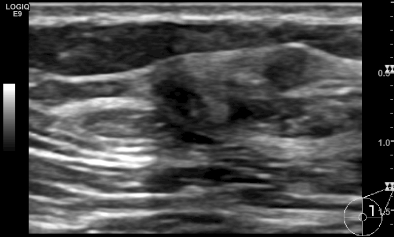
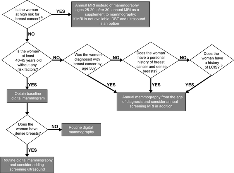
Digital breast tomosynthesis (DBT) has been widely implemented in place of 2D mammography, although it is less effective in women with extremely dense breasts. Breast ultrasound detects additional early-stage, invasive breast cancers when combined with mammography; however, its relevant limitations, including the shortage of trained operators, operator dependence and small field of view, have limited its widespread implementation. Automated breast sonography (ABS) is a promising technique but the time to interpret and false-positive rates need to be improved. Supplemental screening with contrast-enhanced magnetic resonance imaging (MRI) in high-risk women reduces late-stage disease; abbreviated MRI protocols may reduce cost and increase accessibility to women of average risk with dense breasts. Contrast-enhanced digital mammography (CEDM) and molecular breast imaging improve cancer detection but require further validation for screening and direct biopsy guidance should be implemented for any screening modality. This article reviews the status of screening women with dense breasts. KEY POINTS: • The sensitivity of mammography is reduced in women with dense breasts. Supplemental screening with US detects early-stage, invasive breast cancers. • Tomosynthesis reduces recall rate and increases cancer detection rate but is less effective in women with extremely dense breasts. • Screening MRI improves early diagnosis of breast cancer more than ultrasound and is currently recommended for women at high risk. Risk assessment is needed, to include breast density, to ascertain who should start early annual MRI screening.

Keywords: Breast cancer; Breast density; Magnetic resonance imaging; Screening ultrasound; Tomosynthesis.


Conflict of interest statement

Conflict of interest: The authors declare no conflict of interest.

Figures

Fig. 1.
Fig. 1.
Examples of each category of breast composition with (a-b) fatty, (c-d) scattered fibroglandular, (e-f) heterogeneously dense, (g-h) extremely dense breasts; one case where the breast composition appears dense in just one quadrant (i-j) is also shown.
Fig. 1.
Fig. 1.
Examples of each category of breast composition with (a-b) fatty, (c-d) scattered fibroglandular, (e-f) heterogeneously dense, (g-h) extremely dense breasts; one case where the breast composition appears dense in just one quadrant (i-j) is also shown.
Fig. 1.
Fig. 1.
Examples of each category of breast composition with (a-b) fatty, (c-d) scattered fibroglandular, (e-f) heterogeneously dense, (g-h) extremely dense breasts; one case where the breast composition appears dense in just one quadrant (i-j) is also shown.
Fig. 1.
Fig. 1.
Examples of each category of breast composition with (a-b) fatty, (c-d) scattered fibroglandular, (e-f) heterogeneously dense, (g-h) extremely dense breasts; one case where the breast composition appears dense in just one quadrant (i-j) is also shown.
Fig. 1.
Fig. 1.
Examples of each category of breast composition with (a-b) fatty, (c-d) scattered fibroglandular, (e-f) heterogeneously dense, (g-h) extremely dense breasts; one case where the breast composition appears dense in just one quadrant (i-j) is also shown.
Fig. 1.
Fig. 1.
Examples of each category of breast composition with (a-b) fatty, (c-d) scattered fibroglandular, (e-f) heterogeneously dense, (g-h) extremely dense breasts; one case where the breast composition appears dense in just one quadrant (i-j) is also shown.
Fig. 1.
Fig. 1.
Examples of each category of breast composition with (a-b) fatty, (c-d) scattered fibroglandular, (e-f) heterogeneously dense, (g-h) extremely dense breasts; one case where the breast composition appears dense in just one quadrant (i-j) is also shown.
Fig. 1.
Fig. 1.
Examples of each category of breast composition with (a-b) fatty, (c-d) scattered fibroglandular, (e-f) heterogeneously dense, (g-h) extremely dense breasts; one case where the breast composition appears dense in just one quadrant (i-j) is also shown.
Fig. 1.
Fig. 1.
Examples of each category of breast composition with (a-b) fatty, (c-d) scattered fibroglandular, (e-f) heterogeneously dense, (g-h) extremely dense breasts; one case where the breast composition appears dense in just one quadrant (i-j) is also shown.
Fig. 1.
Fig. 1.
Examples of each category of breast composition with (a-b) fatty, (c-d) scattered fibroglandular, (e-f) heterogeneously dense, (g-h) extremely dense breasts; one case where the breast composition appears dense in just one quadrant (i-j) is also shown.
Fig. 2.
Fig. 2.
(a) Right and (b) left mediolateral oblique shows a normal screening mammography in a 47-old woman; menopause occurred at 48 years and four years later the, (c) right and (d) left mediolateral oblique screening mammography shows a decrease in breast density due to normal perimenopausal involutionary changes.
Fig. 2.
Fig. 2.
(a) Right and (b) left mediolateral oblique shows a normal screening mammography in a 47-old woman; menopause occurred at 48 years and four years later the, (c) right and (d) left mediolateral oblique screening mammography shows a decrease in breast density due to normal perimenopausal involutionary changes.
Fig. 3.
Fig. 3.
(a,b) Mediolateral oblique and (c,d) craniocaudal mammography in a 52-year-old woman shows dense breast composition type C. (e) Supplemental screening ultrasound shows an irregular hypoechoic 9 mm mass (yellow arrows) in the 2 o’ clock position of the left breast, adjacent to a cyst (green arrow). Biopsy of the irregular mass revealed an invasive, node-negative, intermediate grade, lobular carcinoma (ER positive, PR negative, HER2 negative, Ki-67<1%).
Fig. 3.
Fig. 3.
(a,b) Mediolateral oblique and (c,d) craniocaudal mammography in a 52-year-old woman shows dense breast composition type C. (e) Supplemental screening ultrasound shows an irregular hypoechoic 9 mm mass (yellow arrows) in the 2 o’ clock position of the left breast, adjacent to a cyst (green arrow). Biopsy of the irregular mass revealed an invasive, node-negative, intermediate grade, lobular carcinoma (ER positive, PR negative, HER2 negative, Ki-67<1%).
Fig. 4.
Fig. 4.
Flow chart illustrating a screening decision support tool according to risk stratification. a High risk is defined as: women with a known or suspected pathogenic mutation in BRCA, TP53, CHEK2, PTEN, ATM, CDH1, STK11, and PALB2; women having a lifetime risk greater than 20% according to acceptable models that determine risk of pathogenic mutations, with Tyrer-Cuzick model the most accurate at the population level (and which includes breast density as a risk factor); women treated with chest or mantle radiation therapy by age 30 and at least 8 years prior. b A personal history of lobular carcinoma in situ confers almost as high a risk as personal history of breast cancer and such women should consider supplemental screening with MRI, especially if the breasts are dense. Atypical lobular hyperplasia (ALH) and atypical ductal hyperplasia (ADH) confer 20–25% lifetime risk as well but there are no studies showing improved cancer detection in women with ALH or ADH who undergo MRI screening in addition to mammography.

References

    1. Oeffinger KC, Fontham ET, Etzioni R et al. (2015) Breast Cancer Screening for Women at Average Risk: 2015 Guideline Update From the American Cancer Society. JAMA 314:1599–1614. - PMC - PubMed
    1. Tabar L, Vitak B, Chen TH et al. (2011) Swedish two-county trial: impact of mammographic screening on breast cancer mortality during 3 decades. Radiology 260:658–663. - PubMed
    1. Coldman A, Phillips N, Wilson C et al. (2014) Pan-Canadian study of mammography screening and mortality from breast cancer. J Natl Cancer Inst 106. - PubMed
    1. Broeders M, Moss S, Nystrom L et al. (2012) The impact of mammographic screening on breast cancer mortality in Europe: a review of observational studies. J Med Screen 19 Suppl 1:14–25. - PubMed
    1. Tabar L, Yen AM, Wu WY et al. (2015) Insights from the breast cancer screening trials: how screening affects the natural history of breast cancer and implications for evaluating service screening programs. Breast J 21:13–20. - PubMed

LinkOut - more resources